You must be signed in to read the rest of this article.
Registration on CDEWorld is free. Sign up today!
Forgot your password? Click Here!
The late Robert Dripps advanced the notion that a satisfactory outcome of anesthesia is largely determined by the quality of preanesthetic and postanesthetic care.1 If we accept this tenet, preoperative assessment of the dental patient must assume paramount importance. Meticulous documentation of the patient’s medical history and flawless baseline vital sign assessment are essential if the dentist is to reasonably estimate the patient’s fitness for the anesthetic and treatment planned.
A system for classifying preoperative physical status was developed by the American Society of Anesthesiologists (ASA) in 1941 and was revised to its current form in 1984 (Table 1).2 Despite its universal acceptance as a standard in preoperative assessment, this system has been shown to lack scientific precision2 and might better be appreciated as a guideline rather than a standard. In addition to the inherent inconsistency of any subjective rating, this particular system does not precisely define variables such as age, obesity, or the duration and nature of the surgery to be performed. For example, a 70-year-old ASA 3 patient undergoing a lengthy neurosurgical procedure would likely be a greater anesthetic risk than a 35-year-old ASA 3 patient about to have three teeth removed.
Despite these shortcomings, the ASA classifications provide a useful basis for decisions regarding the risks for sedation and anesthesia in the office. Class 1 and 2 patients are generally acceptable candidates for in-office sedation and anesthetic care, but Class 4 patients should generally be managed on an inpatient basis. Most class 3 patients who are well controlled by their medication can be safely managed in the office, but those who have questionable stability are better managed in a hospital, especially if a general anesthetic is required.
RECORDING THE MEDICAL HISTORY
Anesthetic History
The patient should be questioned carefully regarding past experiences with local and general anesthetics. Most patients vividly recall any unpleasant experiences, regardless of their true significance. For those who have had little or no experience with any form of anesthesia, questions regarding other family members may be helpful, because the patient may be genetically or psychosocially predisposed to an adverse anesthetic outcome. This is especially true for general anesthetics. Finally, information regarding past hospitalizations will enlighten the examiner regarding the patient’s status.
Current Medications
Information regarding a patient’s medications not only provides insight regarding his or her medical status, but may alert the dentist to possible drug interactions. Careful attention should be paid to any prescribed medications the patient is taking currently or has taken within the past month. In the case of corticosteroids, extended use (ie, greater than 2 weeks) within the previous month or two presents a risk for adrenal atrophy that may indicate a need for glucocorticoid prophylaxis. This is especially true if extensive treatment is planned, or a stormy postoperative course is anticipated. Finally, questioning should be directed to include any medications prescribed but not taken by the patient.
With only a few exceptions, there is little reason to discontinue any medication prescribed for cardiovascular disease. Diuretics can be withheld until after the appointment to minimize the need for micturition. Their long-term use may be associated with hypokalemia and risk for cardiac arrhythmias; any irregularity in the patient’s baseline pulse should be viewed with suspicion. It may be wise to order a serum potassium level if a general anesthetic is planned. Antihypertensive drugs can potentiate the hypotensive influences of sedatives and anesthetics, but there is even greater risk for acute rebound hypertensive episodes if long-term medications other than diuretics are withheld. With the possible exception of angiotensin-converting enzyme (ACE) inhibitors and angiotensin-receptor blockers (ARBs), it is better to continue these medications and give particular attention to intraoperative monitoring and cautious ambulation following postural change. A thorough review of considerations regarding cardiovascular medications was presented in previous continuing education articles in this journal.3,4
All chronically prescribed psychoactive agents should be continued. In most cases, their therapeutic influence requires a steady state serum concentration that has taken several weeks to establish. Furthermore, interrupting sedative/anxiolytics such as benzodiazepines may result in bothersome signs and symptoms of withdrawal. No major interactions with psychoactive drugs are known in anesthetic practice, other than monoamine oxidase inhibitors. The use of meperidine is contraindicated for patients taking this category of antidepressant because the interaction can precipitate seizure and a hypertensive crisis. Putative interactions regarding vasopressors and antidepressants have been overstated. Although indirect-acting sympathomimetics should be avoided, the judicious use of epinephrine or levonordefrin is not contraindicated for patients medicated with this or any of the remaining categories of antidepressants, including tricyclic antidepressants and selective serotonin reuptake inhibitors. A thorough review of considerations regarding psychotropic medications was presented in a previous continuing education article.5
Often we forget to question patients regarding their use of nonprescription drugs, recreational drugs, and homeopathic supplements. Cough, cold, and allergy medications may have a direct impact on vital sign assessment, and they introduce risk for cardiac arrhythmias and drug interactions. The same can be said for appetite suppressants, decongestants, and cocaine, because all are sympathomimetic amines. Recommendations have been provided for managing asymptomatic cocaine abusers,6 and they are reasonable suggestions if there is evidence of excessive use of any sympathomimetic agent. The dentist may wish to avoid outpatient general anesthesia, opting instead for sedation and local anesthesia and, along with monitoring for blood pressure and pulse, electrocardiographic (ECG) monitoring of such patients is encouraged.
Patients who smoke are prone toward hyperactive airways and bothersome episodes of coughing, in addition to well-established compromise of cardiovascular and respiratory function. Heavy smokers have a reduction in oxygen-binding sites on hemoglobin due the presence of carbon monoxide, which also impairs unloading of oxygen from the hemoglobin. This shifts the hemoglobin dissociation curve to the left and negates the accuracy of pulse oximetry to accurately reflect the patient’s actual partial pressure of oxygen in arterial blood (PaO2). Whereas a hemoglobin saturation of 95 by pulse oximetry normally reflects a PaO2 of ~80 mm Hg, oxygen tension will be lower in a patient with elevated carbon monoxide levels. It is wise to oxygenate heavy smokers and to maintain pulse oximeter readings >95. It may also be useful to administer a bronchodilator such as albuterol preoperatively, especially when a general anesthetic is planned.7
The long-term use of opioids or other CNS depressants, either prescribed or abused, should alert the clinician to possible dependence. In such cases, it is advisable to maintain the patient’s current level of use. For those suspected of opioid dependence, it is wise to avoid the use of nalbuphine, pentazocine, or butorphanol, because these particular opioids have mixed agonist–antagonist action that may precipitate a withdrawal syndrome.
History of Allergies
Even when a patient reports an allergic reaction, this does not necessarily preclude use of the particular drug or drug class in question. It is not uncommon for patients to label any adverse drug experience as an ‘‘allergic reaction.’’ This is true not only for reactions to local anesthetics, which more often are attributable to syncope or symptoms related to use of vasopressors, but also for reactions to antibiotics, opioids, and nonsteroidal anti-inflammatory drugs (NSAIDs), which frequently produce nausea and dyspepsia. Any report of allergy should be further questioned to clarify that signs and symptoms were consistent with hypersensitivity reactions (eg, rash, pruritus, urticaria, severe hypotension, airway compromise). Immediate allergic reactions are immunoglobulin (Ig)E mediated and can be confirmed only by testing serum for the presence of reactive antibody. However, other so-called ‘‘pseudoallergic’’ mechanisms can produce identical signs and symptoms of equal consequence. For this reason, any reaction that resembles allergy should be presumed to be at least pseudoallergic in mechanism, pending confirmation by an allergist.
No convincing data have confirmed IgE-mediated reactions to local anesthetics. Nevertheless, patients have occasionally experienced reactions to local anesthetics that are, at the very least, pseudoallergic in nature.8 In the event that an anesthetic is required before medical consultation can be obtained, the wisest choice would be mepivacaine or prilocaine without vasopressors. Conventional wisdom holds that if local anesthetics do indeed produce allergies, esters of benzoic acids would be capable of cross-reacting, but this would not be likely among amide local anesthetics.9,10 Furthermore, by avoiding those solutions containing vasopressors, one avoids bisulfites that are included as antioxidants. Sensitivity to bisulfites is well established, especially among atopic patients.11 An algorithm for managing patients with putative allergy to local anesthetics is provided in Figure 1.
The most common adverse reaction to codeine and its derivatives is nausea. However, IgE antibodies have been detected that react with several opioids, including codeine,12 and nearly all opioids are capable of triggering degranulation of mast cells leading to the direct release of histamine.13,14 Until issues regarding cross-reactivity among opioids are resolved, a prudent approach would be to select alternatives that are molecularly dissimilar. For example, when clinical signs reported for a codeine reaction are allergic in nature, one should select an agent that is dissimilar in structure (eg, meperidine, fentanyl, pentazocine).
Reputed allergic reactions to aspirin or other NSAIDS most likely are related to pseudoallergic mechanisms. Although their molecular structures may be strikingly dissimilar, all NSAIDs inhibit cyclooxygenases from converting arachidonic acid to prostaglandins. This shifts the arachidonic acid pathway toward another group of enzymes, lipoxygenases, which are not inhibited by the NSAIDs. These enzymes convert arachidonic acid into leukotrienes, and even subtle increases in these autacoids may lead to adverse reactions in atopic patients.15,16 It is wise to substitute acetaminophen for patients who claim allergy to any of the NSAIDs.
Penicillins have been confirmed as producing both IgE- and non–IgE-mediated reactions.17 Patients rarely have serologic confirmation that their previous reaction was IgE mediated, leaving the clinician little recourse but to avoid all penicillins when allergy is suspected. Erythromycin and clindamycin are the most conventional alternatives, but if necessary, cephalosporins can be prescribed, provided the reaction to penicillin was only pruritic or maculopapular in nature. A history of urticaria (hives) or anaphylactoid symptoms is more convincing evidence that the patient’s reaction to penicillin was truly IgE mediated, and in this case, one should refrain from prescribing any beta lactam derivative, including cephalosporins.18,19
Recent emphasis on infection control has contributed to an increased number of reports regarding latex allergy. Latex is a milky-white sap obtained from rubber trees (Hevea brasiliensis) and is used in more than 40,000 medical products.20 IgE-mediated reactions to latex have been confirmed, and a surprising number of allergic incidents have been reported, including anaphylaxis and death.21-23 All standard medical history forms should be revised to include an inquiry regarding adverse reactions to rubber products.
PHYSICAL ASSESSMENT
Along with a complete medical history, baseline information regarding vital signs is an essential component of the medical record. It not only aids in assessing the patient’s medical status, but also provides essential reference data during intraoperative monitoring. Ideally, the information is gathered during an interview appointment, when the patient is less likely to be apprehensive concerning eminent treatment. Along with the patient’s age, weight, and height, essential vital signs should include blood pressure, pulse rate, and hemoglobin saturation by pulse oximetry.
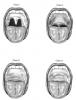
The airway should be examined and the Mallampati classification noted. Airway management is the most important aspect of patient care during sedation or general anesthesia, and examination of the patient’s airway is an essential component of the preoperative assessment. Documentation of a Mallampati airway classification has become a standard of care when any level of sedation is provided (Figure 2). Patients who have Class III and IV airways are more difficult to intubate, more likely to obstruct, and more difficult to artificially ventilate, should this become necessary. Unless one has training and significant experience in advanced airway management, deeper levels of sedation should be avoided in these patients. Patients with additional factors that warn of difficulty in attaining a good mask seal for positive-pressure ventilation include those who are fully bearded or edentulous, or who have a short thyromental distance, and those with a large neck circumference.
Patients should be questioned regarding their physical stamina, including exertional or postural dyspnea and any history of light-headedness or syncopal events. The dentist should note any visual impressions of cardiovascular, respiratory, or neurologic compromise. These might include evidence of distended jugular veins, edema of extremities, or elevated nail beds. For elderly patients, a notation regarding any evidence of dementia will be useful in evaluating any concerns regarding residual influences of sedative or anesthetic agents during the subsequent day or two. A system for assessing functional capacity was introduced by Hlatky et al24 and was modified as a useful adjunct when patients with questionable ASA status are assessed25 (Table 2).
ASSESSMENT OF PREEXISTING MEDICAL CONDITIONS
The proper assessment of preexisting medical conditions requires a basic understanding of the pathophysiologic features of the condition. This will enable the provider to comprehend the patient’s status and to properly design a plan for appropriate management while providing dental care. Cardiovascular diseases represent the most widespread preexisting medical conditions among patients presenting for dental treatment. By convention, the most common disorders will be addressed separately, but patients frequently suffer from combinations of these conditions.
Ischemic Heart Disease
Ischemic heart disease is a condition whereby the myocardium is inadequately supplied with oxygenated blood. The pathogenesis reflects an imbalance between coronary artery supply and myocardial oxygen demand. Atherosclerosis is generally the primary coronary lesion and compromises perfusion through vessel stenosis. However, as this condition progresses, plaque rupture, thrombosis, and/or vasospasm may all be superimposed. If a vessel becomes totally occluded, the normally perfused myocardial cells undergo necrosis (ie, myocardial infarction). Myocardial oxygen demand is determined primarily by heart rate and systolic wall tension (afterload).
Chest pain (angina) is generally the first symptom of coronary artery disease. However, a patient may tolerate varying degrees of coronary stenosis without angina pain provided heart rate and blood pressure do not impose excessive demand. In fact, some patients remain completely asymptomatic, and their disease is discovered during treadmill or other screening examinations. In this case, the condition is labeled silent ischemia. For example, severe diabetes can result in peripheral sensory neuropathy, and these patients may not experience anginal pain during periods of myocardial ischemia or infarction.
The nature and frequency of anginal episodes are indicative of the coronary lesions. When chest pain occurs only during exercise or emotional stress, the condition is called stable or classic angina. Because the pain is triggered by an increase in cardiac strain or myocardial oxygen demand, it is presumed that the coronary lesion is relatively stable and reflects uncomplicated atherosclerosis. It is believed that pain occurs because the sudden increase in demand outstrips the ability of the stenosed vessel to provide adequate oxygenated blood to the heart muscle. In rare cases, chest pain occurs only at rest. In this case, brief periods of coronary spasm are suspected and may be due to heightened excitability of coronary smooth muscle cells. Presumably, the underlying atherosclerosis contributes to this irritability, and the condition is described as Prinzmetal’s or vasospastic angina. When chest pain becomes more frequent and occurs during exercise or rest, it is assumed that atherosclerotic lesions are becoming unstable and are exhibiting plaque rupture and thrombotic events. This is called unstable or preinfarction angina.
Myocardial infarction is the death of cardiac muscle cells attributed to complete occlusion of a primary coronary artery or one of its branches. This occlusion is the result of thrombosis following rupture of an atherosclerotic plaque. Although atherosclerosis produces stenosis of coronary arteries, the actual degree of stenosis does not always predict imminent myocardial infarction. Indeed, some patients who have 80% to 90% stenosis merely present episodes of stable angina and never experience myocardial infarction. This occurs because total occlusion requires thrombosis. The vulnerability of atherosclerotic plaques to rupture is not predicated on the degree of stenosis, but on the portion that comprises cholesterol-rich lipids. At times, this fragile appearance is noticeable on angiographic studies, but otherwise it is presumed on the basis of an unstable pattern of anginal episodes. These episodes are believed to follow embolization of plaque fragments or small thrombi that lodge in small, distal branches and dissolve. They do not occlude coronary perfusion long enough to cause necrosis and, in this regard, resemble the process observed in transient ischemic attacks (TIAs) of the cerebral circulation.
Nevertheless, the risk for myocardial infarction certainly increases as atherosclerosis evolves to produce greater degrees of stenosis. The sequelae that follow infarction depend on the size and location of the necrotic zone. Occlusion of a terminal branch to a small area in the right ventricular wall may result in chest pain only and may heal uneventfully. Necrosis of an isolated papillary muscle may lead to prolapse of the related valve. However, if a large vessel supplying the left ventricle is occluded, necrosis may be so extensive that the ventricle fails to eject adequately or ruptures. In either of these cases, the patient may experience a cardiac arrest, which is the absence of a palpable pulse. Most often, however, cardiac arrest is attributed to an arrhythmia such as ventricular fibrillation that is precipitated by the anoxic insult to neural conduction pathways within the heart.
The nature and frequency of anginal episodes, prescribed medications, and the effectiveness of these medications are key items of information when one is assessing severity and stability of the patient’s ischemic disease. Medications generally include antiplatelet agents, beta blockers, and vasodilators. A system for classification of angina, which was introduced by the Canadian Cardiovascular Society, is useful for patient assessment26 (Table 3). Class 1 or 2 angina reflects a stable pattern and present little cardiovascular risk during dental procedures, provided standard stress reduction protocols are followed. Class 3 or 4 angina reflects severe or unstable patterns, respectively, and presents significant risk for an acute episode of angina or plaque rupture leading to myocardial infarction. For these patients, consultation with the patient’s physician should precede dental treatment. It has been accepted as standard care to postpone elective dental procedures for any patient who has suffered myocardial infarction during the previous 6 months. It has also been reasonable to expand this convention to include those who have undergone any invasive cardiac procedure (eg, angioplasty, stent insertion, coronary artery bypass grafting [CABG]), even if infarction did not occur. However, a recent joint report of the American College of Cardiology and the American Heart Association suggests that after 30 days, in the absence of unstable angina, noncardiac surgery may proceed without further evaluation or testing in the following cases: (1) recent myocardial infarction, (2) balloon angioplasty, and (3) bare-metal stent placement. Continued risk for rethrombosis following drug-eluting stent placement is believed to persist for at least 1 year, and consultation with the cardiologist is wise. Consultation is also encouraged when there is any evidence of decompensated congestive heart failure or significant cardiac dysrhythmias such as advanced heart block or symptomatic tachycardic or bradycardic dysrhythmias.27 If a patient can manage to do four METS of physical activity, routine dental care and even more invasive procedures are likely to be tolerated. (See Table 2 for examples.)
Obviously, the dentist cannot influence the severity of coronary lesions, but it is generally agreed that stress reduction and proper use of sedation can have very beneficial effects by reducing myocardial oxygen demand. Conversely, inadequate anesthesia or the indiscriminate use of vasopressors in local anesthetics will increase heart rate, blood pressure, and subsequent myocardial oxygen demand.28
Heart Failure
Heart failure is the inability of the heart to deliver a supply of oxygenated blood sufficient to meet the metabolic needs of peripheral tissues. Frequently, it is due to a defect in myocardial contractility that can be attributed to a variety of causes, including ischemia from coronary artery disease, primary cardiomyopathies, excessive programmed cell death (apoptosis), and excessive hemodynamic burden imposed by chronic hypertension. In some cases of heart failure, contractility is not impaired and ejection fractions are normal. The defect is related to diminished compliance of the ventricular wall, which prevents it from stretching to accept an adequate filling volume during diastole. This form of heart failure is described as ‘‘diastolic failure’’ to distinguish it from ‘‘systolic failure,’’ attributed to a diminished contractile strength. (More recently, these terms have been revised to heart failure with or without normal ejection fractions.)
Regardless of the fundamental defect, diminished contractility, and/or compliance, heart failure results in two groups of consequences that can be categorized as forward and backward failure. Forward failure is due to diminished cardiac output leading to inadequate perfusion of vital organs. Early clinical manifestations of forward failure include weakness, fatigue, reduced exercise tolerance, and other symptoms of hypoperfusion. Eventually, kidneys begin to fail, vision becomes impaired, and other consequences of end-organ damage begin to appear.
Backward failure relates to consequences of venous backup and congestion due to the inability of the heart to accommodate adequate filling volumes. Clinicians find it useful to describe backward consequences of heart failure as left-sided failure, right-sided failure, or biventricular failure. Elevation of the left atrial pressure results in symptoms and signs of pulmonary congestion, including dyspnea and orthopnea. Patients with orthopnea must elevate their heads on several pillows at night and frequently awaken short of breath or coughing (the so-called nocturnal cough) if their heads slip off the pillows. The patient’s sense of breathlessness is often relieved if he or she sits upright because this position reduces venous return. In advanced cases, orthopnea may become so severe that patients cannot lie down at all and must spend the entire night in a sitting position. Consequences of right-sided failure include symptoms and signs of systemic venous congestion. Pitting edema of the ankles, congestive hepatomegaly, and ascites are prominent. Late in the course of their disease, patients generally present signs and symptoms of biventricular failure, and attempts at characterizing them as right or left sided become artificial.
Drugs used to manage these patients include digoxin, which provides a positive inotropic influence; diuretics, which counter edema; and vasodilators which reduce preload and afterload on the compromised myocardium. Currently, angiotensin-converting enzyme (ACE) inhibitors (eg, captopril [Capoten], lisinopril [Zestril], enalapril [Vasotec]) are regarded as vasodilators of choice. The routine use of digoxin and diuretics has lost favor.29 In recent years, beta blockers have been found useful for managing heart failure. This is a dramatic shift in paradigm, because blocking beta receptors would appear detrimental in that they decrease myocardial contractility. However, the pathogenesis of heart failure has been found to include excessive sympathetic stimulation of the heart leading to hypertrophy, weakening, and loss of compliance. Although initiating beta blockade in these patients is initially met with a mild decline in ejection fraction, over time cardiac function actually improves. In addition, some of the newer beta blockers have mild alpha-blocking action, which produces vasodilation and subsequent reduction of afterload on the failing heart.
The patient with heart failure should be assessed for signs of peripheral edema and symptoms of pulmonary congestion, especially when reclined to a supine position. Careful attention should be given to monitoring blood pressure because sudden elevations can lead to venous backup and pulmonary congestion.
Hypertension
Hypertension is the most common cardiovascular disorder in developed countries. A myriad of conditions confound an understanding of this disorder, and attempts at classification based on etiology are artificial (eg, primary [essential] vs secondary hypertension). Although exceptions have been reported, most untreated adults with hypertension will develop additional increases in arterial pressure over time. Furthermore, it has been demonstrated that untreated hypertension is associated with shortening of life by 10 to 20 years. Morbidity and mortality associated with hypertension are attributed to its deleterious influences on vital organ systems. These influences may be secondary to acceleration in atherosclerosis and subsequent ischemia, or to direct damage caused by sustained pressure on organs. Coronary artery disease, heart failure, renal failure, retinopathy, and stroke are the typical end-organ consequences of chronic hypertension. Of these, cardiovascular consequences are the leading cause of death.
Medical management of hypertensive patients includes the use of diuretics, beta blockers, and vasodilators. The most recent guidelines for diagnosis of hypertension are summarized in Table 4.29,30 Formerly, the severity of hypertension was based on diastolic pressure, but current guidelines use whichever reading is highest—systolic or diastolic. The use of ‘‘stages’’ has replaced dated adjectives such as mild, moderate, and severe. There are no absolute guidelines regarding acceptable limits for elective dental treatment. However, Fleisher31 assessed the association of preoperative stages of hypertension with adverse outcomes during elective noncardiac surgery and found no increase in risk with preoperative blood pressure readings up to 180 systolic blood pressure (SBP) or 110 diastolic blood pressure (DBP). An increase in adverse events was associated with values of 181 to 210 SBP and/or 111 to 120 DBP if patients were also afflicted with coronary artery disease, heart failure, or significant diabetes. SBP >210 and DBP >120 were associated with increased risk, regardless of comorbidities. Based on experience and the results of this study, it would appear reasonable to avoid elective procedures in patients having SBP readings >180 or DBP readings >110. This suggestion does not obviate the requirement for additional judgment, however. Whereas a general anesthetic may aggravate any degree of hypertension, especially during induction and emergence, sedation may actually prove beneficial. Keeping in mind that baseline readings in the dental office are most likely elevated due to stress, it would be reasonable to administer an anxiolytic dose of a sedative and reassess blood pressure before postponing treatment.
Valvular Heart Disease
Valvular heart disease can produce turbulence in blood flow through the heart that is audible on auscultation. This sound is described as a ‘‘murmur.’’ At times, turbulence can be heard under normal conditions, and the murmur is described as ‘‘functional’’ or ‘‘benign,’’ to distinguish it from those attributed to structural or ‘‘organic’’ damage. It is important to understand that a murmur is merely a sign, not a disease.
Three primary conditions affect the heart valves: regurgitation, stenosis, and prolapse. A regurgitant valve allows reflux into the proximal chamber when it closes; stenosis or narrowing of a valve opening impedes flow into the distal chamber. In either case, a murmur can be heard during auscultation. Valve prolapse occurs when leaflets bulge into the proximal chamber upon closing. It does not alter blood flow that results in a murmur, but it may produce a ‘‘clicking’’ sound on closure. Regurgitation may occur alone, or it may accompany valves that are stenotic or prolapsed (eg, mitral prolapse with regurgitation).
There are many causes of valve disease. Lesions may follow injury to supporting structures such as chordae tendineae or papillary muscle, or to the valve leaflets themselves. Most often, valve disease is attributed to congenital defects, excessive strain from chronic hypertension, myocardial infarction, or inflammatory damage following rheumatic fever. Stenosis and regurgitation result in hemodynamic changes that lead to impaired circulation through the heart and strain on muscular chambers. The aortic and mitral valves are most commonly affected and will be examined more closely.
Mitral valve prolapse is a common condition in which the mitral valve leaflets are displaced into the left atrium during ventricular systole. It is more common in women than in men, and, in most cases, mitral valve prolapse represents a benign abnormality unless regurgitation develops.
During acute mitral regurgitation (such as can occur with endocarditis or chordal rupture following ischemia), left atrial and pulmonary venous pressures increase quickly, giving rise to dyspnea and pulmonary edema. In more chronic forms of mitral regurgitation, an increase in left atrial pressure is offset by a concomitant increase in atrial compliance, and symptoms appear late in the course of the disease. Left atrial enlargement predisposes the patient to atrial fibrillation and atrial thromboembolism. In long-standing mitral regurgitation, pulmonary hypertension can develop, which, in turn, leads to tricuspid regurgitation and right-sided heart failure.
Aortic stenosis causes concentric left ventricular hypertrophy as a compensatory mechanism that maintains cardiac output despite the increased pressure gradient across the valve. Eventually, this compensatory mechanism is overcome, causing the left ventricle to dilate and cardiac output to decline. Aortic regurgitation causes a volume overload of the left ventricle. In chronic aortic regurgitation, the volume overload is well tolerated for years. The left ventricle dilates to accommodate the increased volume load and thereby maintains a normal resting cardiac output. Compared with that observed with mitral regurgitation, enlargement of the left ventricle is far more severe. Eventually, compensatory mechanisms fail, and dyspnea follows elevated left atrial and pulmonary venous pressures.
Unless it is injured, endothelium is resistant to thrombus formation and to infection by most bacteria. However, endothelium of the heart can be injured by high-velocity jets produced by stenotic or regurgitant valves. These areas, as well as fibrotic valve leaflets, allow direct infection by virulent organisms or the development of an uninfected platelet-fibrin thrombus called nonbacterial thrombotic endocarditis (NBTE). This vegetation provides a site for bacterial attachment during transient bacteremia and may result in infective endocarditis.
Patients at greatest risk for infective endocarditis are those who have previously experienced endocarditis, which leaves vegetations after healing, and those who have had valve replacements, which may allow vegetations at suture sites. For these patients, antibiotics are administered in conjunction with selected procedures considered to entail a risk for bacteremia and subsequent endocarditis. The benefits of antibiotic prophylaxis have not been proven and in fact may be modest if one considers that most cases of endocarditis do not occur after a procedure. Dental treatments, the procedures most widely accepted as predisposing to endocarditis, are no more frequent during the 3 months preceding this diagnosis than in uninfected matched controls.32 Nevertheless, an expert committee of the American Heart Association has identified procedures that may precipitate bacteremia and suggests antibiotic prophylaxis for those patients at greatest risk for developing endocarditis.33
Valvular heart disease does not have a great impact on sedation and anesthesia, except in the most severe cases. Stenosis compromises filling of the distal chamber, and this may be further diminished with shortened filling time. For this reason, tachycardias should be avoided if patients have significant aortic stenosis, because blood pressure could drop precipitously. Conversely, regurgitant flow is accentuated by slow heart rates and high pressure within the distal chamber. These fundamental pathophysiologic principles spawn two pragmatic caveats: (1) Rapid heart rates should be avoided if patients have aortic or mitral stenosis, and (2) slow rates and hypertension should be avoided if patients have prolapsed or regurgitant valves.34 A cardiology consult should be obtained when there is any evidence of symptomatic aortic or mitral stenosis.27
Cardiac Arrhythmias
The minute output of the heart (cardiac output) is the paramount cardiovascular event required to sustain blood flow throughout the body. The heart must sustain a regular cycle of relaxation and contraction if it is to fulfill this objective. Any abnormality in rate or rhythm is labeled an arrhythmia. A complete analysis of the many supraventricular and ventricular arrhythmias is not within the scope of this article and is not always essential for adequate preoperative assessment. The primary consideration is whether the rhythm is stable enough to allow a cardiac output sufficient to sustain arterial pressure. A history of syncope or dizziness and light-headedness are signs that the rhythm is unstable and a medical consultation is indicated. For procedures using moderate to deep sedation or general anesthesia, monitoring should include continuous ECG tracing and blood pressure assessment every 5 to 10 minutes. Hypotensive episodes may follow both bradycardias and tachycardias. Slow rates may not produce enough cycles to sustain minute output, and tachycardias may reduce time for ventricular filling, which reduces stroke volumes.
For patients with concurrent coronary disease, tachyarrhythmias may precipitate episodes of angina even when blood pressure remains stable. This is due not only to an increase in myocardial oxygen demand, but also to a reduction in coronary perfusion. Coronary perfusion is greatest during diastole, when the myocardium is relaxed and is not compressing the intramyocardial vessels. Tachycardias shorten diastolic periods and compromise time required for adequate flow through the coronary vessels.
Although most arrhythmias are managed with medication, pacemakers are required in refractory cases. Cardiac pacemakers are implanted in more than a million people in the United States each year. They are used most often in patients suffering symptomatic supraventricular arrhythmias and heart blocks unresponsive to drug therapy. Pacemakers have two essential components: (1) a pulse generator powered by a lithium-iodine battery, and (2) pacing leads. These leads are stainless steel wires that most often are introduced via the subclavian or external jugular veins and guided to the cardiac chamber(s), where they are attached to an electrode anchored in the right atrium and/or ventricle. Asynchronous pacemakers generate impulses at a predetermined fixed rate. Synchronous or ‘‘on-demand’’ pacemakers are most common and are activated when the patient’s natural rate falls above or below a preset level. Pacemakers are classified using a three-letter code that indicates the chamber paced, the chamber sensed, and the response delivered. This is summarized in Table 5. (Two additional letters may be included in the code but are not germane for our purposes.) The most versatile and widely used pacemaker uses a DDD code.34,35 Examination of this table will reveal that this particular device paces and senses both atria and ventricles and may trigger or inhibit activity, depending on what is sensed.
Preanesthetic evaluation of patients with pacemakers should include the indication for pacing and a comment regarding its quality of performance. The type, date, and location of the pacemaker implanted should also be recorded. Any history of vertigo or syncope indicates dubious pacemaker function, and the patient should be referred to his or her physician. Monitoring during sedation or anesthesia should reflect the function and integrity of the pacemaker. Ideally, this is provided by continuous electrocardiography and continuous monitoring of pulse by plethysmography using a pulse oximeter.34,35 It is reasonable to consult with the cardiologist regarding any need to temporarily alter the pacing mode.
Modern pacemakers have been improved to resist electromagnetic interference, but the dental office environment may still present some hazard. Ultrasonic scalers, ultrasonic bath cleaners, and electrosurgery units are documented sources of electromagnetic interference.36 Influence from electrocautery units can be minimized by placing the grounding plate as far away from the implanted generator as possible and making sure that the generator is never located between the grounding plate and the electrode tip of the cautery unit. Also, keep the current as low as possible, and limit the pulse duration (1 s) and frequency (1 pulse/10 s).35
Pharmacologic considerations and indications for sedation and anesthesia should not be changed solely because of the presence of a pacemaker. Agents to counter bradyarrhythmias (eg, atropine, epinephrine) should be readily available for use, should pacemaker failure occur. In the unlikely event that defibrillation or cardioversion should become necessary, paddles should not be placed near the implanted generator.
Considerations regarding pacemakers are identical for patients who have implantable cardioverter-defibrillators. Consultation with the cardiologist is encouraged, and a decision can be made to program off of any tachyarrhythmia treatment algorithms during treatment to prevent unwanted shocks due to spurious signals.27,35
About the Author
Dr. Becker is an Associate Director of Education, General Dental Practice Residency, at Miami Valley Hospital in Dayton, Ohio.
REFERENCES
1. Dripps RD, Eckenhoff JE, Vandam LD. Introduction to Anesthesia: The Principles of Safe Practice. Philadelphia: WB Saunders;1988:13.
2. Owens WD, Felts JA, Spitznagel EL. ASA physical status classifications: a study of consistency of ratings. Anesthesiology. 1978;49:239-243.
3. Becker DE. Cardiovascular drugs: implications for dental practice. Part 1B Cardiotonics, diuretics and vasodilators. Anesth Prog. 2007; 54:178-186.
4. Becker DE. Cardiovascular drugs: implications for dental practice. Part 2B Antihyperlipidemics and antithrombotics. Anesth Prog. 2008; 55:49-56.
5. Becker DE. Psychotropic drugs: implications for dental practice. Anesth Prog. 2008; 55:89-99.
6. Pallasch TJ, McCarthy FM, Jastak JT. Cocaine and sudden cardiac death. J Oral Maxillofac Surg. 1989;47:1188-1191.
7. Hata TM, Moyers JR. Preoperative evaluation and management. In: Barash PG, Cullen BF, Stoelting RK, Eds. Clinical Anesthesia. 5th ed. Philadelphia: Lippincott Williams &Wilkins; 2006.
8. Jackson D, Chen AH, Bennett CR. Identifying true lidocaine allergy. J Am Dent Assoc. 1994;125:1362-1366.
9. Anderson JA, Adkinson NF Jr. Allergic reactions to drugs and biologic agents. JAMA. 1987;258:2891-2899.
10. Schatz M. Adverse reactions to local anesthetics. Immunol Allergy Clin North Am. 1992;12:585-609.
11. Schwartz HJ, Gilbert IA, Lenner KA, et al. Metabisulfite sensitivity and local dental anesthesia. Ann Allergy. 1989;62:83-86.
12. Harle DG, Baldo BA, Coroneos NJ, Fisher MM. Anaphylaxis following administration of papaveretum. Case report: implication of IgE antibodies that react with morphine and codeine, and identification of an allergenic determinant. Anesthesiology. 1989;71:489-494.
13. Flacke JW, Flacke WE, Bloor BC, et al. Histamine release by four narcotics: a double-blind study in humans. Anesth Analg. 1987;66:723-730.
14. Weiss ME, Adkinson NF, Hirshman CA. Evaluation of allergic drug reactions in the perioperative period. Anesthesiology. 1989;71:483-486.
15. Babu KS, Salvi SS. Aspirin and asthma. Chest. 2000;118:1470-1476.
16. Weiss ME. Drug allergy. Med Clin North Am. 1992;76:857-882.
17. Erffmeyer JE. Reactions to antibiotics. Immunol Allergy Clin North Am. 1992;12:633-648.
18. Salkind AR, Cuddy PG, Foxworth JW. The rational clinical examination. Is this patient allergic to penicillin? An evidence-based analysis of the likelihood of penicillin allergy. JAMA. 2001; 285:2498-2505.
19. Shepherd GM. Allergy to beta-lactam antibiotics. Immunol Allergy Clin North Am. 1991;11:611-633.
20. Warpinski JR, Folgert J, Cohen M, Bush RK. Allergic reaction to latex: a risk factor for unsuspected anaphylaxis. Allergy Proc. 1991;12:95-102.
21. Holzman RS. Latex allergy: an emerging operating room problem. Anesth Analg. 1993;76:635-641.
22. Kelly KJ, Kurup VP, Reijula KE, Fink JN. The diagnosis of natural rubber latex allergy. J Allergy Clin Immunol. 1994;93:813-816.
23. Misselbeck WJA, Gray KR, Uphold RE. Latex induced anaphylaxis: a case report. Am J Emerg Med. 1994;12:445-447.
24. Hlatky MA, Boineau RE, Higginbotham MB, et al. A brief self-administered questionnaire to determine functional capacity (The Duke Activity Status Index). Am J Cardiol. 1989;64:651-654.
25. Hollenberg SM. Preoperative cardiac risk assessment. Chest. 1999;115:51s-57s.
26. Campeau L. Grading of angina pectoris. Circulation. 1975;54:522.
27. Fleisher LA, Beckman JA, Brown KA, et al. ACC/AHA 2007 Guidelines on perioperative cardiovascular evaluation and care for noncardiac surgery. Circulation. 2007;116:1971-1996.
28. Troullos ES, Goldstein DS, Hargreaves KM, Dionne RA. Plasma epinephrine levels and cardiovascular response to high administered doses of epinephrine contained in local anesthesia. Anesth Prog. 1987;34:10-13.
29. Hunt SA, Abraham WT, Chin MH, et al. 2009 Focused update incorporated into the ACC/AHA 2005 guidelines for the diagnosis and management of heart failure in adults: a report of the American College of Cardiology Foundation/American Heart Association Task Force on Practice Guidelines. Circulation. 2009;19:e391-e479.
30. Chobanian AV, Bakris GL, Black HR, et al. Seventh Report of the Joint National Committee on Prevention, Detection, Evaluation, and Treatment of High Blood Pressure. Hypertension. 2003;42:1206-1252.
31. Fleisher LA. Preoperative evaluation of the patient with hypertension. JAMA. 2002;287:2043-2046.
32. Strom BL, Abrutyn E, Berlin JA, et al. Risk factors for infective endocarditis: oral hygiene and nondental exposures. Circulation. 2000;102:2842-2848.
33. Wilson W, Taubert KA, Gewitz M, et al. Prevention of infective endocarditis: guidelines from the American Heart Association: a guideline from the American Heart Association Rheumatic Fever, Endocarditis, and Kawasaki Disease Committee, Council on Cardiovascular Disease in the Young, and the Council on Clinical Cardiology, Council on Cardiovascular Surgery and Anesthesia, and the Quality of Care and Outcomes Research Interdisciplinary Working Group. Circulation. 2007;116:1736-1754.
34. Morgan GE, Mikhail MS, Murray MJ. Clinical Anesthesiology. 4th ed. New York: Lange Medical Books/McGraw Hill; 2006:463-483, 789-795.
35. Practice advisory for the perioperative management of patients with cardiac rhythm management devices: pacemakers and implantable cardioverter-defibrillators: a report by the American Society of Anesthesiologists Task Force on Perioperative Management of Patients with Cardiac Rhythm Management Devices. Anesthesiology. 2005;103:186-198.
36. Miller CS, Leonelli FM, Latham E. Selective interference with pacemaker activity by electrical dental devices. Oral Surg Oral Med Oral Pathol Oral Radiol Endod. 1998;85:33-36.