You must be signed in to read the rest of this article.
Registration on CDEWorld is free. Sign up today!
Forgot your password? Click Here!
While most implant-based treatment has historically focused on fixed prosthetic tooth replacement,1 the multitude of benefits to the edentulous population from implant overdentures is overwhelming in improved function, emotional stability, physical health, and esthetics. Proper evaluation and treatment planning of the fully edentulous patient has been shown to result in an improved quality of life for patients2,3 and predictable clinical success. The indication for a fixed prosthesis may be limited because of inadequate quantity and structure of the bone. Enhancement of esthetic appearance and facial morphology through replacement of lost hard and soft tissues may be proven easier, if not more effective, with removable overdentures than with conventional fixed prosthesis, with possibly decreased costs and less surgical intervention.
Generally, more implants are required to support a fixed prosthesis than an overdenture. Other factors to consider include the health of the patient, his or her ability to undergo grafting procedures, and cost.4 There has been an abundance of literature presenting a variety of treatment options, case reports, and clinical techniques over the past 20 years, but more recently there has been general agreement about the treatment protocols and long-term documented benefits of implant overdentures. This treatment option has become the most rewarding care provided in this author's clinical practice, and with increasing life expectancy, its full impact on clinical practice is yet to be realized. Although there still remains a lack of consistency of techniques, prosthetic design, and attachment systems, these aspects have been proven less important to successful outcomes than once thought. What are agreed upon are the specific indication for this treatment and the benefit derived. This article presents a simplified approach to patient evaluation, treatment planning decisions, attachment selection, and technique.
First, there is a need to appreciate the sequelae of tooth loss, and associated benefits of implant overdentures, followed by patient evaluation, treatment protocols, and clinical technique. For many years clinicians realized that placement of endosseous osseointegrated implants under a removable prosthesis would provide the definitive advantages of bone preservation,5 prosthetic retention, stability, and a degree of occlusal support resulting in improved function, facial esthetics, and comfort. More recently, comprehensive and ongoing studies conducted at McGill University have documented the improved nutrition, psychosocial status, and quality of life that have been gained through the use of overdenture treatment.6,7 The use of implants also provides a predictable solution for patients and practitioners to the problems associated with conventional dentures by resolving functional and esthetic compromises.
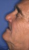
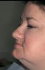
The sequelae of tooth loss and an edentulous arch is residual ridge resorption both in the horizontal and vertical direction. This ongoing loss of hard and soft tissue is most noticeable in the loss of orofacial support: facial esthetics, phonetics, and collapse of vertical dimension. This leads to an aging appearance due to the lack of lip support and decreased facial height (Figure 1 and Figure 2). Concurrent with these changes in facial structures are impaired oral function, pain, insufficient retention, and instability of conventional dentures, as well as nutritional and psychological changes. Many patients seeking resolution to chronic soreness of load-bearing tissues and nonstable or retentive dentures will enjoy increased esthetics, function, comfort, and psychological benefits from implant overdentures, without the need for more extensive fixed restorations.8,9
Patient Evaluation:
Maxillofacial Relationship
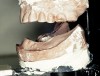
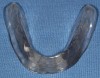
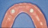
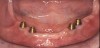
Upon evaluation of the edentulous (or soon to be edentulous) patient, facial esthetics and the amount of extraoral soft tissue support of the lips and associated structures will become an initial guideline to treatment options. If horizontal loss of hard and soft tissue through resorption, disease, or trauma is so advanced that teeth need to be placed far anterior to the residual ridge to provide adequate facial support, then an overdenture (ie, acrylic base and flanges) can provide replacement of these structures (Figure 3). Alternatively, bone grafting procedures can be performed to augment the missing tissues, but limitations must be evaluated. Limiting factors in grafting procedures include adequate blood supply, patient health, and finances. When evaluating vertical loss of hard and soft tissues, the resultant interarch space must be determined to see if excessive crown-to-implant ratio and biomechanical forces will preclude a conventional implant-supported fixed restoration (Figure 4). While grafting procedures have radically changed how clinicians treat patients, there are limitations to the amount of vertical augmentation possible.
Any successful overdenture treatment begins with the understanding that conventional full-denture fabrication principles must be followed. These include ideal border adaptation and extension and full-denture occlusion.10 Through proper articulation and denture tooth setup, all parameters of final treatment success can be evaluated. A try-in of the proposed tooth setup will allow evaluation of esthetics, phonetics, and support, as well as the critical determination of ridge position relative to the proposed prosthesis before surgery. The setup then will be used to guide ideal implant position because the most critical factor in overdenture implant placement is that implants emerge well within the confines of the denture.
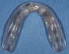
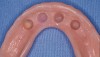
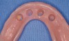
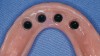
If an existing denture is available and determined to have adequate tooth position, this can become the surgical guide for implant placement. If a denture with adequate occlusal and esthetic parameters is not available, then a new ideal setup is necessary to avoid implant placement in a less-than-ideal or even nonusable position. Often the author duplicates the wax setup in clear acrylic before final processing to serve as a surgical guide (Figure 5A and Figure 5B). The risks of proceeding with implant placement before trying in the tooth setup are compromised space for overdenture attachments, inadequate acrylic thickness, and unfore-seen laboratory and component costs necessary to correct poor angulation.
Evaluation of Ridge
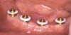
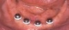
While the majority of patients will realize a wealth of benefits from the two-implant mandibular overdenture, close evaluation of the residual ridge will provide information about the ideal number and position of implants, as well as abutment and attachment selection. Individual, nonsplinted implants can provide ideal retention of the prosthesis to prevent vertical and lateral displacement. However, ideally the majority of occlusal support is provided by the residual ridge and not the implants. The primary goal of any overdenture attachment is to retain the appliance in position with a minimal amount of movement.
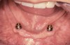
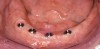
Traditional overdentures are classified as implant-retained and tissue-supported prostheses11 (Figure 6A and Figure 6B). If the patient's residual ridge is inadequate to provide the majority of vertical occlusal support in function, as in cases of extreme "knife-edge" or chronic mucosal soreness because of the nature of the tissues, then more implants or splinting of the implants may be indicated to provide more implant support and decreased loading of the tissues.12 The benefit of placing three to four implants (as opposed to only two) is the ability to ease the load on a less-than-ideal ridge, decreasing mucosal bearing areas during occlusal function. These additional implants also will provide for decreased anterior-posterior movement (rocking) of the appliance. Additional implants also may be more desirable when fixtures of reduced length or diameter are necessary because of limited bone volume.
One benefit of splinting implants (ie, bar restorations) is potential distribution of the forces to more osseointegrated surfaces, thereby sharing the load. Another primary reason for splinting is to enable the laboratory to compensate for significantly malaligned or poorly positioned implants by fabricating a custom substructure with common path of insertion.
Still, the majority of patients will greatly benefit from two to four nonsplinted mandibular implants to provide ideal retention and some level of occlusal support. Numerous studies have shown equal outcomes in long-term implant survival in mandibular overdentures regardless of splinting.13 Many factors prove overwhelmingly in favor of individual nonsplinted implants, such as decreased cost, decreased space requirement inside the denture, and improved access for hygiene. The individual nonsplinted approach provides the most ideal outcome with the greatest cost-effectiveness and greatest efficacy of treatment for the majority of edentulous patients.
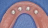
There are several attachments available to provide retention of the prosthesis to the implants. Numerous studies have shown that many designs work well. However, to provide a guide to attachment selection, a number of factors should be considered. All attachments are either rigid or resilient. Rigid attachments restrict rotational movement and provide only a limited path of off-angle insertion, while resilient attachments allow varying amounts of rotation and angulation correction. In situations where implants are even minimally nonparallel, a resilient attachment will consistently show less friction, wear, and breakage. Considering that patients frequently bite appliances into place, this resiliency also will prevent premature wear and breakage. Clearly the major overdenture complication and maintenance concern relates to attachment adjustment and replacement,14,15 as well as fracture from the prosthesis. These issues can be significantly minimized through proper attachment selection and use of resilient attachments.16 A resilient connection between the denture and implants should allow reduced loading of the abutments in so far as the degree of movement takes into account the compressibility (ie, resilience) of the mucosa. The greatest portion of the occlusal forces are thus absorbed directly by the alveolar ridges.17 There are attachment systems that allow angulation correction of up to 20° per implant (40° for two divergent implants) within a resilient range (LOCATOR¬Æ, Zest Anchors, Escondido, CA).
Other factors in attachment selection include height of the attachment to minimize space required inside the denture (to decrease potential fracture caused by inadequate acrylic thickness) and housings with replaceable matrices. The advantage of a housing for the attachment is that with any need to change the retentive component, it is not necessary to re-cure the attachment into the denture base.
Based on the above factors and clinical experience, the author's attachment selection is a resilient, nonsplinted, prefabricated attachment (LOCATOR) of minimal height with easily replaceable retentive components of varying forces that include a selection of cuff heights to emerge through the tissue of a subgingivally placed implant. The use of individual resilient attachments also allows for implant and tooth overdenture abutments to be used in combination, as in the case of a remaining healthy mandibular cuspid that is endodontically treated and placement of a "root" overdenture abutment.
As previously mentioned, the fabrication of a bar allows the laboratory to correct significant implant malalignment, which often is seen in the maxilla because of the resorptive pattern of the basal bone. This suprastructure allows attachments to be cast, soldered, or welded to the bar to provide a common path of insertion, as well as relocate the attachment system within the confines of the denture base because of often buccal-emerging implants. The other potential advantage of splinting implants through the use of bars or telescopic copings is distribution of forces to more implants, so that rotational torque of the implants is resisted under occlusal loading in cases where there is limited buccal bone volume and encroachment on the residual buccal plate.
Maxillary overdenture implants tend to be angled, shorter, and placed in less dense bone because of the nature of resorptive patterns and sinus expansion. More significant to the decreased stress transfer in maxillary implants is the amount of palatal coverage offered by the appliance to provide greater tissue support.15 Maintaining partial palatal coverage to assist in the potential reduction of load to the implants may be recommended in instances of reduced implant support because of the quality of integration, number of implants, or compromised implant positioning and location. This adheres to the primary premise of an "implant-retained and tissue-supported" appliance.
The down side of bars to be considered is the added space required inside the denture,18 hygiene difficulties, and additional cost. Passivity of fit of bar restorations also must be verified to avoid mechanical problems, such as screw loosening and fracture.
As a general rule, four implants are the minimal number in the maxilla to remove partial palatal coverage. While maxillary overdenture implants tend to show a slightly higher risk of failure than observed in the mandible, this clearly appears not to be related to the prosthetic design but to compromised preoperative bone, thereby necessitating a reduced number, length, diameter, and angulation of implants.19,20
Technique
Incorporation of the attachment into the denture can be accomplished either chairside or in the laboratory. The advantage of chairside "pick up" is that the attachment can be made in a passive, loaded (ie, bite force) environment to ensure complete seating of the denture on the underlying tissues. This technique is more technically demanding but enables the incorporation of attachments into an existing denture (Figure 7A through Figure 7I). Laboratory attachment incorporation is less technique sensitive but does not account for the level of muccocompression necessary to ensure full seating on the tissues. It is recommended with laboratory curing of the attachments that the connection of the attachment be made to the base plate before processing the denture at one of the wax rim or set-up try-in appointments.21 This will allow the clinician to evaluate full seating on the tissues and minimize distortion caused by curing of a bulk of acrylic during processing, as well as to evaluate and correct the attachment position before the delivery appointment. Less than ideal attachment position can be corrected at this time through such means as a reline impression in the trial base with the patient in full occlusion to assure intimate contact with the underlying tissues.
Techniques have been developed to simplify chairside incorporation of attachments.22 The most important concerns are blocking out any undercuts that acrylic may flow into (preventing removal of the denture) and ensuring that the prosthesis can fully seat on the tissues without being held up by interference with the attachments.
Design Criteria
The only rationale for incorporation of a metal framework or lingual reinforcing bar is to prevent potential fracture of the appliance caused by minimal acrylic thickness or excessive occlusal forces.21 The downside of this process is the additional cost and laboratory procedures involved. In situations of high potential fracture of the appliance, such as the extreme occlusal forces seen in patients with opposing full-arch implant-supported restorations or areas of minimal acrylic bulk, a metal frame will serve to resist flexure and potential fracture. An important consideration for the laboratory is to allow open space in the framework for incorporation of the attachments.
Surgical Considerations
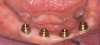
While two individual implants provide outstanding, well-documented clinical results, three or four implants will provide greater retention and level of implant support and minimize anterior-posterior rocking from the unsupported long extension of the denture base. Extreme distal implant placement will decrease anterior support by allowing an anterior lever arm in function (Figure 8). A more stable result will be obtained through implant placement with greater anterior-posterior spread that can decrease rocking, or through the placement of an additional implant in the anterior segment. While the cuspid position has traditionally been the site of choice for the two-implant overdenture, there is much merit in placing the implants closer to the lateral incisor position to minimize rock and allow for potential additional implant placement in the future. In a larger ridge, consideration of two cuspid and one additional incisor implant-for a total of three implants-is more favorable to prevent the anterior-posterior rock and provide a tripod of support. The implants can be placed in a one-stage or nonsubmerged approach because esthetic soft-tissue contours are not critical in overdenture therapy. This also will allow a more incisal position of the implant–abutment connection and hygiene access. The specific arch position in relation to tooth and embrasure location also is not critical with a removable prosthesis.
Conclusion
Through proper patient evaluation, adherence to conventional denture techniques, and ideal communication among surgical, laboratory, and restorative colleagues, implant overdentures provide simple, predictable, and cost-effective treatment to edentulous patients. Additionally, they provide the benefits of esthetics, phonetics, bone preservation, increased comfort, better psychosocial state, and enhanced nutrition, all resulting in an improved quality of life (Figure 9).
References
1. Branemark PI, Zarb GA, Albrektsson T. Tissue-Integrated Prostheses: Osseointegration in Clinical Dentistry. Chicago, IL: Quintessence; 1985.
2. Awad MA, Locker D, Korner-Bitensky N, et al. Measuring the effect of intra-oral implant rehabilitation on health-related quality of life in a randomized controlled clinical trial. J Dent Res. 2000;79(9):1659-1663.
3. Awad MA, Lund JP, Shapiro SH, et al. Oral health status and treatment satisfaction with mandibular implant overdentures and conventional dentures: a randomized clinical trial in a senior population. Int J Prosthodont. 2003;16(4):390-396.
4. Mericske-Stern R. Prosthodontic management of maxillary and mandibular overdentures. In: Feine JS and Carlsson GE, eds. Implant Overdentures: The Standard of Care for Edentulous Patients. Chicago, IL: Quintessence Pub. Co.; 2003:83-98.
5. Jemt T, Chai J, Harnett J, et al. A 5-year prospective multicenter follow-up report on overdentures supported by osseointegrated implants. Int J Oral Maxillofac Implants. 1996;11(3):291-298.
6. Feine JS, Carlsson GE. Implant Overdentures: The Standard of Care for Edentulous Patients. Chicago, IL: Quintessence Pub. Co.; 2003.
7. Morais JA, Heydecke G, Pawliuk J, et al. The effects of mandibular two-implant overdentures on nutrition in elderly edentulous individuals. J Dent Res. 2003;82(1):53-58.
8. Quirynen M, Alsaadi G, Pauwels M, et al. Microbiological and clinical outcomes and patient satisfaction for two treatment options in the edentulous lower jaw after 10 years of function. Clin Oral Implants Res. 2005;16(3):277-287.
9. Zitzmann NU, Marinello CP. Treatment outcomes of fixed or removable implant-supported prostheses in the edentulous maxilla. Part I: patients' assessments. J Prosthet Dent. 2000;83(4):424-433.
10. Kim Y, Oh TJ, Misch CE, et al. Occlusal considerations in implant therapy: clinical guidelines with biomechanical rationale. Clin Oral Implants Res. 2005(1);16:26-35.
11. Zitzmann NU, Marinello CP. A review of clinical and technical considerations for fixed and removable implant prostheses in the edentulous mandible. Int J Prosthodont. 2002(1);15:65-72.
12. Mericske-Stern R, Assal P, Buergin W. Simultaneous force measurements in 3 dimensions on oral endosseous implants in vitro and in vivo. A methodological study. Clin Oral Implants Res. 1996;7(4):378-386.
13. Gulizio MP, Agar JR, Kelly JR, et al. Effect of implant angulation upon retention of overdenture attachments. J Prosthodont. 2005;14(1):3-11.
14. Watson GK, Payne AG, Purton DG, et al. Mandibular overdentures: comparative evaluation of prosthodontic maintenance of three different implant systems during the first year of service. Int J Prosthodont. 2002;15(3):259-266.
15. Ochiai KT, Williams BH, Hojo S, et al. Photoelastic analysis of the effect of palatal support on various implant-supported overdenture designs. J Prosthet Dent. 2004;91(5):421-427.
16. Krennmair G, Weinlander M, Krainhofner M, et al. Implant-supported mandibular overdentures retained with ball or telescopic crown attachments: a 3-year prospective study. Int J Prosthodont. 2006;19(2):164-170.
17. Besimo C. Removable Partial Dentures on Osseintegrated Implants: Principles of Treatment Planning and Prosthetic Rehabilitation in Edentulous Mandible. Chicago, IL: Quintessence Pub. Co.;1998.
18. Phillips K, Wong KM. Space requirements for implant-retained bar-and-clip overdentures. Compend Contin Educ Dent. 2001;
22(6):516-522.
19. Bryant SR, MacDonald-Jankowski D, Kim K. Does the type of implant prosthesis affect outcomes for the completely edentulous arch? Int J Oral Maxillofacial Implants. 2007;22(Suppl):117-139.
20. Jemt T, Lekholm U. Implant treatment in the edentulous maxillae: a 5-year follow-up report on patients with different degrees of jaw resorption. Int J Oral Maxillofac Implants. 1995;10(3):303-311.
21. Shor A, Goto Y, Shor K. Mandibular two-implant-retained overdenture: prosthetic design and fabrication protocol. Compend Contin Educ Dent. 2007;28(2):80-88.
22. Vogel R. Clinical technique to simplify overdenture success. Implant Realities. 2006;1:19-20.
About the Author
Robert C. Vogel, DDS
Private Practice
Palm Beach Gardens, Florida