You must be signed in to read the rest of this article.
Registration on CDEWorld is free. Sign up today!
Forgot your password? Click Here!
Methods of Retraction
Depending on case requirements, there are four retraction techniques in general use today: cord; electrosurgery; soft-tissue laser; and paste. The retraction method used may be influenced by clinician familiarity with the technique; the location, quality, and condition of the soft tissue; the clinician's skill level; or the complexity of the case.
Cord Techniques
The cord-packing technique is the most popular method of retraction and is completed using twisted, knitted, woven, or braided cord. A variety of natural and synthetic fibers is used in making gingival retraction cords, including wool yarn, cotton, and silk. The cords are available commercially plain, impregnated, or pretreated with hemostatic medicaments.
Tissue management and achieving hemostasis are important factors in ensuring a high-quality impression and, in turn, a properly fitting restoration. Although it may be possible to accomplish both tasks with a single treatment modality, this is not always the case. This article discusses various retraction techniques and their respective advantages and disadvantages. The importance of achieving excellent hemostasis before making the final impression-and methods for doing so-also are addressed.
When using a cord-packing technique, an appropriately sized cord is gently placed into the gingival sulcus with the intent of mechanically displacing the soft tissues from the tooth and margin of the preparation. In general, it is best to use the smallest cord possible because larger cords sometimes can tear delicate gingival tissue, increase hemorrhage, and damage the sulcular epithelium. Fortunately, the newer impression materials can capture excellent marginal detail within relatively small gingival spaces.
Retraction cords are typically placed after tooth preparation is complete and then removed immediately before the impression tray is seated. Cords can be packed with many different types of hand instruments. The specific instrument that works best often depends on the type of cord used. For example, a serrated instrument usually will work best with firmer cords and the tightly knitted or braided varieties. A smooth-tipped packer tends to work better with loosely twisted or braided varieties because the packer's smooth tip is less likely to catch on the fibers and separate them.
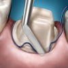
Cords always should be packed by angling the instrument tip toward the starting point of the cord (Figure 1 and Figure 2). Packing away from the starting point tends to dislodge or pull the cord out of the sulcus. With all cord techniques, it is extremely important for the clinician to carefully check that all cords, stray fibers, and impression material remnants are removed completely from the gingival sulcus before the patient is dismissed. Residual materials will be treated by the body as a foreign object and can lead to infection, inflammation, and other periodontal problems. As with all retraction methods, try to expose 0.5 mm to 1 mm of tooth structure, which leads to the creation of a restoration with the proper emergence profile.
Single-Cord
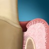
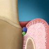
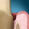
The single-cord technique is the most popular and commonly used. In this technique, the tooth is prepared completely (Figure 3) and then a single cord is packed into the gingival sulcus to achieve retraction (Figure 4). The packed cord is removed just before making the final impression (Figure 5).
The single cord can be wrapped once around the circumference of the preparation, or it can be wrapped multiple times. A single wrap is acceptable when the depth of the sulcus is nearly identical to the diameter of the cord. However, a single wrap with a single cord may not produce adequate retraction when the cord diameter is significantly smaller than the depth of the sulcus. This tends to produce a teardrop-shaped retraction that allows the tissue to collapse over the top of the cord or only a very thin margin of impression material that can easily tear on removal. Therefore, wrapping the cord multiple times may help to achieve proper retraction and compensate for an undersized cord. An excellent, though less popular, alternative to the single-cord technique is the double-cord technique.
Double-Cord
This technique is especially good for impressions where a deeper gingival sulcus is present (Figure 6). As its name implies, two cords are used. The first of the two cords is usually smaller, packed into the bottom of the sulcus, and generally used to help control fluids and hemorrhage. It can be packed before or after the preparation is completed. The first cord may be left in during the impression or removed immediately before it is made. If left in, it must be located below the preparation margin. The second of the two cords is larger and packed directly on top of the smaller first cord (Figure 7). This tends to produce a V-shaped retraction (Figure 8), allowing better access for the impression material and contributing to an impression margin with greater bulk.
Electrosurgery
Electrosurgical retraction—or electrosurgery—works by surgically removing a small portion of the epithelial lining of the gingival sulcus to create room for the impression material. Typically, a thin wire electrode is inserted into the sulcus, and an alternating electrical current above 100 kHz is passed through the tip, simultaneously removing the desired layer of tissue and cauterizing the surgical site.When properly used, electrosurgery provides excellent retraction, as well as hemostasis, and the epithelial tissues heal rapidly. Electrosurgery also can be a valuable adjunctive treatment modality when retraction cords alone are not effective.
However, electrosurgery is somewhat technique-sensitive. Some of the significant risks of improper use include dentinal and cementum burns and damage to the periodontal attachment. This ultimately can lead to the formation of periodontal defects and gingival recession that can compromise the esthetics and longevity of the final restoration. To reduce the risks, electrosurgery should be avoided in deep gingival areas where visibility is poor and the risk of injury to the tooth or other tissues is high.
More recently, units operating at frequencies higher than 2,500 kHz (radio frequencies) have been found safer and more efficient. These are referred to as radiosurgery devices and should be distinguished from the older electrosurgery units, which operate at lower frequencies. Because there is concern regarding their use for patients with artificial pacemakers—or when gaseous anesthetics are used—electrosurgery remains a less popular method of retraction.
Soft-Tissue Laser
Soft-tissue lasers create surgical retraction in much the same way as electrosurgery. Lasers are generally considered safer than electrosurgery because they use a high-intensity form of light, rather than an electrical current, to remove the tissue. The laser light is typically delivered into the surgical area via a thin glass fiber or fiber-optic bundle. Lasers tend to produce a more shallow cellular necrotic burn in the tissues adjacent to the epithelial layer, so healing is faster and more predictable than with electrosurgery.
Although lasers also can cause burn damage to the dentinal, cementum, and attachment tissues, the risks are lower. Lasers are also safe for patients with pacemakers or when gaseous anesthetics are used.
Depending on the type and wavelength of the laser, it may be either useful or totally ineffective in assisting with hemostasis. Lasers most often are recommended in cases where margins are unexpectedly deep or there is excessive bleeding.
Paste Retraction
A recent addition to available retraction techniques is usually referred to as paste retraction. In this technique, retraction paste material is placed directly into the sulcus and left for a period of 1 to 2 minutes. Then, the paste is removed before making the final impression. Retraction paste physically displaces the tissue, creating space between the tooth and the tissue, much like a retraction cord. Because the paste is applied with little or no pressure, there appears to be reduced potential for either damaging the epithelial lining of the sulcus or rupturing the periodontal attachment.
Overview of Hemostasis
Thorough and effective hemostasis is an important part of preparing for an impression because blood and moisture can negatively affect the performance of most impression materials, resulting in a compromised impression. While some hemostasis can result from the pressure of a retraction cord within the gingival sulcus, most often chemical agents are used to improve hemostatic effects. Likewise, some astringent chemicals used for hemostasis also will facilitate effective retraction.
In most cases, the application of chemical hemostatic agents (Table) is accomplished through the use of retraction cords impregnated with a medicament. However, cords also can be purchased plain and treated chairside with different medicaments immediately before use. The use of impregnated cords may be slightly more expensive, but it ensures a more exact dosing, or concentration, of chemical.

As with any medication, assistants need to be familiar with the risks and benefits of each medicament. It is essential to take a good medical history of each patient, including a history of any past allergic or adverse reactions to any chemical agents or materials. For example, epinephrine should be avoided with patients who report previous sensitivity or may have extensively lacerated tissues. For these patients, the use of epinephrine may result in excessive systemic uptake and lead to an adverse syncopal-like reaction (ie, epinephrine syndrome).
Certain medicaments also may impact the setting reaction of the impression material. For example, the presence of sulfur in aluminum and ferric sulfate has been reported to inhibit the setting of some A-silicone impression materials.
Exceeding the recommended treatment times of any hemostatic agent should be avoided. This can lead to delayed tissue healing, as well as damage to the periodontal attachment.
Mixing different agents can be synergistic in some cases. This is usually accomplished by dipping a cord impregnated with one medicament into another type of liquid medicament before packing. However, epinephrine and ferric sulfate should never be combined because they generate a dark precipitate that is extremely difficult to remove from the preparation. When medicaments are contraindicated, electrosurgery and lasers may assist in achieving hemostasis.
Conclusion
Tissue management and achieving hemostasis are important factors during the impression-making process. Each task represents an independent objective, even though it may be possible to accomplish both with a single treatment modality (eg, impregnated retraction cord). Although the choice of retraction and hemostasis used may depend on dentist preference, dental assistants need to learn the advantages and disadvantages of the four types of retraction methods currently in use in general practice, aswell as methods of achieving excellent hemostasis. Understanding each technique allows assistants to become active partners in their patients' health and satisfaction.
Disclosure
Mark Pitel, DMD, serves as the director of scientific and clinical affairs for Heraeus Kulzer, Inc.
References
1. Albers HF. Impressions—A Text for Selection of Materials and Techniques. 1st ed., Santa Rosa, CA: Alto Books; 1990.
2. Anusavice KJ. Phillips' Science of Dental Materials. 10th ed., Philadelphia, PA: WB Saunders Company; 1996.
3. Craig RG, Powers JM. Restorative Dental Materials. 11th ed., St. Louis, MO: Mosby, Inc; 2001.
4. deWaal H, Castellucci G. The importance of restorative margin placement to the biologic width and periodontal health. Part II. Int J Periodontics Restorative Dent. 1994;14(1):70-83.
5. Hamilton J. Troubleshooting impression material problems. Office of Continuing Dental Education, University of Michigan, School of Dentistry; Ann Arbor, MI. Course No. 2003.
6. Hoos JC. Impression taking troubleshooting guide. In: Collaborative Techniques., Mahwah, NJ: Montage Media; 2001.
7. O'Brien WJ. Dental Materials and Their Selection. 3rd ed., Carol Stream, IL: Quintessence Publishing Co, Inc; 2002.
This article is reprinted and adapted with permission from its respective section in the continuing education book, Successful Impression Taking. First Time. Every Time. Inside Dental Assisting gratefully acknowledges Dr. Mark Pitel and Heraeus Kulzer, Inc (Armonk, NY) for their permission to repurpose this pertinent information.