You must be signed in to read the rest of this article.
Registration on CDEWorld is free. Sign up today!
Forgot your password? Click Here!
Before the first brick of a new foundation is laid, evaluation of the soil conditions must first be completed. If the soil cannot support the foundation, the entire structure is placed in jeopardy. If engineering faults are discovered, they are resolved before the construction begins.
This same level of evaluation should be followed in restorative dentistry. A solid foundation is often the key to long-term success. If edentulous alveolar ridges are less than ideal and have excessive tissue, the final dentures will lack functional stability. If a direct restoration is placed adjacent to weak tooth structure, the filling will fail as soon as the remaining tooth breaks or leaks. And if multiple units of prosthodontic work are completed with disregard to the health of the joints and muscles, their longevity may be in jeopardy as well.
Success in restorative dentistry is influenced by numerous factors. In the same vein, lack of success is often the result of missing signs and symptoms before a bur ever touches a tooth. This article will outline a strategy to pre-restoratively evaluate the system in which prosthodontic restorations will function. Placing new restorations on a weak or dysfunctional foundation can leave a lot to chance.
Moving Forward with Treatment?
One of the first decisions to be made is whether or not a patient may be safe or “risky” for the clinician to begin restorative treatment. For this author, a “risky” patient will exhibit three characteristics. The first characteristic is a patient suffering from active temporomandibular dysfunction (TMD). A patient with active TMD usually has a joint or joints that are not functioning with their proper condyle disk assemblies. It is common to think of the joint having disk problems; however, it may be better to think of these as disk position problems. Over time, the lack of muscular coordination and the inability of the joint to seat fully into the glenoid fossa lead to alteration of the disk shape. When the natural anatomical shape of the disk is lost, the ability for the disk to naturally reposition becomes compromised and displacements occur. If the posterior joint anatomy becomes compromised, allowing the disk to anteriorly displace further, permanent change in the joint can occur. Although this topic is beyond the scope of this article, the point to not miss is that TMD patients often have altered and/or changing joint anatomy.
A second situation to be concerned about is a severe bruxer. Bruxism can be the result of numerous causes, many of which are multidimensional. It can be difficult to distinguish between parafunction, conscious habits, and the degree of neurological involvement related to a bruxism issue.1 The truth is that these people have destroyed their teeth, and can certainly destroy any dentistry done on their teeth. If a patient’s problems are beyond the scope of a clinician’s ability, caution is warranted.
And lastly, people with psychological issues should raise a note of concern. The clinician’s review of a patient’s medical and dental history should include screening for psychological functioning and distress. This could include screening patients for a history of seeing multiple dentists, self-image issues, sleep disorders, fatigue, and patients with uncompleted care in their mouth.2 Identifying such issues can help the clinician to determine whether or not a patient is capable of committing to the treatment and able to tolerate the stressors associated with care. Making changes to a person with image issues can often lead to a path of failure, as no change may be good enough. Patients with uncompleted treatment in their mouth may signal their lack of ability to fully commit to a provider. Of course, the above issues are not reason in themselves to deny care, but they may provide trigger points to research their history deeper and inform and educate the patient further. Success in dentistry involves a committed and educated patient ready to place full trust in the doctor and the outcome.
The good news is that most patients are safe to restore. And when given a complete examination, any conditions of concern that arise are often capable of resolution before commencing treatment. The purpose of completing a thorough examination before treatment is to recognize whether patients have instability in their teeth, muscle, or in the temporomandibular joint (TMJ). When instabilities are recognized, treatment options can be created to achieve resolution.
Recognizing Instability in the System
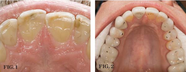
The signs of instability in the system would include wear that is beyond normal, tooth movement that is not wanted, broken teeth, and teeth with excessive mobility. The normal amount of wear for teeth is a slow process and follows a minimally progressive course. In an adult, tooth wear averages 10.7 µm per year. Exposed dentin is not normal and should be evaluated for the cause (Figure 1). Dentin wears at a rate far greater than enamel and this can cause wear and breakdown to accelerate.3 Tooth movement that is unwanted is the result of excessive forces on a tooth or breakdown of a tooth’s supporting structures. Normal teeth have 50 µm to 100 µm of mobility that varies by time of day and amount of forces applied. Numerous factors can lead to increased tooth mobility. Periodontal disease is a common cause of mobility; however, excessive load and lateral forces found in occlusal trauma can also cause this condition to exist.4 Thankfully, resolution of the mobility can resolve to pre-trauma levels once the cause is removed.5 Patients can exhibit varying degrees of some or all the signs of instability.
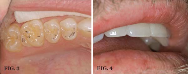
Just as it is important to recognize the signs of instability during an examination, it is equally important to recognize the signs of stability. A patient may exhibit numerous signs of stability and only a few signs of instability or vice versa. The signs of stability are even, stable holding contacts on all teeth with the joints in centric relation (Figure 2), anterior guidance that immediately discludes the posterior teeth in excursive and protrusive movements, stable bilateral temporomandibular joints, and teeth in harmony with the neutral zone.6 By knowing the signs of stability, they can be incorporated into future restorative treatment.
 CENTRIC RELATION (1.) Exposed dentin such as seen in these incisors is not normal. (2.) Stable, even holding contacts on all teeth when the condyles are in CR is a primary sign of occlusal stability. |
By having even tooth contact (no contact on cusp inclines) with the TMJs in centric relation, true stability can be maintained or reintroduced. To quote Dr. Peter Dawson, “Centric relation is the only condylar position that allows an interference free occlusion.” Centric relation is a natural, stable axial position from which the jaw can anatomically function. When the elevator muscles contract and the lateral pterygoids release, this stable axial position will be reached. This fixed position is in the most superior part of the glenoid fossa with the condyles being braced by their medial poles.7 A tripod of stability is reached when both condyles are fully seated and even anterior contact exists. It is from this relationship that an interference-free occlusion can be derived. It becomes clear that a clinician must also verify a lack of pathology or alteration in the joint structures to allow proper joint function. Centric relation is a position achieved only with the proper condyle disc assembly in proper position.
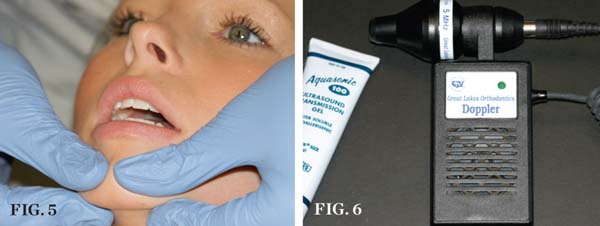
The effect of proper anterior guidance on muscle activity has been well documented in the literature for decades. By having immediate disclusion of the posterior teeth in excursive and protrusive movements the elevator muscles are shut down, which lessens the force on the teeth.8-11 Posterior teeth should only be loaded along the long axis of their roots.12 When posterior teeth remain in contact during excursive movement (working and non-working side interferences) the elevator muscles remain active (Figure 3). This causes muscular functional disharmony between the muscles of mastication. Proper guidance eliminates lateral forces on posterior teeth, thus reducing wear to normal levels.
Teeth respond to force by changing their position unless there is an equal balancing force. For this reason, teeth must be restored or placed in harmony with the neutral zone. The neutral zone is a perioral complex established facially by the muscles of the lips and soft tissue and lingually by the muscles of the tongue. Teeth will move naturally to find a neutral spot between these forces (Figure 4).13
 FUNCTIONAL HARMONY (3.) Numerous examples of incline interferences exist in this picture; note the large “hit and slide” evident on tooth No 5. (4.)These restorations are in balance between the muscles of the lips and the tongue; this positional balance is referred to as the neutral zone. |
Determining Restorability of the TMJ
It is necessary to examine the temporomandibular joint to establish its current condition and what affect this may have on proposed treatment. Information may be gathered through physical examination, oral history, or radiographically. Discussion of a patient’s history will often reveal information helpful in diagnosis of the TMJ. Helpful questions would include any history of injury such as blows to the face, sports injuries, or automobile accidents. Is the patient ever experiencing pain in the joints, noise such as clicking or popping, or is there a history of the jaw locking in any position? The physical examination would include palpation of the muscles of mastication, palpation of the joint capsule, evaluation of range of motion, and load testing of the joint.
With examination of the muscles, is there soreness to palpation? Is the patient suffering aches in any of the muscles? And if so, what is the intensity, duration, and frequency? Is there cramping or knotting present? Does there appear to be hypertrophy in any of the masticatory muscles? A yes to any of these questions gives reason to diagnostically dig further and helps to paint an overall picture of stomatognathic health.
The joints must be capable of loading under firm pressure in centric relation without tenderness, tension, or pain (Figure 5).14 If this response is positive, further investigation must be undertaken as the joint is not in centric relation with the proper disc condyle assembly. Tenderness and pain may indicate displacement of the disc from its proper position. Tension may indicate a lack of muscular release preventing the condyle from seating in the glenoid fossa.
Another useful tool to aid in joint diagnosis is Doppler auscultation. It offers a fast, reliable method to listen for joint derangement, and when combined with load testing it can be highly diagnostic (Figure 6).15 From a radiologic standpoint, the gold standard of imaging is magnetic resonance imaging (MRI). As opposed to standard radiographs or computer-assisted tomography (CAT) scans, the disk position can be imaged and determined with certainty. Because of the expense associated with MRI imaging, it is often solely employed for patients in severe dysfunction or pain. For this reason, a clinician must be able to reasonably determine the health of the TMJ using less expensive and invasive diagnostic tools.
 DIAGNOSIS (5.) Using bi-manual manipulation to load the joints in centric relation, this patient is having her TMJs load-tested to determine their health. (6.) A Doppler auscultation unit offers an excellent method to listen to the function of the joints and determine their current condition. |
Although an in-depth discussion of the possible TMJ joint conditions, which is quite specific, is well beyond the scope of this article, the joint will be in three possible conditions with varying degrees of derangement. The first condition would be structurally intact. This would be a healthy, unaltered joint with the condyle disk assembly in its proper anatomical position. Restorative care for these patients is very predictable. The second condition is altered at the lateral pole. Muscular incoordination has resulted in the lateral pole of the disk to displace anteriorly. Depending on the severity of the disk alteration, this condition may be reducing or non-reducing (regaining its proper position on the condyle). The third condition is altered at the medial pole. In this case, the derangement has progressed to the point that even the medial pole of the disk is anteriorly displaced. This can be a painful situation for the patient, especially one with initial medial pole issues. But the severity of the discomfort can decrease over time, making the diagnosis less predictable, yet the joint stability remains even less reliable.16 The pertinent point not to be missed can be summed up by this quote from Dr. Peter Dawson, “If the TMJs are not stable, the occlusion will not be stable, so it is a risky proposition to undertake occlusal changes without knowing the condition of the TMJs.”
Examination of the Teeth
The teeth themselves often can be full of diagnostic information. Is there excessive tooth wear (Figure 7 through Figure 9)? Is there excessive mobility in any teeth? Is there unwanted migration of teeth? Is there a centric relation/maximum intercuspation discrepancy (hit on inclines and slide into full intercuspation) (Figure 3)? If there is a discrepancy, is the deviation in the arc of closure or line of closure? Is the patient able to chew all types of food on both sides without pain? Does the patient feel like their bite is changing or unstable? In addition, are there functional patterns evident by the wear present on the teeth. For instance, horizontal bruxers (Figure 10) often have flat tabletop wear present. Vertical, constricted wear patterns (Figure 11) often have anterior chipping and lingual wear of maxillary anterior teeth.
 CLINICAL EXAMPLES (7.) Teeth Nos. 4 through 6 were loaded excessively in lateral movements; tooth No. 5 was the first tooth to hit in the CR arc of closure. (8.) An example of excessive lingual tooth wear in a patient with a restricted envelope of function. |
 CLINICAL EXAMPLES (9.) Posterior dentinal exposure is a sign of occlusal instability; this exposure will only get worse if the cause is not resolved and the tooth restored. (10.) An example of a horizontal functioning patient; note the flat “table-top” wear patterns. |
After all of this data is collected it may beg the question, “What does one do with it?” When the signs or symptoms of instability are recognized in the teeth, joints, or muscles, further investigation is warranted before beginning restorative care. That would include patients with a positive load test, muscular symptoms, tooth wear or mobility that is excessive, and a hit-and-slide into maximum intercuspation. These patients will undergo a further diagnostic evaluation that includes facebow-mounted diagnostic casts mounted in CR if possible (Figure 12). Further evaluation of the joint may include additional radiographic examination, pharmacological therapy, splint therapy, or physical therapy.
 CLINICAL EXAMPLES (11.) An example of a vertical wear patient; note the chipping and wear confined to the anterior teeth. (12.) An open CR bite record should be recorded with a non-compressible material and mounted with a facebow record, on a semi-adjustable articulator. |
Solutions to Occlusal Problems
Once stability issues in the joints are resolved, restoration of the teeth can begin. In addition, many times an occlusal problem will be diagnosed in a patient and the TMJ health is in a condition to begin restoration. In these cases, three solutions exist to correct the occlusal discrepancy. The first option would be equilibration. The goal of equilibration is to reorganize the posterior occlusion to remove deflective posterior contacts, and to refine anterior guidance to provide posterior disclusion and equal holding contacts on all teeth in centric relation. A second option would be orthodontic therapy or surgical movement of all or portions of the alveolar ridges. The goal would be to move teeth into a position for ideal occlusal function. The third option would involve restorative treatment of the teeth. The need for this would seem obvious, as restorative care is the only way to restore worn teeth back to their normal functional contours. The reality is that many cases will require a combination of the above three options to solve occlusal problems.
Patients with no signs of instability will be restored in their current maximum intercuspation. Care then must be taken to not introduce instability (posterior incline interferences) into the system with the planned restorations, and to restore so that anterior protected occlusion will be maintained and or refined.
Conclusion
Achieving success in restorative dentistry is an ever-evolving challenge. No two patients are alike, and everyone will have their own combinations of signs and symptoms. It is the clinician’s role to properly examine and diagnose in order to make sound restorative decisions. This begins with a complete examination that will provide the necessary data to determine the current condition of the stomatognathic system. By following a simple yet uncompromised diagnostic approach, predictability can be achieved.
References
1. Lobbezoo F, Naeije M. Etiology of bruxism: morphological, pathophysiological and psychological factors. Ned Tijdschr Tandheelkd. 2000;107(7):275-280.
2. Burris J, Evans D, Carlson C. Psychological correlates of medical comorbidities in patients with temporomandibular disorders. J Am Dent Assoc. 2010;141(1):22-31.
3. Larson TD. Tooth wear: when to treat, why, and how. Part I. Northwest Dent. 2009;
88(5):31-38.
4. Carranza F. Clinical Periodontology. Philadelphia, Pa: Saunders; 2002:438-439.
5. Niedermeier W. Parameters of tooth mobility in cases of normal function and functional disorders of the masticatory system. J Oral Rehab. 2007;20(2):189-202.
6. Dawson P. Functional Occlusion: From TMJ to Smile Design. St. Louis, Mo: Mosby; 2006:345-349.
7. Hess L. The relevance of occlusion in the golden age of esthetics. Inside Dentistry. 2008;4(2):36-44.
8. Okano N, Baba K, Igarashi Y. Influence of altered occlusal guidance on masticatory muscle activity during clenching. J Oral Rehabil. 2007;34(9):679-684.
9. Williamson EH, Lundquist DO. Anterior guidance: its effect on electromyographic activity of the temporal and masseter muscles. J Prosth Dent. 1983:49(6):816-823.
10. Manns A, Chan C, Miralles R. Influence of group function and canine guidance on electromyographic activity of elevator muscles. J Prosthet Dent. 1987;57(4):494-501.
11. Shingoaya T, Kimura M, Matsumoto M. Effects of occlusal contacts on the level of mandibular elevator muscle activity during maximal clenching in lateral positions. J Med Dent Sci. 1997;44(4):105-112.
12. Carranza F. Clinical Periodontology. Philadelphia, Pa: Saunders; 2002:700.
13. Cranham J. The horizontal position of the maxillary incisal edge: the key to optimum esthetics, phonetics, and function. Contemporary Esthetics. 2006;10(2):22-24.
14. Dawson P. Evaluation, Diagnosis and Treatment of Occlusal Problems. 2nd ed. St. Louis, Mo: Mosby; 1989:92-106.
15. Puri P, Kambylafkas P, Kyrkanides S. Comparison of doppler sonography to magnetic resonance imaging and clinical examination for disc displacement. Angle Ortho. 2006;76(5):824-829.
16. Dawson P. Functional Occlusion: From TMJ to Smile Design. St. Louis, Mo: Mosby; 278-294.
About the Author
Leonard A. Hess, DDS
Associate Faculty
The Dawson Academy
St. Petersburg, Florida
Private Practice
Monroe, North Carolina


