You must be signed in to read the rest of this article.
Registration on CDEWorld is free. Sign up today!
Forgot your password? Click Here!
Intraoral scanners (IOSs) have become pivotal in the advancement of digital dentistry. IOSs enable the capture of detailed intraoral optical impressions (IOIs), which are rendered as high-precision virtual models. This technology has improved both the efficiency of dental workflows and the overall patient experience, offering a more comfortable alternative to conventional impression-taking methods.
The significance of IOSs, however, goes beyond replacing conventional impressions. While their most common use in clinical practice continues to be in the creation of single-unit restorations through computer-aided design/computer-aided manufacturing (CAD/CAM) workflows, IOSs now integrate with a range of advanced diagnostic tools, such as cone-beam computed tomography (CBCT) and 3-dimensional (3D) facial scanning. These integrations enhance the accuracy and personalization of treatment plans.
The capabilities of IOSs have advanced significantly in recent years. With the rapid pace of technological innovation in this field, this article aims to provide a concise overview of both the established and emerging applications of IOSs in modern dentistry.
Advances in Functionality, User-Friendliness
Recent technological advancements have enhanced the functionality and user-friendliness of IOSs.1 Modern IOSs feature faster scanning capabilities, eliminating the need for scanning powder and providing high-resolution color images. These improvements not only optimize the workflow but also improve the visual assessment during dental procedures. IOSs now boast sophisticated software algorithms for seamless image stitching, which enhances the scanning process and accuracy.
Hardware developments have also been noteworthy, with many IOSs moving toward ergonomic, wireless designs that offer greater mobility and ease of use.2 However, these advancements come with challenges such as reliance on battery life and potential connectivity issues.
On the software side, IOS systems have undergone major innovations, extending their functionality beyond simple IOI capture. Advanced software now transforms IOSs into comprehensive platforms that support diagnostics, treatment planning, patient communication, and educational use. Additionally, many IOS manufacturers have embraced open data interfaces, which improve interoperability with a wide range of digital platforms. These open interfaces allow for data export in one or more standard imaging formats, providing dental practitioners with greater flexibility and more customization options.
Clinical Applications and Versatility of Intraoral Scanners
IOSs have found extensive applications across various dental specialties.1 In restorative dentistry, they are primarily used for fabricating dental restorations through CAD/CAM workflows, which remains by far their most widespread use (Figure 1 through Figure 5).2 The accuracy of IOSs enables dental professionals to produce restorations with precision that is on par with conventional methods.3 In addition to restorative dentistry, IOSs are being utilized for various applications in prosthodontics, orthodontics, forensic dentistry, and oral and maxillofacial surgery.
In oral and maxillofacial surgery, the advantages and versatility of IOSs are particularly notable. IOSs are instrumental in the treatment of conditions such as cleft lip and palate, which often requires impression-taking in young children, including neonates, both awake and under general anesthesia.4,5 The use of IOSs in these procedures enhances airway safety. Additionally, IOSs are invaluable for diagnostic purposes, such as assessing upper lip scarring and symmetry, as well as for treatment planning and the fabrication of orthopedic plates.6 The integration of IOS technology streamlines workflows and facilitates the incorporation of other digital tools, further augmenting their clinical utility.
Advanced Treatment Planning Technologies
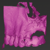
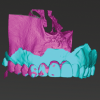
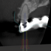
The use of IOSs in combination with other imaging modalities, such as CBCT and 3D facial scans, is playing a crucial role in advancing treatment planning in modern dentistry. This integration enables the creation of so-called "digital twins," ie, 3D models that precisely capture anatomical details and their spatial relationships. These digital twins enhance the precision of various dental interventions. When IOI data and data from facial scans are combined, they facilitate techniques such as digital smile design.7 Additionally, digital twins generated from the integration of IOI and CBCT support both static and dynamic navigation methods.8,9 This approach is increasingly being adopted across a range of clinical applications, including dental implant placement, orthodontic mini-implant placement, and endodontic access cavity preparation (Figure 6 through Figure 9).
Automated Tooth Segmentation and Orthodontic Landmarking
Accurate segmentation of individual teeth from IOI data is essential for effective diagnosis and treatment planning in orthodontics. However, automatic boundary detection between teeth and gingiva remains a formidable challenge. Deep neural network-based approaches are now delivering promising results, offering the potential for more efficient and reliable tooth segmentation.10
The manual placement of landmarks on dental models, crucial for diagnostic purposes and outcome assessments, is both labor-intensive and prone to inaccuracies. Recently developed software leverages a combination of machine learning and linear programming to automatically recognize and label each tooth and its landmarks, facilitating rapid and precise identification.11 Although human verification remains necessary, this technology represents an advancement toward the automation of routine orthodontic evaluations.
Enhancing the Patient and User Experience
The integration of IOSs into dental practice has enhanced patient comfort, particularly for impression-taking. Patients appreciate the quicker, more comfortable procedures compared to conventional techniques, while dental practitioners benefit from the efficiency that IOSs provide.1,12 In spite of these benefits, the pace of IOS technology adoption in large dental institutions has been relatively slow.13 Challenges include compatibility with existing health record systems, data storage management, and the initial costs of adoption.
Precision and Reliability of Intraoral Scanners
IOSs need to be both highly reliable and accurate to be effective in clinical settings.1 While IOSs achieve clinically acceptable precision when scanning complete dentate arches, they face challenges in edentulous areas, where conventional methods still frequently outperform them in terms of detail. The difficulty for IOSs lies in accurately capturing the soft-tissue movements and details that are essential for fabricating complete dentures (Figure 10 and Figure 11). In cases where the denture-bearing ridges are firm and supported by attached mucosa, IOSs can achieve accuracy similar to conventional impressions. However, when soft-tissue mobility or movement is a factor, such as in complete denture cases, IOSs often struggle to produce the necessary level of detail.14
For dental implants, IOSs generally meet the high precision standards required, particularly for single- and multiple-implant restorations. However, when it comes to capturing implant positions in cases with full-arch edentulism, improvements in IOS reliability are still needed. Conventional methods often provide superior accuracy in these more complex restorations. Recent studies have explored innovations such as the shift from vertically positioned scan bodies to horizontally positioned scan gauges.15 These gauges help minimize positional errors by reducing the need for image stitching during scanning, leading to improved precision. While these developments show promise, full-arch implant restorations still highlight the limitations of current IOS technology.
Studies comparing IOS and conventional impressions show comparable marginal accuracy for crowns and no significant differences in clinical outcomes for fixed dental prostheses.3 Although IOS accuracy has generally improved over time, a recent systematic review revealed that not all newer models offer better precision than their predecessors.16 It is also worth noting that significant variations in performance persist among different IOS systems.16
Expanding Diagnostic Capabilities
Beyond their use in CAD/CAM, IOSs are increasingly recognized for their diagnostic capabilities.1 Using superimposition tools based on best-fit alignment, IOSs can effectively assist in evaluating soft-tissue dimensions and monitoring tooth wear over time.17,18
IOSs are emerging as effective tools for accurately assessing and monitoring tooth wear. Tooth wear assessments using intraoral scanners have a discrimination threshold within the range of 70 µm to 80 µm.19 Measurements beyond this threshold accurately capture true tooth wear. IOS software solutions can thus play a valuable role in detecting tooth wear and tracking its progression over time.17,20 Periodic IOS assessments provide dental practitioners with critical information on levels of tooth wear, helping them make informed decisions about appropriate management strategies.
For caries detection, some IOSs are equipped with fluorescence or near-infrared imaging technologies.21 These devices have demonstrated potential in identifying proximal, occlusal, or both types of carious lesions.22,23 However, recent evidence suggests that IOS-based caries detection has lower diagnostic accuracy compared with conventional visual examination.24 This limitation raises concerns about the potential for missed diagnoses of early carious lesions and an underestimation of lesion severity.24 Ongoing challenges in diagnostic accuracy notwithstanding, these features hold promise for facilitating the detection of carious lesions in the near future, potentially reducing reliance on radiographic imaging.1
Some IOSs with color imaging capabilities are used for tooth shade determination.25 While IOSs can provide accuracy comparable to visual shade selection, a systematic review found that they often fall short in shade matching accuracy compared with spectrophotometers.26 This discrepancy arises from IOSs' inability to maintain optimal reading angles and uniform illumination.26 This leads to the recommendation that contemporary IOSs be used only as supplementary tools for tooth shade determination.1
Future Directions in IOS Technology
Looking forward, the potential of IOSs is expected to continue to expand with advancements in technology. Research is focused on improving the integration of IOSs with other digital tools such as CBCT and 3D facial scanning. This integration will likely facilitate more sophisticated and personalized treatment planning.
The emerging role of IOSs in teledentistry is particularly promising. In teledentistry, high-resolution, approximate true-color IOIs can be transmitted to dental practitioners in real-time or stored for subsequent review, allowing assessment from a remote location. These IOIs, when combined with supplementary data such as radiographs, facilitate monitoring of changes over time, identification of potential issues, and support in treatment planning. Currently, teledentistry assessments utilizing IOIs demonstrate effectiveness in detecting various dental conditions; however, their accuracy in evaluating periodontal health remains insufficient for clinical purposes.27 Advances in digital imaging and analysis are expected to enhance teledentistry capabilities, enabling practitioners to provide preliminary diagnoses and recommendations without the need for in-person visits. Such progress could expand access to dental care, particularly for individuals in underserved or remote locations.
Furthermore, IOSs are showing potential in forensic dentistry, where they can aid in human identification by capturing detailed dental and palatal morphology.28,29
Ongoing software innovations, many leveraging machine learning, are further boosting IOS capabilities, enhancing their use in computer-assisted diagnosis, patient communication, and treatment planning. These advancements are transforming IOSs into multifunctional platforms that support a broad range of clinical applications.
Summary
IOSs are at the forefront of digital dentistry. The integration of IOS technology into dental practices has transformed various procedures, from CAD/CAM restorations to intricate orthodontic and surgical applications. However, challenges persist in terms of technology implementation, cost, and the precision required in complex dental situations. Continuous advancements in both hardware and software are addressing these issues and expanding the potential of IOSs. Innovations in IOSs with built-in diagnostic tools and digital treatment planning platforms are solidifying IOSs as a cornerstone of modern dental care. As digital dentistry continues to evolve, IOSs are set to play an increasingly vital role in both routine dental care and specialized treatments.
Acknowledgment
The authors thank Drs. Jeronim Esati, Wadim Leontiev, and Lucia K. Zaugg for their invaluable contributions to the preparation of the illustrations.
About the Authors
Florin Eggmann, Dr. med. dent.
Research Fellow, Department of Preventive and Restorative Sciences, University of Pennsylvania School of Dental Medicine, Philadelphia, Pennsylvania; Assistant Professor, Department of Periodontology, Endodontology, and Cariology, University Center for Dental Medicine Basel UZB, University of Basel, Basel, Switzerland
Markus B. Blatz, DMD, PhD
Professor of Restorative Dentistry, Chair, Department of Preventive and Restorative Sciences, and Assistant Dean, Digital Innovation and Professional Development, University of Pennsylvania School of Dental Medicine, Philadelphia, Pennsylvania
Queries to the author regarding this course may be submitted to authorqueries@conexiant.com.
References
1. Eggmann F, Blatz MB. Recent advances in intraoral scanners. J Dent Res. 2024: doi: 10.1177/00220345241271937. Online ahead of print.
2. Revilla-Leon M, Frazier K, da Costa JB, et al. Intraoral scanners: an American Dental Association Clinical Evaluators Panel survey. J Am Dent Assoc. 2021;152(8):669-670.e2.
3. Bandiaky ON, Le Bars P, Gaudin A, et al. Comparative assessment of complete-coverage, fixed tooth-supported prostheses fabricated from digital scans or conventional impressions: a systematic review and meta-analysis. J Prosthet Dent. 2022;127(1):71-79.
4. Benitez BK, Brudnicki A, Surowiec Z, et al. Digital impressions from newborns to preschoolers with cleft lip and palate: a two-centers experience. J Plast Reconstr Aesthet Surg.2022;75(11):4233-4242.
5. Shanbhag G, Pandey S, Mehta N, et al. A virtual noninvasive way of constructing a nasoalveolar molding plate for cleft babies, using intraoral scanners, CAD, and prosthetic milling. Cleft Palate Craniofac J.2020;57(2):263-266.
6. Meyer S, Benitez BK, Thieringer FM, Mueller AA. Three-dimensional printable open-source cleft lip and palate impression trays: a single-impression-workflow. Plast Reconstr Surg.2024;153(2):462-465.
7. Lassmann Ł, Calamita MA, Blatz MB. The "smile design and space" concept for altering vertical dimension of occlusion and esthetic restorative material selection. J Esthet Restor Dent.2024. doi: 10.1111/jerd.13317.
8. Connert T, Leontiev W, Dagassan-Berndt D, et al. Real-time guided endodontics with a miniaturized dynamic navigation system versus conventional freehand endodontic access cavity preparation: substance loss and procedure time. J Endod. 2021;47(10):1651-1656.
9. Struwe M, Leontiev W, Connert T, et al. Accuracy of a dynamic navigation system for dental implantation with two different workflows and intraoral markers compared to static-guided implant surgery: an in-vitro study. Clin Oral Implants Res. 2023;34(3):196-208.
10. Hao J, Liao W, Zhang YL, et al. Toward clinically applicable 3-dimensional tooth segmentation via deep learning. J Dent Res.2022;101(3):304-311.
11. Woodsend B, Koufoudaki E, Mossey PA, Lin P. Automatic recognition of landmarks on digital dental models. Comput Biol Med. 2021;137:104819.
12. Serrano-Velasco D, Martín-Vacas A, Paz-Cortés MM, et al. Intraoral scanners in children: evaluation of the patient perception, reliability and reproducibility, and chairside time - a systematic review. Front Pediatr. 2023;11:1213072.
13. Jahangiri L, Akiva G, Lakhia S, Turkyilmaz I. Understanding the complexities of digital dentistry integration in high-volume dental institutions. Br Dent J.2020;229(3):166-168.
14. Rasaie V, Abduo J, Hashemi S. Accuracy of intraoral scanners for recording the denture-bearing areas: a systematic review. J Prosthodont. 2021;30(6):520-539.
15. Giglio GD, Giglio AB, Tarnow DP. A paradigm shift using scan bodies to record the position of a complete arch of implants in a digital workflow. Int J Periodontics Restorative Dent. 2024;44(1):115-126.
16. Vitai V, Németh A, Sólyom E, et al. Evaluation of the accuracy of intraoral scanners for complete-arch scanning: a systematic review and network meta-analysis. J Dent. 2023;137:104636.
17. Schlenz MA, Schlenz MB, Wöstmann B, et al. Intraoral scanner-based monitoring of tooth wear in young adults: 36-month results. Clin Oral Investig. 2024;28(6):350.
18. Kuralt M, Gašperšič R, Fidler A. Methods and parameters for digital evaluation of gingival recession: a critical review. J Dent.2022;118:103793.
19. Charalambous P, O'Toole S, Austin R, Bartlett D. The threshold of an intra oral scanner to measure lesion depth on natural unpolished teeth. Dent Mater. 2022;38(8):1354-1361.
20. Bronkhorst H, Bronkhorst E, Kalaykova S, et al. Inter- and intra-variability in tooth wear progression at surface-, tooth- and patient-level over a period of three years: a cohort study: inter- and intra-variation in tooth wear progression. J Dent.2023;138:104693.
21. Ntovas P, Michou S, Benetti AR, et al. Occlusal caries detection on 3D models obtained with an intraoral scanner. A validation study. J Dent. 2023;131:104457.
22. Michou S, Tsakanikou A, Bakhshandeh A, et al. Occlusal caries detection and monitoring using a 3D intraoral scanner system. An in vivo assessment. J Dent.2024;143:104900.
23. Kanar Ö, Tağtekin D, Korkut B. Accuracy of an intraoral scanner with near-infrared imaging feature in detection of interproximal caries of permanent teeth: an in vivo validation. J Esthet Restor Dent. 2024;36(6):845-857.
24. Jones B, Chen T, Michou S, et al. Diagnostic agreement between visual examination and an automated scanner system with fluorescence for detecting and classifying occlusal carious lesions in primary teeth. J Dent. 2024;149:105279.
25. Akl MA, Mansour DE, Zheng F. The role of intraoral scanners in the shade matching process: a systematic review. J Prosthodont.2023;32(3):196-203.
26. Tabatabaian F, Namdari M, Mahshid M, et al. Accuracy and precision of intraoral scanners for shade matching: a systematic review. J Prosthet Dent. 2024;132(4):714-725.
27. Steinmeier S, Wiedemeier D, Hämmerle CHF, Mühlemann S. Accuracy of remote diagnoses using intraoral scans captured in approximate true color: a pilot and validation study in teledentistry. BMC Oral Health. 2020;20(1):266.
28. Bae EJ, Woo EJ. Quantitative and qualitative evaluation on the accuracy of three intraoral scanners for human identification in forensic odontology. Anat Cell Biol. 2022;55(1):72-78.
29. Mikolicz A, Simon B, Gáspár O, et al. Reproducibility of the digital palate in forensic investigations: a two-year retrospective cohort study on twins. J Dent. 2023;135:104562.


