You must be signed in to read the rest of this article.
Registration on CDEWorld is free. Sign up today!
Forgot your password? Click Here!
In the past, when amalgam restorations were the gold standard in restorative dentistry, major concerns were expressed about the ability to achieve tight contacts with composite materials. Over the past 40 years, however, significant advances in composite restorations have been made. In recent years, light-cured restorations have allowed clinicians to create tight contacts and restorations with new interproximal matrices, rings, and anatomical wedges, rather than the traditional Tofflemire matrix with universal retainers, slim Jims, or wooden wedges. At the same time, digital technologies have been rapidly transforming dentistry across all dental specialties. Among the many advances in diagnosis and treatment that digital dentistry has fostered, new ideal tooth positions through restorative and orthodontic procedures have been made possible. Whereas in the past, models mounted on articulators were the standard of care, today digital technologies allow clinicians to immediately view digital scans to obtain information that can be shared chairside and with laboratories. With these technologies, clinicians have the opportunity to see a magnified view of the teeth in the dental arches and to observe how these teeth occlude. Internal views are also possible by rotating the digital image, enabling the best position of each tooth to be determined. This allows evaluation of the proximal contacts, as the clinician can view the occlusion of the opposing tooth and how it articulates with the opposing contacts. This article examines how proximal contacts can be restored using orthodontic movements and selected restorative techniques and materials.
ORTHODONTIC MOVEMENTS
Using aligner therapy, orthodontic movements can be induced that create healthier proximal contacts. In addition, orthodontic movements may be able to achieve tight contacts (eg, by uprighting the tooth and creating mesial movements) and thus help avoid the need for additive restorative material, which is a benefit for the longevity of the tooth. Uprighting and aligning a tooth helps direct better vertical forces with occlusion. This tooth movement is also indicated if the patient has a pre-existing laboratory-made restoration that can be preserved without reconstruction, thus helping maintain the vitality of the tooth.1
Ultimately, orthodontics is the least invasive form of dentistry, provided that the gingival and bony structure will support the orthodontic movements. When there are multiple open contacts within an arch, comprising mostly 0.1-mm to 0.3-mm spaces, and the marginal ridges are aligned, a series of overcorrection clear aligner trays can be used to move the teeth, most often mesially, to create tight proximal contacts.
It should be noted, however, that not all contacts need to be closed. A proximal contact does not need to be closed if the space is easily cleansable and does not retain food, and if the interproximal tissue is healthy with no bleeding and with periodontal probe readings equal to or less than 3 mm or 4 mm when sounding to bone.
POSTERIOR RESTORATIVE TECHNIQUES
In the presence of a pre-existing lab-processed restoration, the most predictable solution to create a tight contact is to remove the restoration and recreate a new restoration with a wide and tight interface in the proximal surfaces.
The adage "extension for prevention,"2 by G. V. Black is no longer advocated for restorative procedures, as etching, bonding, matrix systems, and composite materials are now utilized to achieve a minimally invasive approach. The type of restoration depends on the amount of tooth structure available. The presence and degree of caries dictate the preparation of healthy tooth structure as well as the position of the individual tooth intraorally. Interproximal restorations are not as strong as occlusal restorations, as they do not have a 360° interface with enamel.
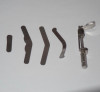
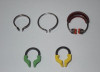
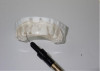
If the interproximal restoration extends onto the buccal and lingual walls, often the traditional Tofflemire matrixor freehand techniques can be used to help rebuild the external walls. To complete the restoration, a sectional matrix band can then be placed with a wedge that matches the interproximal space. Standard wooden wedges have now been replaced with new anatomical interproximal wedgesthat help create a more ideal contoured restoration (Figure 1 through Figure 3).
Upon preparation, the tooth requiring restoration needs isolation with the aid of a dry angle, cotton rolls, retractors, or a rubber dam.
The preparation ideally needs to be as minimally invasive as possible beyond the decay or pre-existing restoration. Ideally, finishing the restoration on enamel walls will allow the strongest bonding.3 However, if the pre-existing restoration is very large, then preparation into the existing composite is viable. Micro-etching with air abrasion particles will allow ideal bonding, as the surfaces are cleaned of saliva and contamination.3 Following this, suitable liners are applied to the dentin.4
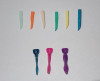
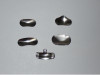
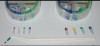
Matrices are available in various thicknesses, widths, and heights (Figure 4). The ideal matrix will adapt in four directions, slightly beyond the restoration at both the gingival seat and above the occlusal marginal ridge. The matrix must also be able to extend beyond the buccal and lingual walls to have the best marginal adaptation. Chairside matrix systems including specialized matrix ring forceps can be created to aid chairside efficiency (Figure 5 and Figure 6). Matrix thickness varies significantly, from 0.001 inch (dead soft) and upwards. If the width of the proximal box is wide, the use of a thicker matrix allows the adaptation to sustain an idealized contour for the composite.
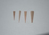
Wedges are then chosen to adapt at the gingival seat of the restoration. The wedge needs to be wide enough to fully engage the matrix at the gingival seat with no visible space between the tooth structure and the matrix. The anatomical wedges are best to allow an ideal convex emergence profile of the restoration at the gingival seatwith no voids between the matrix and the gingival floor of the tooth.
A matrix clamp can then be used that is tight and engages both sides of the matrix band and that allows the burnishing of the matrix against the proximal surface of the adjacent tooth.The ideal contact is a wide and broad contact to allow an unwaxed fine piece of floss to "lightly snap" through the contact with resistance.
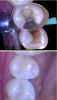
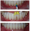
The increased thickness of a matrix allows a stronger capability to retain shape when the distance to the mesial or distal contact point of the adjacent tooth is large. If the space is significant, using a traditional Tofflemire band in the Tofflemire universal retainer can often prove effective. After applying the composite, and if the contact is light or not ideal, a dovetail preparation can be created within the new restoration to allow additional restorative materials to be built upon, thus creating the ideal tight contact with a different matrix. Practitioners can choose from among various matrices for their chairside experience and for achieving tight contacts. Some clinicians can create tight contacts with composite materials using the Tofflemire bands, whereas other clinicians have an ideal matrix that allows them to repeatedly create their ideal contact with little contouring beyond polishing buccal and lingual walls, defining the marginal ridge, and adjusting the occlusion (Figure 7 through Figure 9).
If the preparations are deep, the restoration can be accomplished with flowable material at the gingival seat, and then packable restorative material is placed in 2-mm increments and light cured. This restoration process is often referred to as a "sandwich" technique. Bulk filling is another technique that can be utilized curing 4 mm in depth with the ability to self-cure.5,6For crowns and onlays that are lab processed, the same parameters of proximal contacts are utilized as discussed above.
Longevity of restorations often depends on the shared occlusal forces applied and the patient's awareness of daytime grinding and clenching. The consumption of specific foods can exert greater force on the teeth, adding additional stress to the restoration interface and potentially leading to a break in the restoration. For instance, the force applied with chewing ice and whole almonds can be extremely detrimental to virgin teeth and dental work.7 It is important for clinicians to educate their patients about habits that impact oral health such as smoking and grinding/clenching,and about the benefits of a diet free of concentrated sugars and dehydrated fruits, as these also affect the longevity of the dental work. Grinding and clenching at night are brain mediated, and extreme pressure can be applied during sleep bruxism that is not duplicated during the day. An occlusal night guard will help distribute occlusal forces on the teeth.8 It has been estimated that nighttime pressure during sleep can yield six times the force generated during the day.9-11Encouraging home exercises to release facial muscle tension and educating patients to keep "lips together, teeth apart, and tongue in place" will help prevent occlusal wear during the hours when the patient is awake.12
ANTERIOR RESTORATIVE TECHNIQUES AND BLACK TRIANGLES
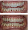
Tooth shape can be improved or modified when there is not ideal anatomical form. The appearance of "black triangles" often develops between the interproximal contacts and the gingival margin. These spaces develop for a variety of reasons. Upon completion of orthodontic movement, the teeth may display tooth imperfections or spaces beyond the interproximal contact. Gingival recession can occur over time as a result of many different factors, and often abfractions result. Thick gingival tissue is more stable, whereas recession is more prevalent with a genetic thin phenotype of gingival tissue, which appears almost transparent against the tooth and through which the periodontal probe can be seen when probing through the tissues.13 Gingival recession can also result from excessive mechanical brushing techniques, the use of hard toothbrush bristles, or labial placement of the teeth within the supporting bone. Grinding or clenching (daytime or nighttime/brain mediated) also contribute to gingival recession. A constricted chewing pattern, where the lower anterior teeth have been forced to adapt behind the upper anteriors, create man-made incisal tooth wear, and the pressure and vibration on the teeth can create gingival recession. Postorthodontic movement can also cause black triangles to develop between the anterior teeth.14 Crowding can "strangulate" the gingival papilla,and often a space is created between the gingival and interproximal contact of the teeth when aligned. If the space is not visible or does not bother the patient, then often it is unnecessary to modify the tooth or perform restorative treatment beyond retention for the future. To address black triangles, restoration of the tooth is indicated if a wide interproximal space allows food retention below the contact or if the esthetics are not acceptable to the patient (Figure 10).
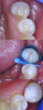
Relapse of anterior teeth, especially lower incisors, is more likely to occur with light interproximal contacts,resulting in rotation or movement toward its original position. Memory patterns exist, and with light proximal contacts there is little interface between the teeth to help stabilize a new position. For these proximal contacts, restoration of the tooth is indicated and may help the papilla regenerate, provided that the periodontal complex is ideal. If the distance from the contact point to the crest of the bone measures 4 mmand the roots are 2 mm apart, then the likelihood that the papilla will regenerate is 100%.15-17 Today clinicians have anatomical matrix systems that permit easier adaptation of restorative filling with flowable composite resin to allow optimal movement of material prior to curing (Figure 11 and Figure 12).18 To prevent staining at the interface of the restoration over time, it is prudent to first clean the surface of the tooth with micro-etching to ensure optimal adaptation of the additive restorative material.
Injection molding is another technique utilized to help create ideal interproximal contacts for both anterior and posterior teeth. A matrix is designed around a printed model from a digital design of the preferred tooth anatomy (Figure 13). Individual isolation of each tooth that is being modified is important for ease of finishing. Often, two matrices are made for chairside usage and ease of fabrication. The first matrix allows for the interproximal addition to be made on every other tooth, and the second matrix is fabricated from the final/ideal model.19
Proper color matching for the anterior teeth is vital for esthetic success.20,21If whitening is planned, the clinician should make certain that the restorative procedure is performed at least 1 week after whitening to match the color and achieve the best esthetics. If the color of the tooth is not ideal or the tooth is badly broken down, veneers or crowns are possible long-term solutions.
CONCLUSION
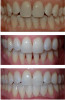
Optimizing occlusion and arch forms often changes proximal contacts. To achieve healthy proximal contracts, treatment that incorporates orthodontic movements and finishing with restorative additive procedures is minimally invasive and gratifying to both the patient and dentist.22 Stability and ideal arch form with vertical occlusal forces and no anterior constriction allow easier dental hygiene and temporomandibular joint comfort for the patient. In the patient shown in Figure 14, aligner therapy followed by the use of additive restorative materials was used to close interproximal spaces.
Today, digital technologies are instrumental not only in diagnosis and treatment in restorative dentistry, but in helping educate our patients on the condition of their dentition. Digital technology enables a comprehensive view of ideal future tooth anatomy, while it allows the patient to see imaging displayed on a screen and thus be involved in the smile design process and make more informeddecisions regarding their treatment.23,24
Digital technologies also allow accurate evaluation of the proximal contacts, as the clinician can use digital imaging to view the occlusion of the opposing tooth and how it articulates with the opposing contacts. Therefore, in optimizing treatment for patients with suboptimal proximal contacts, it is essential for dental clinicians to be comfortable with digital technology to recreate direct and indirect ideal interproximal contacts, which ultimately will help improve their patients' oral health.
References
1. Pulp necrosis. Cleveland Clinic website. https://my.clevelandclinic.org/health/diseases/23573-pulp-necrosis. Updated July 18, 2022. Accessed February 6, 2024.
2. Frencken JE, Peters MC, Manton DJ, Leal SC, Gordon VV, Eden EE. Minimal intervention dentistry for managing dental caries - a review: report of a FDI task group. Int Dent J. 2012;62(5):223-243.
3. Huang C-T, Kim J, Arce C, Lawson NC. Intraoral air abrasion: a review of devices, materials, evidence, and clinical applications in restorative dentistry. Compend Contin Educ Dent. 2019;40(8):508-513.
4. King K, Simon J, de Rijk W. Dentin bonding. CDEWorld website. https://cdeworld.com/courses/4348-dentin-bonding?&q=dentin+liners. May 2009.
5. Snyder TC. Understanding today's bulk-fill posterior composite restoration techniques. CDEWorld website. https://cdeworld.com/courses/20835-understanding-today-s-bulk-fill-posterior-composite-restoration-techniques. October 2017.
6. Roetzer P. New advancements in flowable dual-cured bulk-fill composites. CDEWorld website. https://cdeworld.com/courses/22776-new-advancements-in-flowable-dual-cured-bulk-fill-composites?&q=bulk+filling. May 2023.
7. Chewing ice puts teeth at risk. California Association of Orthodontists website. https://caortho.org › chewing-ice-puts-teeth-at-risk. Published June 30, 2020. Accessed February 6, 2024.
8. Holmgren K, Sheikholesman A, Riise C. Effect of a full-arch maxillary occlusal splint on parafunctional activity during sleep in patients with nocturnal bruxism and signs and symptoms of craniomandibular disorders. J Prosthet Dent. 1993;69(3):293-297.
9. Lavigne G, Kato T. Usual and unusual orofacial motor activities associated with tooth wear. Int J Prosthodont. 2005;18(4):291-292.
10. Lavigne GJ, Goulet J, Zuconni M, Morrison F, Lobbezoo F. Sleep disorders and the dental patient: an overview. Oral Surg Oral Med Oral Pathol Oral Radiol Endod. 1999;88(3):257-272.
11. Lavinge GJ, Kato T, Kolta A, Sessie BJ. Neurobiological mechanisms involved in sleep bruxism. Crit Rev Oral Biol Med. 2003;14(1):30-46.
12. Colson DG. Your Mouth: The Gateway to a Healthier You. DJC Corp; 2011:59-71.
13. da Costa FA, Perussolo J, Dias DR, Araujo MG. Identification of thin and thick gingival phenotypes by two transparency methods: a diagnostic accuracy study. J Periodontal.
2023;94(5):673-682.
14. Jati AS, Furquim LZ, Consolaro A. Gingival recession: its causes and types, and the importance of orthodontic treatment. Dental Press J Orthod. 2016;21(3):18-29.
15. Kois J. "The gingiva is red around my crowns"--a differential diagnosis. Dent Econ. 1993;83(4):101-102.
16. Tarnow DP, Magne AW, Fletcher P. The effect of the distance from the contact point to the crest of bone on the presence or absence of the interproximal dental papilla. J Periodontol. 1992;63(12):995-996.
17. Savani F, Weisgold AS, Rose LF. Biologic width and its relation to periodontal biotypes.
J Esthetic Dent. 1998;10(3):157-163.
18. Clark D. Restoratively driven papilla regeneration: correcting the dreaded "black triangle." Tex Dent J. 2008;125(11):112-115.
19. Hulac S. A transitional full mouth rehabilitation. Inside Dentistry. 2021;17(4):28-31.
20. Yamaguchi S, Karaer O, Lee C, Sakai T, Imazato S. Color matching ability of resin composites incorporating supra-nano spherical filler producing structural color. Dent Mater. 2021;37(5):e269-e275.
21. Daud A, Gray G, Lynch CD, et al. A randomised controlled study on the use of finishing and polishing systems on different resin composites using 3D contact optical profilometry and scanning electron microscopy. J Dent. 2018;71:25-30.
22. Kois JC, Diagnostically driven interdisciplinary treatment planning. Seattle Study Club J. 2002:6(4):28-34
23. Georg R. Digital smile design: utilizing novel technologies for ultimate esthetics. Compend Contin Educ Dent. 2023;44(10):567-573.
24. Simon H, Magne P. Clinically based diagnostic wax-up for optimal esthetics: the diagnostic mock-up. J Calif Dent Assoc. 2008;36(5):355-362.