You must be signed in to read the rest of this article.
Registration on CDEWorld is free. Sign up today!
Forgot your password? Click Here!
Osseointegration, the direct connection between living bone and a dental implant, was originally described by Brånemark et al in the mid-1960s.1,2 At that time there was ongoing research into the topic of bone healing. The recognition of the achievement of osseointegration was a somewhat serendipitous occurrence rather than the result of a research initiative directed specifically toward dental implants.
Early History
The Brånemark research group was using titanium cylinders that were made to surround a microscopic lens that was placed into bone to observe the healing of the bone that occurred after placement of this apparatus. As reorganization of the living bone occurred, primarily in the form of neovascularization adjacent to the optical lens, there was a simultaneous bone response to the oxidized surface of the titanium, which made removal of the implant and its optical lens difficult. Fortunately, members of the research team suggested that this bond between the titanium housing of the optical lens and the bone may demonstrate other therapeutic opportunities beyond the realm of basic science.3,4
The transition from basic bone research to translational research represented a huge advance. Dental implants, before this time, had been unpredictable at best. The primary reason for the lack of predictability was that there was little or no soft-tissue barrier to the ingress of bacteria that eventually caused a breakdown at the interface of oral mucosa and bone. In contrast, the biocompatible material of oxidized titanium represented a favorable surface for bone and soft-tissue connection to the implanted device.5-7
The first generation of osseointegrated implant bore little resemblance to the physical features that are predominant today.2,8 The early implants were placed beneath the oral mucosa and allowed to heal in that position, undisturbed from the prosthesis above it. Healing in the mandibular arch was generally more rapid than was healing in the maxilla, primarily because of the increased bone density in the mandible.9
Over time, however, it was recognized that bone could heal to endosseous implants in either jaw and that placement below the oral mucosa was not essential to achieve osseointegration. Albrektsson et al described the occurrence of four essential events to achieve osseointegration.Those events involved the identification of a suitable host into whom a biocompatible implant could be placed by an oral surgeon using gentle surgical techniques and then meticulously placed into masticatory function by a prosthodontist.8 Considering these events, it was likely that further investigation would eventually lead to improvements in implant design and materials that would transform the dental implant into a very suitable facsimile of a natural tooth root.10
Mandible vs Maxilla
Originally, osseointegration was used to replace only the mandibular teeth, using a fixed, implant-retained, full-arch prosthesis.The treatment was initiated in the mandible because the mandible was associated with unfavorable support and stability for a removable dental prosthesis. In contrast, the anatomy of the maxilla was much more favorable, with the presence of the palate for support and the residual ridges that resorb far more slowly than those in the mandible. The maxilla generally provided a more favorable environment for a removable dental prosthesis. Consequently, the use of a maxillary complete denture with a mandibular fixed, implant-supported prosthesis provided patients with a comfortable and highly functional dentition. However, with the success in the mandibular arch, there was an increasing interest in the use of maxillary implants, which would offer the patient the opportunity to have full-arch restorations in both jaws.11,12
In the mid-1980s, the planning for replacement of all teeth in each jaw was relatively straightforward. According to the plan at the time, patients underwent removal of all remaining teeth, and implants were placed in a delayed fashion to allow placement in healed bone. Some clinicians placed implants immediately after tooth removal, but these implants were "submerged" to provide undisturbed healing.13,14 The completely edentulous ridges before the use of dental implants allowed the vertical placement of the implants, which would achieve bicortical stabilization simply by placing the implant through the superior cortical bone and extending it into the inferior bone cortex. The plan was to place implants in the posterior position in a way that would allow six implants to support the final prosthesis. The posterior position might be compromised somewhat by the inferior alveolar canal; however, with six implants, it was rarely a problem. Oral surgeons understood the requirements of tooth position and, working with prosthodontists, were routinely able to achieve favorable esthetic and functional outcomes.13,14
Improvement of Surgical Guides
Surgical guidance at that time was simple. A wax trial prosthesis was fabricated on the diagnostic cast. It was duplicated with acrylic, and the facial-incisal limit was established. Similarly, the lingual position was identified. General boundaries were established to ensure that an implant was placed in a position to "hide" the implant components within the boundaries of the surgical guide and, subsequently, within the confines of the prosthesis.15,16
Eventually the plan for placement of six implants in the edentulous mandible was reduced to the placement of five and eventually four implants that would be used to support a full arch of teeth. At this point, the verbal communication of intended implant locations was no longer adequate, primarily because of the increasing complexity of the restorative design. The simple forms of implant placement guides were gradually replaced by more restrictive guides that would ensure favorable placement for implants.17-20
As implants increased in sophistication, the demand for more ideal implant placement grew. Guidance of the surgical positioning of implants was thought to be essential. With the introduction of 3D imaging, it became increasingly possible to design and create a dental prosthesis before the natural teeth were removed. In addition, the ability to accurately predict the location for implants led to prostheses that were true facsimiles of the natural dentition.
In the edentulous patient, such guidance was provided to ensure that implants were in relatively acceptable positions that demonstrated favorable distribution of the implants. Implant placement into the first molar areas and the canine areas in conjunction with a single implant near the midline was thought to be acceptable. The first molar implants would rarely cause cosmetic concerns, thereby limiting the esthetic implants to the canine tooth position, and the central incisor would be placed in the most favorable bone. The fabrication of surgical guides that provided the surgeon with a large amount of freedom toward implant placement was thought to be generally acceptable. The use of full-arch surgical guides further increased in complexity as the goal of treatment increased demands on definitive cosmetic prostheses.
Stability of the surgical guide remained as a primary area of concern. Often surgeons would use horizontal mini-screws to secure the guide. When placing implants for use as support for single tooth replacement, these devices would be secured by friction, or possibly by a provisional luting agent.21-23 When all teeth were removed, there was little support that could be provided to the surgical guide.
Surgical guides have often been more beneficial in the partially edentulous jaw, primarily because the implant position is framed by the natural teeth in the arch. The guides, along with the natural teeth, could be used to determine the position of the implant.24 Depending on the location of the implant, force distribution to the implant and its surrounding bone would likely be different than is the case when only natural teeth are involved. When considering forces down the long axis of an implant in comparison with forces down the long axis of an adjacent tooth, there will be physiologic differences between the two. Finite element analysis has been used in the past to define this difference in force application. With the near ubiquitous nature of cone-beam computed tomography, the angle of the crown to the long axis of the teeth or implants is better visualized.
The apparent mismatch of the natural tooth crown to the natural tooth root or to the implant "root" was thought to be a concern originally; however, it has not presented itself as such. Patients rarely comment on the difference in angulation between the natural tooth and the implant-supported crown. It is not clear whether the patient can feel the difference or whether no difference exists. If a difference does exist, it is likely that the patient will grow accustomed to it over time.
The natural teeth, depending on their number and location, could surround a single implant, creating a so-called tooth-bounded edentulous space. Alternatively, if the missing teeth are distal to the natural dentition, unbounded edentulous space may be created. Depending on the jaw relation of the patient and the angle of the natural teeth in the jawbone, it is certainly feasible that a difference in tooth arrangement may not be perceived. This is an event that should be discussed thoroughly with the patient before implant placement.
Today, the use of 3D-generated surgical guides has become common. Such guides may be provided for the patient with rigid screw fixation to afford more accurate placement of implants. In the partially edentulous patient, the remaining natural teeth may provide adequate security, once in position, to stabilize the guide without any further measures of affixing it to the remaining teeth.
As always, the goal of implant treatment is to provide support, retention, and stability that will work in concert to create a more effective and functionally improved prosthesis. Identification of the correct number of implants in the appropriate locations will provide a much greater opportunity for treatment success.
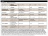
Currently there is no one surgical guide that will work for every patient in every situation (Table 1). Personal preference on the part of the surgeon is likely to be the standard rather than the exception. The preference of the surgeon will relate primarily to the ability to stabilize the surgical guide and subsequently remove it at the appropriate time. Because no surgical guide has been demonstrated to place multiple implants with absolute certainty that the fit of the prosthesis will not be compromised, remembering the 10-µm "allowable" error, the search for the ideal solution will continue.
Surgical guides have been created in the past with all the attributes referred to above. They were stabilized in position by horizontal retention screws, and definitive vertical stops allowed the implant to go into position only to a prescribed depth and, once achieved, be left alone. Although elegant in theory, the practicality of such a requisite level of precision remains elusive. The response of the implant manufacturers has been to create adjustable abutments that give an abutment some amount of freedom for the prosthesis to fit to it. Unfortunately, as of this writing, such prostheses do not appear to be gaining acceptance within the marketplace.25-29
Treatment of Residual Anatomy
One of the complications that may be faced may be related to a compromise in the amount of residual alveolar bone. Natural teeth are rarely lost all at once. Consequently, there are different levels of exposure relative to time at risk affecting the residual anatomy. Several treatment options can be considered for these situations.
One treatment option is to consider contouring the alveolar ridge to create a favorable 3D architecture of the residual ridges. This is particularly effective when patients have lost teeth at an earlier age and still have sufficient residual alveolar anatomy. A second option is to use pink gingival ceramics to create the illusion of natural gingiva. Both suggestions achieve the greatest success when the patient has relatively robust residual alveolar architecture.
When the situation differs and the residual anatomy is already deficient, the choice of grafting becomes more appropriate. Up until now there has been no definitive grafting solution that addresses all types of residual anatomy. The situation is complicated by the fact that there are many interchangeable terms, many interchangeable techniques, and a great number of therapeutic options. Dental literature is replete with scientific investigations to demonstrate the superiority of one technique, material, or device that would be usable regardless of the presenting condition. Sadly, all fall short of the goal.
For example, the term graft usually has modifiers that determine what type of graft is being used. An autogenous graft is the same as an autograft, but both differ from an allograft that comes from the same species but not the same individual. An alloplastic graft is the same as an alloplast. The source of different graft material can be perplexing. A human cadaver may be the source of an allograft, such as for a burn victim. A xenograft, which may come from equine, bovine, porcine, ovine, or caprine sources, may not always be prepared the same way even though it may be used in a similar fashion. Bone, for example, could be deproteinized or demineralized. It could be lyophilized, but it would be unlikely to be homogenized, at least in its use in dentistry.
The "gold standard" for grafting is a term that has been applied to numerous situations (Table 2). At this time the author doubts that anyone would consider that a gold standard has truly been established, although the term is used frequently. Part of the problem is that assigning patients to one treatment arm or another in a randomized controlled clinical trial is exceedingly dependent on the skill of the surgeon, and the preference of the surgeon is often related to individual surgical skills. The discussion appears to be circular in nature.
Conclusion
Although the field of implant dentistry has advanced greatly, it will continue to evolve and offer an expanding array of options. For example, there are many approaches to the grafting of the residual maxilla or mandible. Due to various difficulties in the treatment of either arch, it is not unusual for a surgeon to treat one jaw with an ideal arrangement and the opposing jaw with a more predictable but less artistic solution. Although it would appear that dentistry is making enormous progress in identification of appropriate guides and grafts, it is likely that more innovative solutions will be developed to address continuing concerns.
About the Author
Steven E. Eckert, DDS, MS
Professor Emeritus
Mayo Clinic
Rochester, Minnesota
Private Practice
ClearChoice Dental Implant Center
Edina, Minnesota
References
1. Brånemark PI. Osseointegration and its experimental background. J Prosthet Dent. 1983;50(3):399-410.
2. Brånemark PI, Adell R, Breine U, et al. Intra-osseous anchorage of dental prostheses. I. Experimental studies. Scand J Plastic Reconstr Surg. 1969;3(2):81-100.
3. Brånemark PI. Intravital microscopy. Its present status and its potentialities. Med Biol Illus. 1966;16(2):100-108.
4. Adell R, Lekholm U, Rockler B, Brånemark PI. A 15-year study of osseointegrated implants in the treatment of the edentulous jaw. Int J Oral Surg. 1981;10(6):387-416.
5. Salinas TJ, Sadan A. Establishing soft tissue integration with natural tooth-shaped abutments. Pract Periodontics Aesthet Dent. 1998;10(1):35-42.
6. Hermann JS, Buser D, Schenk RK, et al. Biologic width around titanium implants. A physiologically formed and stable dimension over time. Clin Oral Implants Res. 2000;11(1):1-11.
7. Linkevicius T, Apse P. Biologic width around implants. An evidence-based review. Stomatologija. 2008;10(1):27-35.
8. Albrektsson T, Zarb G, Worthington P, Eriksson AR. The long-term efficacy of currently used dental implants: a review and proposed criteria of success. Int J Oral Maxillofac Implants. 1986;1(1):11-25.
9. Ahlqvist J, Borg K, Gunne J, et al. Osseointegrated implants in edentulous jaws: a 2-year longitudinal study. Int J Oral Maxillofac Implants. 1990;5(2):155-163.
10. Albrektsson T, Jansson T, Lekholm U. Osseointegrated dental implants. Dent Clin North Am. 1986;30(1):151-174.
11. Palmqvist S, Sondell K, Swartz B, Svenson B. Marginal bone levels around maxillary implants supporting overdentures or fixed prostheses: a comparative study using detailed narrow-beam radiographs. Int J Oral Maxillofac Implants. 1996;11(2):223-227.
12. Becker W, Becker BE, Israelson H, et al. One-step surgical placement of Brånemark implants: a prospective multicenter clinical study. Int J Oral Maxillofac Implants. 1997;12(4):454-462.
13. Buser D, Mericske-Stern R, Bernard JP, et al. Long-term evaluation of non-submerged ITI implants. Part 1: 8-year life table analysis of a prospective multi-center study with 2359 implants. Clin Oral Implants Res. 1997;8(3):161-172.
14. Buser D, Mericske-Stern R, Dula K, Lang NP. Clinical experience with one-stage, non-submerged dental implants. Adv Dent Res. 1999;13:153-161.
15. Becker CM, Kaiser DA. Surgical guide for dental implant placement. J Prosthet Dent. 2000;83(2):248-251.
16. Cehreli MC, Sahin S. Fabrication of a dual-purpose surgical template for correct labiopalatal positioning of dental implants. Int J Oral Maxillofac Implants. 2000;15(2):278-282.
17. Louvrier A, Marty P, Barrabé A, et al. How useful is 3D printing in maxillofacial surgery? J Stomatol Oral Maxillofac Surg. 2017;118(4):206-212.
18. Tan KB. The use of multiplanar reformatted computerised tomography in the surgical-prosthodontic planning of implant placement. Ann Acad Med Singapore. 1995;24(1):68-75.
19. Balshi SF, Wolfinger GJ, Balshi TJ. Surgical planning and prosthesis construction using computer technology and medical imaging for immediate loading of implants in the pterygomaxillary region. Int J Periodontics Restorative Dent. 2006;26(3):239-247.
20. Kupeyan HK, Shaffner M, Armstrong J. Definitive CAD/CAM-guided prosthesis for immediate loading of bone-grafted maxilla: a case report. Clin Implant Dent Relat Res. 2006;8(3):161-167.
21. Sarment DP, Sukovic P, Clinthorne N. Accuracy of implant placement with a stereolithographic surgical guide. Int J Oral Maxillofac Implants. 2003;18(4):571-577.
22. Nokar S, Moslehifard E, Bahman T, et al. Accuracy of implant placement using a CAD/CAM surgical guide: an in vitro study. Int J Oral Maxillofac Implants. 2011;26(3):520-526.
23. Guichet D. Digitally enhanced dentistry: the power of digital design. J Calif Dent Assoc. 2015;43(3):135-141.
24. Tanasić IV, Tihacek-Sojić LĐ, Milić-Lemić AM. Prevalence and clinical effects of certain therapy concepts among partially edentulous Serbian elderly. J Prosthodont. 2015;24(8):610-614.
25. Yong LT, Moy PK. Complications of computer-aided-design/computer-aided-machining-guided (NobelGuide) surgical implant placement: an evaluation of early clinical results. Clin Implant Dent Relat Res. 2008;10(3):123-127.
26. Lima SA, Murad MA, Moyne C, Stemmelen D. Electro-osmosis in kaolinite with pH-dependent surface charge modelling by homogenization. An Acad Bras Cienc. 2010;82(1):223-242.
27. Pozzi A, Moy PK. Minimally invasive transcrestal guided sinus lift (TGSL): a clinical prospective proof-of-concept cohort study up to 52 months. Clin Implant Dent Relat Res. 2014;16(4):582-593.
28. Marchack CB. An immediately loaded CAD/CAM-guided definitive prosthesis: a clinical report. J Prosthet Dent. 2005;93(1):8-12.
29. Marchack CB. CAD/CAM-guided implant surgery and fabrication of an immediately loaded prosthesis for a partially edentulous patient. J Prosthet Dent. 2007;97(6):389-394.