You must be signed in to read the rest of this article.
Registration on CDEWorld is free. Sign up today!
Forgot your password? Click Here!
Bite mark analysis and identification combine as the scientific link between a bite mark and the potential biter. Bite mark evidence is accepted by legal systems worldwide in cases of sexual assault, rape, murder, abuse (child, partner or elder), and less frequently, cases of robbery.1 Methods of interpretation are often under scrutiny by law officials, particularly those in defense of the presumed biter, and it remains a highly controversial topic among all disciplines of forensic science.2 The dental hygienist is on the front line for bite mark identification and documentation in patients suffering abuse. Dental hygienists, along with other members of the dental team, should be familiar with bite mark recognition and documentation.3
When an injury presents features that suggest a bite mark, initial considerations should be identification as a bite injury and differentiation between human and animal bites.2
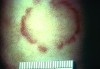
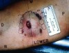
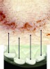
Human bite marks present as diffuse or specific bruises, depending on if there is a single bite or multiple, overlapping bites. Bites may be abrasions or lacerations and may or may not completely avulse the soft tissue (common in combination bites). Typically, a human bite mark comprises two opposing U-shaped arches separated by open spaces (Figure 1). A hematoma may occupy the center space of the bite mark, caused by soft tissue compression during the biting action (Figure 2 and Figure 3).4
Occasionally, bite marks display unique characteristics of contacting tooth surfaces or details related to palatal or lingual tooth surfaces leaving imprints on soft tissues. Mandibular arches naturally create a more defined impression because of the small anterior, in relation to larger maxillary anterior teeth; the mandibular jaw is also movable in relation to the maxilla, remaining stationary. Sextant five teeth have a smaller surface area and stress (defined as force exerted by unit area) in inverse proportion to surface area. Likewise, dentitions with fewer teeth will inflict more soft tissue stress, leaving a larger mandibular imprint and more significant hematoma. Impressions may present within the bite mark, maxillary and mandibular lines, as positive or negative features. Positive features include rotations or incisal fractures; missing teeth or diastemas provide negative features of a bite mark (Figure 4, Figure 5 and Figure 6).5
In comparison, bites of dogs or other carnivorous animals tend to tear skin and expose underlying tissue.4 Animal bites leave several deep puncture wounds and ragged irregular edges to the bite wound that result during shaking, tearing and avulsion of human skin during the bite. This irregular border resembles a human dental arch outline. Dog bites leave two defined punctures (from canine teeth) and a narrower ‘U’ shaped bite impression because of the narrow anterior dental arch. Dog bites are also usually more conical in shape and longer than a human bite mark. Feline bites are almost always accompanied by parallel claw scratch marks. They are shorter than dog bites and have multiple small punctures present in the dental arch impression. Rodent bites, usually noticed on a discovered corpse several days postmortem, are rarely seen in living victims; however, they might be found on crib-bound children living in unsanitary housing conditions. Rodent bites are distinct because of long chisel-shaped impressions from a rodent’s prominent, thick incisors.6
Other circular or elliptical impressions on tissues may clinically resemble human bite marks, such as: door knobs, heel impressions or ECG electrodes. The health care provider should carefully interview the patient and consider their medical history (past and present) to eliminate hematomas caused by preexisting systemic conditions (such as hemophilia) or medications (such as anticoagulants) that increase bruising incidence, as well as skin diseases such as psoriasis, Lyme disease or scabies that may present as bite marks.2
Bite mark analysis is based on the assumptions that human dentition is individually unique and that uniqueness is replicated on the bitten surface.5 Even identical twins do not present with identical dental arches or tooth forms. The effect of natural wear, environmental factors exposure, dental treatments and dental diseases will lead to dentition changes in each individual.7 In a 2003 survey of forensic odontologists, 91 percent of respondents found the human dentition unique and 78 percent believed that the unique dentition would replicate on human skin — findings echoed by diplomats of the American Board of Forensic Odontology (ABFO).8
Determining the anatomical position of a bite mark is crucial to its forensic analysis. Bite marks have been recorded on nearly all anatomical parts of the human body. Bite locations are specific to types of crimes committed against the victim, victim’s gender, and age of the victim. Women are 50 percent more likely to be bitten than males. Women are most likely to present with bite marks to the breasts, arms and legs, while males are more commonly bitten on the arms, shoulders/backs and faces. Children are usually bitten on the face, arms, legs and genitals.1 Children are also more likely to present with multiple bites in one anatomical location, while adults tend to display only one bite mark. Thorough examination of these bite marks is necessary and may require additional review by medical colleagues also trained in bite mark recognition and abuse reporting. Dental hygienists, as well as other dental team members, must be vigilant to recognize human bite injuries, especially in juvenile patients, because bites may be the result of accident or abuse.2 No matter where a bite is located, timely recognition is crucial for a bite victim, both for documentation and analysis and for potential medical treatment. Just like animal bites, human bites may potentially transmit Herpes Simplex Virus, Hepatitis B, Hepatitis C, syphilis, tuberculosis and actinomycosis.6
Human skin, as a bite registration material, is highly variable in its response to trauma. Skin is also variable in terms of anatomical location, underlying muscle tissue and fat, curvature, tautness or adherence to underlying tissues.6 Skin’s highly viscoelastic properties allow stretching to occur during the biting process or during evidence collection. This is because of elastic collagen fibers in the dermis, which distort under pressure and then recoil to their original position. The degree to which this stretching occurs depends on age of the victim, anatomical location of the bite and body position during biting. Every bite mark on skin displays some degree of distortion due to stretching. Changes in bite mark appearance and distortion will likely increase as the injury grows older and begins to heal.5 There is considerable healing and change in appearance in the first 24 hours following a bite, which continue for the following five days at a slower rate. Stress applied to the skin during biting is sufficient to produce a chemical-histological alteration in collagen fibers and influences the presentation of bite marks by anatomical location.9
Bite mark distortion is divided into two categories: primary and secondary distortion. Primary distortion occurs at the time of biting; secondary distortion afterward. The main components of primary distortion are the dynamics of the biting process and amount of detail left on the bitten surface; these are referred to as dynamic distortion and tissue distortion, respectively.10 The two distortive processes occur simultaneously and may vary considerably between bite marks. Dynamic distortion occurs uniquely depending on the movements of the victim and biter, making it possible for one biter to leave multiple bites with multiple degrees of tissue distortion. Components of secondary distortion are time, posture and photographic distortion. Changes that occur in a bite mark from the time of injury to the time of examination are referred to as time distortion; healing significantly alters the appearance and dimensions of a bite mark, making timely evidence collection pertinent for adequate and accurate analysis. Posture distortion occurs when position of the body when bitten differs from position of the body during examination and documentation. Photographic distortion happens during evidence collection when the camera utilized is not positioned at an angle 90 degrees from the bite. When examining a bite mark, it is essential to account for body positioning during biting process. Nearly nothing may be done to prevent primary distortion, so examiners should take precautionary steps to reduce all possible secondary distortions.10
The dental hygienist should be familiar with bite mark recognition and documentation procedures, since many bites are noticeable on a patient’s hands, arms, head, face and neck during the extraoral-intraoral examination.11 When noticing what may be a bite mark, the dental hygienist should maintain a relaxing and secure office environment and keep discussion neutral, creating an environment of trust and allowing the patient to freely discuss suspected abuse. The dental hygienist should always document these injuries, including location, size and traumatic nature. Photographs may also be taken, preferably with the use of a millimetric scale reference. Law enforcement should always be notified immediately.
It is ideal that the bitten patient receive a medical examination within 72 hours to ensure samples that carry the best evidentiary potential. During that examination, bite mark evidence is collected based on recommendations from ABFO. The bite is photographed with a ABFO no. 2 ruler, with the victim posing in several different positions, attempting to recreate the position in which the body was bitten, possibly eliminating secondary distortions. Three swabs are then taken of the bite injuries in the hope of producing DNA evidence. The first sterile cotton swab is dampened with distilled water and air-dried prior to placement in a specimen tube. A second sterile swab is used to wipe the area and wrapped for specimen. A third sterile swab serves as a control and is swabbed against an uninjured area of the patient’s skin. Polyvinyl siloxane impressions of the bitten area are taken following swab samples. These impressions provide a three-dimensional model of the bite mark and may be scanned for computer analysis and evidence comparison. The next step requires the forensic photographer, who photographs and documents the bite mark impression for the next three days to track the healing progress and the bite mark evolution.3
With the field of bite mark forensics expanding, dental professionals require training and experience in recognition and documentation of injuries. Bite mark analysis involves more than simply matching a skin imprint to a potential biter’s dentition.2 Partner, elder and child abuse patients often present to the dental office with visible skin patterns resulting from human or animal bites. The dental hygienist is frequently the first line of contact for an abused patient. The dental hygienist should approach these special situations with caution and without judgment, maintain a relaxed environment and allow the patient to openly discuss their abusive situation, if any. Dental hygienists should always uphold the highest ethical principles and legal requirements, document all observations and patient statements, and report suspected abuse cases to law enforcement officials for further investigation.1
References
1. Pretty IA. Forensic dentistry: 2. Bite marks and bite injuries. Dental Update. 2008; 35: 48-61.
2. Hinchliffe J. Forensic odontology, part 4. Human bite marks. Br Dent J. 2010; 8: 363-8.
3. De Rosa S, Di Vella G, Marcario V, et al. Child abuse and dental neglect: the dental team’s role in identification and prevention. Int J Dent Hyg. 2009; 96-101.
4. Lotter K. Forensic bite mark analysis —how reliable is the evidence? American Society of Forensic Odontology. 2008. Available at: https://asfo.org/news_content.asp?NewsID=143&MemberID=. Accessed Jul. 17, 2012.
5. Pretty IA. The barriers to achieving an evidence base for bite mark analysis. Forensic Sci Int. 2006; 159S: S110-S120.
6. Jones R. Bitemarks. forensic medicine for medical students. 2012. Available at: www.forensicmed.co.uk/wounds/bitemarks/. Accessed Sept. 27, 2012.
7. Gratt BM, Nguyen NB, Rawson RD, Sognnaes RF. Computer comparison of bite mark patterns in identical twins. J Am Dent Assoc. 1982; 105:449-51.
8. Pretty IA. A web-based survey of odontologist’s opinions concerning bite mark analysis. J Forensic Sci. 2003; 48: 1117-20.
9. Harvey W. Dental identification and forensic odontology. London: Kimpton Publishers; 1975.
10. MacDonald DG, Sheasby DR. A forensic classification of distortion in human bite marks. Forensic Sci Int. 2001; 122: 75-8.
11. Pretty IA, Sweet D. Anatomical location of bite mark and associated findings in 101 cases from United States. J Forensic Sci. 2000; 45: 812.
About the Authors
Kimberly A. Erdman, RDH, PHDHP, is a Pennsylvania registered dental hygienist, public health dental hygiene practitioner currently seeking a master’s degree in dental hygiene, public health emphasis through the University of Bridgeport. Since 2010, she has volunteered with a forensic odontology team.
Lt Col José E. Colón, DMD, DMSc, is an oral and maxillofacial pathologist and forensic odontologist. He currently serves as the assistant to the director of dental services for the Veterans Health Administration, Central Office, Washington, D.C.