You must be signed in to read the rest of this article.
Registration on CDEWorld is free. Sign up today!
Forgot your password? Click Here!
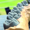
The aim of this study is to investigate and understand the shape, anatomy, and functional characteristics of teeth, with a special focus on the relationship between balancing and working cusps and occlusal stability. The study of the dental morphology cannot be done without analyzing the function of teeth because anatomical characteristics of each tooth are closely linked to its function. A clear example of shape-function relationship is found by examining molar cusps and incisor edges. The relationship between the upper (maxilla) and lower jaw (mandible) are shown mounted on an articulator (Figure 1 and Figure 2).
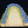
We must bear in mind that creating a manmade tooth must combine shape, function, and esthetics. In the vestibular cavity, we can identify characteristics of anterior and posterior teeth. Incisors (whose name comes from the fact that they are suited to cut or bite food) have a relatively sharp edge. The edge of mandibular incisors makes contact with the palatine middle-third of anterior maxillary teeth. Canines have only one cusp, thanks to which they can tear, pierce, or apprehend food boli-their main functions. The main morphological aspect of posterior crowns is their cuboidal shape, whose occlusal face has three to five cusps, divided by fissures, grooves, and pits that are linked together by ridges (Figure 3). Cusps can be located in the vestibular or oral facing side of the tooth; the primary groove that divides these two cusp groups is called the central sulcus groove. Depending on how cusps make contact with fossae of the opposing teeth, we can identify: 1) stamping or centric holding cusps (palatal cusps of maxillary posterior teeth and vestibular or buccal cusps of mandibular posterior teeth), or 2) shearing or guiding cusps (lingual cusps of mandibular posterior teeth and vestibular or buccal cusps of maxillary teeth) (Figure 4 and Figure 5). Stamping cusps are particularly relevant for the stability of the occlusion because they prevent unwanted parafunctional horizontal movements thanks to their contact with inclining ridge surfaces in opposing fossae. Unfortunately, especially in the case of fixed dental prostheses shaped with wax, dental technologists tend to drastically reduce the size of cusps and types of contact, transferring some of their functions to shearing cusps (which are less suited to bear occlusal loads). Their relevance is clear also when we analyze the distribution of biting loads. As mentioned, maxillary shearing cusps are located in the vestibular cavity while mandibular shearing cusps are lingually oriented.
Looking at the occlusal surface of a molar, we see grooves, fissures, and pits. The grooves can be differentiated as primary or secondary (Figure 6). Primary grooves divide the cusps. Premolar teeth have only a primary groove, their central sulcus groove, which divides the tooth into a vestibular and a lingual half and divides stamping cusps from shearing cusps. Teeth with more than two cusps have a primary groove, but they also have one or more intercuspal grooves. These grooves originate from the central sulcus groove and radiate toward the vestibular or the lingual area, dividing cusps of the same side. Secondary or supplemental grooves are found on the inclined planes of cusps and are usually curved in nature. The direction of grooves depends on mandibular kinematics, because grooves are the approach and the getaway of moving cusps. For this reason, they have a crucial role in ensuring an immediate disclosure of the teeth.
We can identify protrusive grooves, working grooves, and non-working grooves. Protrusive grooves are part of the central sulcus groove; they head from the central pits toward the mesial region in maxillary or upper teeth, toward the distal region in mandibular or lower teeth. They facilitate the transition of centric opposing cusps during the protrusive movement. Working grooves are vestibular in the maxillary teeth and lingual in the mandibular teeth; their direction converges toward the central pits. They offer access on the working side for centric opposing cusps when they move toward the pits. Non-working grooves are mesial-palatal in the maxillary or upper jaw and distal-vestibular in the mandibular or lower jaw, so they go outward following an oblique trajectory. They are a mutual gateway on the balancing side for the centric cusps when they leave the pits (Figure 7). Pits are points of convergence for primary grooves/fissures; in molars, they are the origin of eccentric movements of opposing cusps. Usually, but not always, they receive opposing cusps in occlusion.
Finally, we have ridges, convex projections seen on cusps and on their edges. We can identify marginal ridges and triangular ridges. Marginal ridges link together cusps and bind the outside of the occlusal surface. Triangular ridges start from the top of the cusps and reach the central sulcus groove. Oblique ridges are found on the occlusal surfaces of maxillary molars. The canine ridge is very important for occlusion and the lateral and protrusive movements.
Occlusion is the third anatomical and functional part of the dental apparatus, which is composed of: TMJ, muscles of mastication (chewing muscles), and occlusal table. There are two distinct notions of occlusion: a physiologic occlusion and a therapeutic occlusion. An optimal occlusion is not always the physiologic occlusion; likewise, a physiologic occlusion is not always the optimal one. An essential requirement for balanced occlusion is stability over time with or without compensatory adjustments of the other two parts of the dental apparatus. The functional prosthetic restoration of the occlusion poses different challenges for both clinician and technician. We must know that every minimal anomalous contact, whether during maximum intercuspation or during its function, can cause dysfunctions of the chewing system; for this reason, it is essential to know and to track the function of teeth both in static and dynamic occlusion. Each restoration can influence static and dynamic occlusal balance.
From a clinical and technical point of view, there are two different cases: simple cases (where the occlusion is stable and must be maintained) and complex cases (in which the occlusion is not stable or there is no occlusion due to elements lacking in the intermandibular relationship). The majority of patients display a balanced and reliable occlusion in their normal mandibular position, which is the maximum intercuspation position. In more complex cases, when there are no occlusal references or when the occlusion is not physiological and not balanced, it is necessary to create a new occlusal scheme. The new scheme must be stable in order to have a correct and precise intercuspation—both static and dynamic—and the centric relationship will be used as the new standard mandibular position (whether it is obtained instrumentally or clinically, with adequate moves).
In order to restore a stable and balanced occlusion, it is essential to recreate occlusal tables that anatomically respect:
• cusp-pit relationships,
• precise grooves/fissures of release,
• contact points (not contact areas),
• tripodal disposition in posterior areas and less intense contacts in anterior areas,
• canine guides (opening on the non-working side and they do not have interferences in the working side), and
• anterior guides (in protrusive, they ensure a progressive disclosure of the diatoric areas).
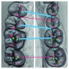
Figure 8 through Figure 12 demonstrate this. Correct visual identification can be carried out in either the laboratory or the clinic using occlusion foils that gradually change their color.
The chewing surfaces of every single tooth are not located in a horizontal plane, but they are located in an imaginary curve called the Curve of Spee (Figure 13). If we observe the arch laterally, this curve is convex in the maxillary arch, while it is concave in the mandibular arch. Anterior teeth are not involved in the Curve of Spee. The Curve of Spee is essential for the disclosure of posterior teeth during the protrusive movement, that is to say when the mandible moves forward because, thanks to a curved line, we have larger disclosure space for posterior teeth compared to the one we could have with a horizontal line. There is also another curve called the Curve of Wilson, or superior concavity, located in the frontal plane and that runs on the tips of vestibular and lingual cusps of both sides (Figure 14). It is determined by the lingual inclination of mandibular molars and premolars and by the vestibular inclination of maxillary molars and premolars. Figure 15 and Figure 16 show the correct cusp-pit relationship.
Conclusion
The concepts described in this article are very important for both the fixed prosthesis and the removable prosthesis. With a thorough understanding of these concepts, the dental team is better equipped to provide the correct occlusion and function for the chewing planes.
About the Author
Gabriele Amos Mainetti
Owner
Teeth and Texture Mainetti
Morbegno, Italy


