You must be signed in to read the rest of this article.
Registration on CDEWorld is free. Sign up today!
Forgot your password? Click Here!
A pontic is an artificial tooth used to fill the space left by a missing tooth in a fixed dental prosthesis (FDP). Extracting a tooth or teeth will result in changes to the edentulous area and the adjacent teeth, and pontics should be designed based on these changes.1 There are different types of pontics used to restore anterior and posterior missing teeth, and it is important to choose a design that satisfies the function, hygiene maintenance, and esthetics of the patient, especially those with high smile lines or poor oral hygiene.2
Basic Pontic Designs
The literature has described four basic pontic designs used over the years in clinical practice: hygienic or sanitary, ridge lap (also known as full ridge lap or total ridge lap), modified ridge lap, and ovate pontics.3
The hygienic (sanitary) pontic design does not contact the soft tissue underneath (Figure 1), which provides easy access for oral hygiene and therefore improves the health of the tissue in the pontic area. However, the gap between the alveolar ridge and the pontic is prone to food impaction. This type of pontic should only be used in the posterior region of the mandible because it compromises the phonetics and esthetics when used while restoring the maxillary FDPs.1
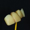
The ridge lap pontic design achieves good esthetic result but can also increase food impaction as the basal contour remains concave (Figure 2 through Figure 4), making it difficult for adaptive contact with dental floss and thereby making it problematic for the patient to achieve hygiene maintenance.1 This design also shows the most histologic changes in the tissue underneath; therefore it should be avoided when designing implant- or tooth-supported FDPs.4 Although both the sanitary and ridge lap designs' original purpose was for hygiene, allowing food to pass through the area between natural tissue and the pontic, many clinicians have moved away from these designs as they can have the reverse effect when designed improperly or introduce changes to the existing tissue.
The modified ridge lap pontic design has a convex basal surface that rests on a small buccal area of the alveolar ridge (Figure 5). The convexity is designed by removing the palatal aspect of the pontic, resulting in a palatal gap that can be an area for food impaction (Figure 6 and Figure 7). The patient has to make sure the palatal gap is always clean. Although the design offers easy access for cleaning in the anterior region, it has some limitations in terms of esthetics, function, and phonetics if the patient exhibits severe ridge resorption.1
Unlike the modified ridge lap pontic, the ovate pontic comes in contact with a large area of soft tissue, applying pressure (Figure 8 and Figure 9). The ovate pontic design is highly functional, provides proper phonetics, and offers pleasing esthetic results in the anterior region because it produces an emergence profile similar to that of a natural tooth. It does, however, offer a very small risk of food impaction.1 Nonetheless, the edentulous area being restored must exhibit a concavity and a healthy amount of soft tissue to support an ovate pontic. Therefore this design is most suitable for immediate provisionalization after extraction-the immediate pontic technique.5 Otherwise, the pontic site will require clinical preparation to accept the ovate pontic by gingival grafting or gradual gingival molding with the provisional (Figure 10).
Pontic Design Considerations
As explained above, there are different pontic designs that can be chosen for FDPs. However, there are multiple factors that also need to be considered when planning the tissue surface of pontics in any FDP.
Function
Although it is recommended to minimize the tissue surface contacts, this should not jeopardize the strength of the FDP under occlusal forces. In short abutment heights, which usually happen in the posterior region (Figure 11 and Figure 12), minimizing the tissue surface contact can lead to weakening of the pontic area. This is especially true in all-ceramic FDPs, where minimal dimensions in connector areas are recommended in order to maintain strength and avoid porcelain fracture.6 In these cases, other interventional approaches such as gingivoplasty, alveoloplasty, or the use of stronger materials for the FDP should be considered.
Hygiene
Some studies indicated that if the tissue surface of the pontic was a glazed/polished material, the tissue response would be minimal. Therefore, porcelain was the material of choice for the tissue-surface of pontics due to its low plaque accumulation. Further studies have now proven that the material used for pontics is irrelevant in relation to the tissue response. There is no significant change in the tissue whether the materials used are glazed or unglazed porcelain, gold, or acrylic resin.7 (Regardless of material selected, a highly polished surface is recommended to eliminate plaque accumulation.) It has been shown in the literature that the design, shape, and hygienic measurements of the pontic play a more crucial role in avoiding any inflammatory reaction of the edentulous area than the material itself. Pontic designs should be convex on the basal tissue surface in order to be cleansable for the patient (Figure 13 through Figure 14). Concave pontic designs on the tissue surface are difficult to clean with dental floss or interproximal brushes (Figure 15 and Figure 16) and therefore make hygiene problematic for patients to maintain. Among all the pontic designs that were mentioned, modified ridge lap and ovate pontic designs demonstrate convex cleansable surfaces. R. Sheldon Stein, DMD, was the first to introduce ovate and modified ridge lap designs.4 These two pontic designs are favorable for hygiene maintenance due to the fact that dental floss can adapt to the basal surface of the pontic for cleaning. The modified ridge lap design can be used primarily in posterior FDPs, and ovate pontics are best used in anterior cases.
Esthetics
Esthetics is among the important factors that need to be considered when it comes to designing pontics in FDPs, especially in anterior cases. Pontics can play a critical role in preserving incisive papillae or the remaining gingiva, particularly in patients exhibiting a high smile line. A slight gingival pressure of the modified ridge lap design or, preferably, the ovate pontic helps maintain the interproximal papillae after extraction or the papillae can be molded gradually by the provisional. Furthermore, the coronal length and position should be designed to flow in line with the adjacent teeth or follow esthetic practice concepts. Adjusting or modifying the provisional facilitates the evaluation of the esthetics for the patient. Provisionals are not only a prototype for the esthetics, incisal-edge positions, phonetics, speech, etc, of the final restoration but also help in evaluating the pontic design to assess the space/anatomic limitations for the most desirable design.
Pontic Designs in a Full-Arch Implant-Supported Prosthesis
Restoring the full maxillary or mandibular edentulous arch with a fixed implant-supported prosthesis can be more challenging due to bone resorption patterns and the uneven remaining alveolar ridge.8 When designing pontics in such cases, the esthetic, functional, and most importantly, hygienic demands for the patient must be considered.9 There are some complications regarding fixed implant-supported prostheses in the edentulous maxilla. The most common challenge is poor phonetics, usually because air escapes through the cervical embrasures.8 This could be improved by adding porcelain to the gingival area of the prosthesis, filling the interproximal spaces between the implants, which would also improve the esthetics. Adding porcelain to the basal surface of the prosthesis can lead to a concave basal surface (Figure 16). Therefore, after sealing the gaps and improving the phonetics and esthetics, it is recommended to evaluate the basal surface of the implant-supported prosthesis to recontour and convert any concavities to convexities, which make it easier for the patient to clean (Figure 15). Designing cleansable pontics on implant-supported prostheses is critical. Tissue inflammation from poor oral hygiene can easily be directed towards the implant and the marginal bone level, causing the implant to move towards an ailing and failing diagnosis.
Conclusion
Although the abovementioned factors were explained separately in this article, they should be considered simultaneously since they are intertwined; ignoring one can jeopardize the others. For instance, uncleansable poor pontic design can lead to inflammation of the underlying tissue, which in turn can lead to bone resorption and gingival recession, resulting in poor esthetics and compromising the prognosis of the abutments. In pontic design, as everywhere in dentistry, an inch of prevention is worth a pound of cure.
Acknowledgements:
The authors would like to acknowledge Steve Pigliacelli, MDT, CDT, of Marotta Dental Studio for providing some of the pictures.
About the Authors
Siamak Najafi-Abrandabadi, DDS
Clinical Assistant Professor, Department of Prosthodontics
New York University College
of Dentistry
New York, NY
Maria Claudia Alvarado, DDS
Advanced Programs for International Dentists in
Esthetic Dentistry
New York University College of Dentistry
New York, NY
References
1. Howard W, Ueno H, Pruitt C. Standards of pontic designs. J Prothet Dent. 1982;47(5):493-5.
2. Edelhoff D, Spiekermann H, Yildirim M. A review of esthetic pontic design options. J Prothodont. 2002;33(10):736-46.
3. Tae Hyung K, Domenico C, Alena K. simulated tissue using a unique pontic design: A clinical report. J Prosthet Dent. 2009;102(4):205-10.
4. Stein RS. Pontic-residual ridge relationship: A research report. J Prosthet Dent. 1966;16:251.
5. Spear F. Maintenance of interdental papilla following anterior tooth removal. Pract Periodont Aesthet Dent. 1999;11(1):21-8.
6. Mahmood DJ, Linderoth EH, Vult Von Steyern P. The influence of support properties and complexity on fracture strength and fracture mode of all-ceramic fixed dental prostheses. Acta Odontol Scand. 2011;69(4):229-37.
7. Podshadley AG. Gingival response to pontics. Journal of Prosthodontics. 1968;19(1):51-7.
8. Del Castillo R, Ercoli C, Delgado JC, Alcaraz J. An alternative multiple pontic design for a fixed implant-supported prosthesis. J of Prosthet Dent. 2011;105:198-203.
9. Garber DA, Rosenberg ES. The edentulous ridge in fixed prosthodontics. Compend Contin Educ Dent.1981;2:212-23.


