You must be signed in to read the rest of this article.
Registration on CDEWorld is free. Sign up today!
Forgot your password? Click Here!
Today periodontal probing is the best diagnostic tool to gather information regarding the health status and attachment level of periodontal tissues. Periodontal probing requires special skills as well as an understanding of the tissues being examined, the probing procedure, and the use of an appropriately designed instrument. Periodontal probing seeks to complement the initial visual assessment of the periodontal tissue. It has multiple roles: to assess the hemorrhagic response to physical pressure; to determine the presence of etiologic factors such as calculus, defective dental restorations, and root erosion; to locate the cementoenamel junction (CEJ); and to determine the pocket dimensions. While it remains the best way to measure probing depths and status of the clinical attachment level during clinical examinations, periodontal probing has several drawbacks when used to monitor periodontal status longitudinally. Despite its lack of accuracy in determining sulcus or pocket depth, probing provides the clinician with a useful estimate of the location of the coronal insertion of intact connective-tissue fibers into the root. Although the true anatomic measurement of the pocket can be accomplished solely through histologic examination,1 periodontal probing depth (PPD) is still an important clinical measurement because the depth of the pocket and degree of attachment loss may influence the course of the disease.2
To determine the degree of periodontal breakdown accurately, the tip of the probe must be located at the most coronal intact connective-tissue fibers. In other words, the “true” pocket depth must be measured. In many cases, however, the recorded measurements do not correspond with true pocket depth measurements. This discrepancy may be caused by anatomic or pathologic characteristics of the pocket tissues or those surrounding the pocket, individual characteristics of the probe used, or operator factors such as probing force, probe placement, angulation, manual dexterity, and accuracy of observation.3,4 Factors that may influence the precision of periodontal probing are related to design and handling facility of the instrument and health of the gingival tissues, as well as experience of the clinician.5,6
This review summarizes various aspects of PPD measurement. The importance of periodontal health relative to probe-tip penetration also is reviewed. Different factors that influence the PPD measurement are discussed in detail in conjunction with their effect on accuracy and reproducibility of the PPD measurements. Three generations of periodontal probes also are discussed.
Periodontal Health
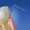
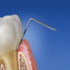
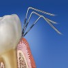
A primary aim of periodontal probing is to locate the most coronal level of the connective-tissue attachment. However, this generally is not attainable, as penetration of the probe tip in the pocket or the lining soft tissues correlates with periodontal health.7,8 It has been established that the extent of probe penetration is influenced by the inflammatory status of the tissues.7,9-12 In most instances when healthy tissues are examined, the probe tip stops coronal to the apical termination of the junctional epithelium (Figure 1), whereas at inflamed sites the probe tip frequently passes apical to this point (Figure 2). The depth of probe penetration partially depends on the extent to which the gingival connective tissue has been lysed or infiltrated by inflammatory cells. In other words, intact connective tissue underlying the crevicular epithelium is an important factor resisting probe penetration. Spray et al13 suggested that the state of health of the underlying connective-tissue fibers influences probing measurements. There is a “hammock effect” in health, where healthy fibers act as a barrier and prevent apical movement of the instrument, while inflamed connective tissue offers less resistance to penetration. With reduction in inflammation, an accurate estimate of the sulcus depth is more likely to be obtained. The probe penetration is significantly greater in the presence of visible inflammation, but not where there was bleeding after probing.8 These results suggest that the location of the inflamed connective tissue may be a critical factor. Anderson et al14 determined the correlation between clinical and histologic inflammation and probe-tip penetration of the pocket tissues in dogs. A strong correlation was found between probe penetration and degree of inflammation, and the difference in mean inflammation scores between sites where probes were located coronal or apical to the epithelium was statistically significant.
Anatomically, the gingival sulcus is defined as the distance from the gingival margin to the coronal extension of the junctional epithelium.15 However, the ability of the periodontal probe to measure this distance accurately is questionable. Results of human studies performed by Sivertson and Burgett16 indicate that the periodontal probe routinely penetrates to the coronal level of the connective tissue attachment of untreated periodontal pockets. Armitage et al7 found that, in healthy specimens, the probe failed to reach the apical termination of the junctional epithelium. In cases with experimental gingivitis, however, most probes came closer to the apical termination of the junctional epithelium, but on the average still fell short. In periodontitis specimens, the probes consistently went past the most apical cells of the junctional epithelium. A significant relationship between the degree of inflammation and level of probe penetration was found. Saglie et al17 noted that probing depths measured in the laboratory were always shallower than those recorded clinically. The authors attributed this discrepancy to the presence of a zone of completely and partially destroyed periodontal fibers, which allowed the probe to extend apically to the coronal level of connective-tissue attachment. The results of these studies illustrate that periodontal probes do not precisely measure, and often overestimate, the true histologic sulcus depth, and that inflammation has a significant influence on probe penetration. This has important implications regarding how measurements taken with periodontal probes are interpreted. Because probes rarely stop at the exact location of the most apical cells of the junctional epithelium, probing measurements are clearly not precise assessments of the actual level of connective-tissue attachment. PPD measurements overestimate connective-tissue attachment loss at inflamed sites and underestimate it at noninflamed sites. An increased probing depth is a sign of reduced tissue resistance to probing, which in turn can be interpreted as an indication of the presence of an inflammatory cell infiltrate in the gingival tissue.11 Most research has shown that the tendency for penetration of the probe into the tissues at the base of pocket resulting in an overestimate of probing depth is greater at inflamed sites7,10,11 and in nonsmokers.18
Periodontal Probes
The periodontal probe continues to be one of the more useful diagnostic tools to determine the presence and severity of periodontal lesions. An ideal periodontal probe should possess specific characteristics:
1. It should be tissue-friendly and not traumatize periodontal tissues during probing.
2. It should be suitable as a measuring instrument.
3. It should be standardized to ensure reproducibility, particularly with respect to recommended pressure.
4. It should be suitable both for use in the clinical setting where precise data documentation is required on an individual patient basis, and for screening purposes, as in epidemiology.
5. It should be easy and simple to use and read.
Over the years, the shape, design, and function of probes have changed to enhance accuracy and reproducibility. Three generations of probes have been suggested by Philström19: first generation—conventional handheld instruments; second generation—force application during measurement; third generation—force application using automated measurement and computerized data capture. The conventional handheld probes most commonly are preferred for their ease and simplicity in application. However, the use of second- and third-generation probes also is common, especially in the field of research where variables such as pressure or force on probing, reproducibility, and accuracy are investigated. Various studies considering these different probes and their characteristics also are found in the literature. Samuel et al20 have published an in vitro study testing the accuracy and reproducibility of automated and conventional probes. In that study automated probes were reported to offer increased accuracy over conventional probes, and the reproducibility of both Florida pocket-depth and disk probes was found to be comparable with that of the conventional probes. Buduneli et al21 in an in vitro model investigated the accuracy and reproducibility of two manual probes and concluded that overall accuracy was higher with the WHO probe compared with the Williams probe. This study also revealed better reproducibility percentages for the WHO probe in comparison with the Williams probe.
Probe Characteristics
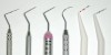
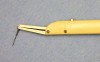
Characteristics of the probe, such as its diameter at the tip and the calibration, can influence PPD measurement. Different probes, such as Michigan, Williams, Marquis, Goldman-Fox, and Nabers probes, have different dimensions and a different diameter at the tip. The tip diameters range from 0.28 mm for the Michigan “O” probe to 0.7 mm for the Williams probe. Moreover, the widths of probe markings in the painted bands differ by as much as 0.7 mm between probes because of manufacturing errors. Figure 3 illustrates different manual probes. Van der Zee et al5 evaluated the accuracy of probe markings in a variety of probes and noted that probes from the same batch from the same production line could differ by more than 0.5 mm in calibration and the mean tip diameter ranged from 0.28 mm to 0.7 mm. They concluded that probe-tip diameter and calibration should be considered in addition to other variables of periodontal probing. Standardization of tine characteristics and avoidance of the use of different types or batches in a single study should enhance the accuracy and reproducibility of periodontal probe-dependent measurements.
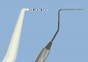
Atassi et al22 compared a parallel-sided probe to a tapered probe (Figure 4). Results indicated that the parallel-sided tine tended to yield a deeper reading when a difference occurred. Garnick and Silverstein23 reviewed the effect of the probe-tip diameter on accurate probe placement and recommended a probe-tip diameter of 0.6 mm and a 20-g force to measure a reduction in the clinical probing depth after therapy. Quirynen et al24 found interexaminer variability was dependent upon probe type. The study compared a conventional periodontal probe with an automatic, computerized, constant-force, electronic probe in vivo and found that PPD measurements recorded with the manual probe were consistently deeper than those recorded with the electronic probe. Wang et al4 evaluated intra- and interexaminer reproducibility for conventional and electronic probes and found that reproducibility may not necessarily be higher with an electronic, force-controlled periodontal probe than with a conventional manual probe. In an attempt to overcome some of the technical challenges associated with conventional manual periodontal probes, numerous electronic periodontal probes have been developed that permit probe insertion with a controlled force.9
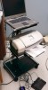
The controlled-force probe that has achieved the most widespread use is the Florida Probe® (Florida Probe Corp, Gainesville, FL) (Figure 5A and Figure 5B). This computer-linked device has in vitro resolution of 0.1 mm and is capable of recording probing depths and relative attachment levels.25-31 Clinical measurements obtained with conventional manual probes are consistently greater than those obtained with controlled-force probes.24,32-37 One of the possible reasons for this is reduced tactile sensitivity associated with the use of controlled-force probes. This is especially true in patients with untreated periodontitis for whom the presence of subgingival calculus can interfere with probe insertion. With conventional probes, it generally is easier for the operator to manipulate the probe tip past subgingival calculus deposits. A definite advantage of computer-linked probes is that they can record probe readings automatically. Some systems allow voice-activated data entry.38 The usefulness of controlled-force probes in day-to-day clinical practice has not yet been demonstrated.9
One possible reason for the lack of widespread acceptance of controlled-force electronic probes by practitioners might be increased patient discomfort when these devices are used, particularly around the anterior teeth. During probing with conventional manual probes, the operator can decrease the insertion force rapidly if the patient shows any early signs of discomfort. With controlled-force probes, this patient–dentist feedback is not possible because the probe is inserted into the pocket in one motion and with fixed or predetermined force.
Probing Force
Probing force can influence probing measurement and has been studied widely. Force-controlled periodontal probes have been introduced to increase the reliability of probing measurements. Van der Velden et al3 used probing forces of 0.15 N, 0.25 N, 0.5 N, and 0.75 N and found a significant difference between PPD recordings with a low and a high probing force. As a result, they recommended a probing force of 0.75 N as optimal, with probes of 0.63 mm in diameter. Chamberlain et al6 indicated that higher probing forces are more reproducible. In their study, the probe tip extended to the most coronal connective-tissue attachment in health and disease, using a force of 0.75 N. Reproducibility is important, however, because increasing PPDs or attachment-level changes may be indicative of disease activity. Reproducibility of repeated measurements has been considered a good indicator of reliability.33,39
Osborn et al34 compared the intra- and interexaminer measurement error of the Florida pocket-depth probe, the Florida disk probe, and the conventional manual probe in subjects with moderate to severe periodontitis. At the site level, the mean intraexaminer standard deviations of differences in repeated relative attachment-level measurements using the Florida disk probe and the conventional probe ranged from 0.55 mm to 0.82 mm and 0.62 mm to 1.14 mm respectively. Intraexaminer standard deviations of differences in probing depth measurements using the Florida pocket-depth probe ranged from 0.6 mm to 0.93 mm (differences in relative attachment levels for the Florida pocket-depth probe was not reported). Hassel et al40 noted a substantial variation in probing force exerted by six clinicians who probed four surfaces of 30 teeth in five patients. Moreover, they found a poor correlation between PPD measured by the probe and the probe force applied. They concluded that probing forces had only a moderate influence on the depth of measurements and that the probing technique was the more critical factor in PPD measurement than the pressure applied to the probe. Hassel et al40 reported that probing technique was important if clinical evaluations were to be correlated to the condition in reality, and favored a slow, deliberate searching style of probing for each area of the pocket. They also reported that it was the pocket topography rather than the absolute PPD that was important. Furthermore, they stated that application of heavy force is contraindicated and does not lead to greater precision.
Sites of Measurement
Persson41 compared line-angle measurements with midproximal measurements in untreated sites and found that the mean PPD was 1 mm greater with midproximal measurements than with line-angle measurements. This finding implies that clinical and epidemiologic studies using line-angle measurements may underestimate PPD and, thus, the true level of disease. From one visit to another, it is difficult to duplicate precisely the insertion force and to reproduce exactly the site and angulation of probe insertion. PPD appears to influence the reproducibility of PPD measurements, and the general consensus is that measurements of shallow pockets are more reproducible than those of deep pockets.42 Kalkwarf and Kaldahl43 determined that the pressure-controlled technique produced significantly deeper clinical probing measurements on the direct facial and lingual aspects of teeth regardless of the stage of periodontal therapy that had been completed, and manual probing obtained deeper measurements on the distolingual aspects of teeth in the posterior regions, which had not received surgical therapy. Control of vertical force during probing may provide a more objective method of monitoring periodontal status during longitudinal trials. In shallow pockets (1 mm to 3 mm) the difference was smallest, and the discrepancy increased with increasing PPD regardless of sites, stage of therapy, or location in the mouth. As well as varying by PPD, examiner reproducibility for any given method also may vary among tooth types, tooth surfaces, and PPD.39 The higher degree of reproducibility for buccal surfaces probably is explained by better visibility and by facilitated reproducibility of probe placement within the shallower pockets on these surfaces. Furthermore, in this study, access, visibility, as well as cooperation during measurements, varied between patients. Figure 6 illustrates proper probe insertion technique, highlighting possible outcomes based on variations in the angle of insertion.
Conclusion
For more than a century, the periodontal probe has played an integral part in the periodontal examination and the detection of periodontal diseases. Its use not only enables treatment to be planned appropriately, but also facilitates longitudinal monitoring so that the response to treatment may be assessed and sites of possible disease progression identified. Yet, periodontal probing is an imprecise technique with several potential sources of error.44-46 This review has described the effect of a number of these variables on PPD measurement. Because of the different factors and technical challenges affecting the PPD measurements, it is generally expected that the consecutive readings of PPD at a given site may vary by up to 1 mm as a function of the limited sensitivity of this system of measurement.9,42 Despite these problems and challenges, properly used periodontal probes provide critically important information regarding the periodontal status of patients. Measurements obtained with periodontal probes are the best way to assess damage caused by periodontal infections and are essential for longitudinally monitoring the response to treatment.
Acknowledgments
The authors thank Dr. E. B. Hancock and Dr. S. B. Blanchard, Indiana University School of Dentistry, for their support and feedback. The authors also thank Dr. Sarah Fitzpatrick, University of Florida, for her assistance with the Florida Probe images.
References
1. Listgarten MA. Periodontal probing: what does it mean? J Clin Periodontol. 1980;7(3):165-176.
2. Carlos JP, Brunelle JA, Wolfe MD. Attachment loss vs. pocket depth as indicators of periodontal disease: a methodologic note. J Periodontal Res. 1987;22(6):524-525.
3. van der Velden U, de Vries JH. Introduction of new periodontal probe: the pressure probe. J Clin Periodontol. 1978;5(3):188-197.
4. Wang SF, Leknes KN, Zimmerman GJ, et al. Intra- and inter-examiner reproducibility in constant force probing. J Clin Periodontol. 1995;22(12):918-922.
5. van der Zee E, Davies EH, Newman HN. Marking width, calibration from tip and tine diameter of periodontal probes. J Clin Periodontol. 1991;18(7):516-520.
6. Chamberlain AD, Renvert S, Garrett S, et al. Significance of probing force for evaluation of healing following periodontal therapy. J Clin Periodontol. 1985;12(4):306-311.
7. Armitage GC, Svanberg GK, Löe H. Microscopic evaluation of clinical measurements of connective tissue attachment levels. J Clin Periodontol. 1977;4(3):173-190.
8. Caton J, Greenstein G, Polson AM. Depth of periodontal probe penetration related to clinical and histologic signs of gingival inflammation. J Periodontol. 1981;52(10):626-629.
9. Armitage GC. Periodontal diseases: diagnosis. Ann Periodontol. 1996;1(1):37-215.
10. Robinson PJ, Vitek RM. The relationship between gingival inflammation and resistance to probe penetration. J Periodontal Res. 1979;14(3):239-243.
11. Fowler C, Garrett S, Crigger M, et al. Histologic probe position in treated and untreated human periodontal tissues. J Clin Periodontol. 1982;9(5):373-385.
12. Tessier JF, Ellen RP, Birek P, et al. Relationship between periodontal probing velocity and gingival inflammation in human subjects. J Clin Periodontol. 1993;20(1):41-48.
13. Spray JR, Garnick JJ, Doles LR, et al. Microscopic demonstration of the position of periodontal probes. J Periodontol. 1978;49(3):148-152.
14. Anderson GB, Caffesse RG, Nasjleti CE, et al. Correlation of periodontal probe penetration and degree of inflammation. Am J Dent. 1991;4(4):177-183.
15. Listgarten M. Ultrastructure of the dento-gingival junction after gingivectomy. J Periodontal Res. 1972;7(2):151-160.
16. Sivertson JF, Burgett FG. Probing of pockets related to the attachment level. J Periodontol. 1976;47(5):281-286.
17. Saglie R, Johansen JR, Flotra L. The zone of completely and partially destructed periodontal fibers in pathological pockets. J Clin Periodontol. 1975;2(4):198-202.
18. Biddel AJ, Palmer RM, Wilson RF, et al. Comparison of the validity of periodontal probing measurements in smokers and non-smokers. J Clin Periodontol. 2001;28(8):806-812.
19. Philström LB. Measurement of attachment level in clinical trials: probing methods. J Periodontol. 1992;63(12 Suppl):1072-1077.
20. Samuel ED, Griffiths GS, Petrie A. In vitro accuracy and reproducibility of automated and conventional periodontal probes. J Clin Periodontol. 1997;24(5):340-345.
21. Buduneli E, Aksoy O, Köse T, et al. Accuracy and reproducibility of two manual periodontal probes. An in vitro study. J Clin Periodontol. 2004;31(10):815-819.
22. Atassi F, Newman HN, Bulman JS. Probe tine diameter and probing depth. J Clin Periodontol. 1992;19(5):301-304.
23. Garnick JJ, Silverstein L. Periodontal probing: probe tip diameter. J Periodontol. 2000;71(1):96-103.
24. Quirynen M, Callens A, van Steenberghe D, et al. Clinical evaluation of a constant force electronic probe. J Periodontol. 1993;64(1):35-39.
25. Gibbs CH, Hirshfeld JW, Lee JG, et al. Description and clinical evaluation of a new computerized periodontal probe—The Florida Probe. J Clin Periodontol. 1988;15(2):137-144.
26. Magnusson I, Fuller WW, Heins PJ, et al. Correlation between electronic and visual readings of pocket depths with a newly developed constant force probe. J Clin Periodontol. 1988;15(3):180-184.
27. Magnusson I, Clark WB, Marks RG, et al. Attachment level measurements with a constant force electronic probe. J Clin Periodontol. 1988;15(3):185-188.
28. Marks RG, Low SB, Taylor M, et al. Reproducibility of attachment level measurements with two models of Florida Probe. J Clin Periodontol. 1991;18(10):780-784.
29. Yang MCK, Marks RG, Magnusson I, et al. Reproducibility of an electronic probe in relative attachment level measurements. J Clin Periodontol. 1992;19(8):541-548.
30. Clark WB, Yang MCK, Magnusson I. Measuring clinical attachment: reproducibility of relative measurements with an electronic probe. J Periodontol. 1992;63(10):831-838.
31. Clark WB, Magnusson I, Namgung YY, et al. The strategy and advantage in use of an electronic probe for attachment measurement. Adv Dent Res. 1993;7(2):152-157.
32. Galgut PN, Waite IM. A comparison between measurements with a conventional periodontal pocket probe, an electronic pressure probe and measurements made at surgery. Int Dent J. 1990;40(6):333-338.
33. Badersten A, Nilvéus R, Egelberg J. Reproducibility of probing attachment level measurements. J Clin Periodontol. 1984;11(7):475-485.
34. Osborn JB, Stoltenberg JL, Huso BA, et al. Comparison of measurement variability in subjects with moderate periodontitis using a conventional and constant force periodontal probe. J Periodontol. 1993;63(4):283-289.
35. Rams TE, Slots J. Comparison of two pressure-sensitive periodontal probes and a manual probe in shallow and deep pockets. Int J Periodontics Restorative Dent. 1993;13(6):521-529.
36. Perry DA, Taggart EJ, Leung A, et al. Comparison of a conventional probe with electronic and manual pressure regulated probes. J Periodontol. 1994;65(10):908-913.
37. Hull PS, Clerehugh V, Ghassemi-Aval A. An assessment of the validity of a constant force electronic probe in measuring probing depths. J Periodontol. 1995;66(10):848-851.
38. Mintzer RE, Derdivanis JP. Automated periodontal probing and recording. In: Yukna RA, Newman MG, Williams RC, eds. Current Opinion in Periodontology. Philadelphia, PA: Current Science; 1993.
39. Osborn J, Stoltenberg J, Huso B, et al. Comparison of measurement variability using a standard and constant force periodontal probe. J Periodontol. 1990;61(8):497-503.
40. Hassell TM, Germann MA, Saxer UP. Periodontal probing: interinvestigator discrepancies and correlations between probing force and recorded depth. Helv Odontol Acta. 1973;17(1):38-42.
41. Persson GR. Effects of line-angle versus midproximal periodontal probing measurements on prevalence estimates of periodontal disease. J Periodontal Res. 1991;26(6):527-529.
42. Glavind L, Löe H. Errors in the clinical assessment of periodontal destruction. J Periodontal Res. 1967;2(3):180-184.
43. Kalkwarf KL, Kaldahl WB, Patil KD. Comparison of manual and pressure-controlled periodontal probing. J Periodontol. 1986;57(8):467-471.
44. Watts T. Constant force probing with and without a stent in untreated periodontal disease: the clinical reproducibility problem and possible sources of error. J Clin Periodontol. 1987;14(7):407-411.
45. Watts TL. Visual and tactile observational error: comparative probing reliability with recession and cementoenamel junction measurements. Community Dent Oral Epidemiol. 1989;17(6):310-312.
46. Watts TL. Probing site configuration in patients with untreated periodontitis. A study of horizontal positional error. J Clin Periodontol. 1989;16(8):529-533.
About the Authors
Saba Khan, DDS, MSD, Clinical Assistant Professor, Department of Periodontology, University of Illinois, College of Dentistry, Chicago, Illinois
Leyvee Lynn Cabanilla, DDS, MSD, Assistant Professor, Department of Periodontology and Dental Hygiene, University of Detroit Mercy School of Dentistry, Detroit, Michigan