You must be signed in to read the rest of this article.
Registration on CDEWorld is free. Sign up today!
Forgot your password? Click Here!
Restoring function and esthetics for the partially edentulous patient is rewarding for both the patient and dentist. Both anterior and posterior treatment options are similar. However, the esthetic zone presents with it the challenge of achieving acceptable cosmetic outcomes. Meeting patient expectations may, therefore, present with it another set of challengers. The focus of this article is to present single-tooth replacement options for the esthetic zone. Although this topic has been described elsewhere,1 the approach here will be from a treatment outcome perspective: functionally, biologically, esthetically, and cost-effectively.
When faced with a missing anterior tooth, there are essentially five options available:
- No treatment. Leave as is.
- Removable partial denture (RPD) therapy: acrylic or cast-metal frame.
- Resin-bonded fixed partial denture (FPD): 3-unit or 2-unit cantilever.
- Conventional FPD: 3-unit or 2-unit cantilever.
- Single implant-supported restoration.
NO TREATMENT—LEAVE AS IS
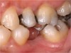
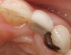
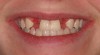
Patients rarely choose to do nothing. With culturally high esthetic demands, this option is socially undesirable.2 In fact, patients with missing teeth that are not replaced have inferior psycho-social outcomes when compared to restored patients.3 From a functional standpoint, a single missing tooth can lead to additional long-term problems, such as tooth drift, both interproximally and vertically, resulting in space collapse, and the development of possible occlusal irregularities (Figure 1). However, patients are not seeking consultations in dental offices for this option.
RPD THERAPY
Although not high on a patient’s wish-list for tooth replacement therapy, this option presents several advantages that are difficult to ignore. Advantages include4:
- It is biologically conservative, as alteration of adjacent tooth structure, if required, is usually minimal.
- It is removable for ease of oral hygiene and cleansability.
- Low financial commitment compared to other treatment modalities.
Disadvantages include4,5:
- It is removable. Some patients may not find this a desirable feature.
- Due to increased surface area, it collects significant plaque and requires constant cleaning.
- Wearing an RPD 24 hours a day may cause problems of soft tissue inflammation, tooth decay, or adverse force transfer to abutments during nocturnal parafunctional activity.
- When the protocol for use and hygiene are not followed by the patient, there is a high probability of associated irreversible abutment teeth problems.
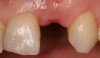
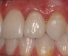
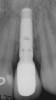
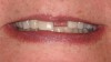
Depending on the patient’s treatment objectives, this option is rarely viewed as a long-term solution to the problem at hand. However, as an interim short-term tooth replacement option, this treatment modality may prove to be a useful solution allowing the patient the necessary time to prepare both financially and/or psychologically for a more definitive treatment solution (Figure 2).
RESIN BONDED FPD: 3-UNIT OR 2-UNIT CANTILEVER
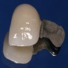
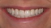
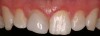
The conservative resin-bonded winged FPD approach (also known as the Maryland Bridge) presents with it the attractive option of biological conservation of tooth structure (Figure 3). This appeal is significant, warranting serious consideration for this treatment approach.6,7 Unfortunately, as appealing as it is, the long-term functionality of this option has proved to be less than ideal. Other problems related to this treatment modality include discoloration of retainer/support teeth due to the metal wing, debonding, and removal of lingual tooth structure to accommodate the wing retainers.8
CONVENTIONAL FPD: 3-UNIT OR 2-UNIT CANTILEVER
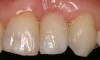
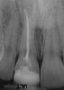
Before the introduction of root-form dental implant therapy into the North American marketplace,9this option was considered the standard of care for fixed tooth replacement in the esthetic zone; either as a 3-unit end abutment FPD or 2-unit cantilever FPD when indicated (Figure 4).10 Material choices have recently expanded from the porcelain-fused-to-metal prostheses to various all ceramic solutions.11,12 However, for conventional FDPs, the fundamental disadvantage that this option has over all others presented to date include the need for significant tooth reduction/removal to accommodate the retainer full-coverage/partial-coverage restorations. Consequently, when compared to all of the other options presented, including dental implant therapy yet to be discussed, it can be argued that conventional FPDs are the most biologically invasive.
SINGLE IMPLANT-SUPPORTED RESTORATION
For dental implant therapy to be possible, the anterior missing tooth site requires sufficient bone volume for the placement of a conventional root form dental implant. Also, to achieve suitable esthetic outcomes, the soft-tissue profile requires similar anatomy to the contra-lateral side.13-15 If bone and/or soft-tissue bulk is inadequate, then various surgical hard and soft tissue augmentation procedures need to be considered, either as a separate procedure or, if indicated, at the time of implant placement.13,14
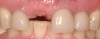
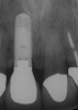
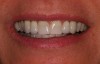
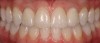
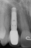
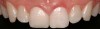
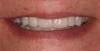
Of all the options presented, when treatment planned and performed to recommended implant surgical and prosthetic guidelines, this option is the most conservative from biological standpoint. The esthetic outcome can be as natural as the tooth it is otherwise replacing (Figure 5).13-15 Patient satisfaction for such successful treatment outcomes is high.16 However, not all patients who are missing a single anterior tooth are ideal candidates for implant therapy. There may be medical contraindications, significant site preparation required, and/or financial constraints. Dental providers may find the resin-bonded or conventional FPD option a more suitable treatment modality with the patient accepting the associated disadvantages and limitations. If this is the case, it is important to have a wider overall treatment outcome picture when comparing the different options.17 From a long-term perspective, few patients perceive real value to either doing nothing or choosing an RPD. Consequently, this leaves the two main contenders: conventional FPD therapy versus single-tooth implant therapy.
FPD THERAPY VERSUS SINGLE-TOOTH IMPLANT THERAPY
One approach when comparing the two treatment modalities is to evaluate and compare treatment outcome survival rates. For FPDs, the areas commonly evaluated in the literature include: pulp vitality, root canal therapy, post/core treatments, short-span versus long-span FPDs, and cantilevers.
Pulp Vitality
- In a recent study by Cheng et al18 evaluating 10 years of collective data in 122 single crowns and 77 FPD groups, survival rates for pulp vitality
- were 84.4% for single crowns and 70.8% for FPDs. Maxillary anterior teeth used as FPD abutments had the highest rate of pulpal necrosis. The
- authors concluded that “the survival of the vital pulp in teeth restored with a single crown was significantly higher than those serving as an
- abutment of a fixed partial denture.”
Abutment Teeth with Root Canal Treated Post/Core Foundations
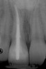
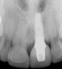
De Backer et al19 recently evaluated data collected over a 16- to 20-year period from the undergraduate dental clinics at Ghent University, Belgium. For the single-crown group, similar survival rates were found in vital (74.9%) and root canal treated (RCT) groups (79.4%) (Figure 6). At 20 years, the FPD was 77.4% for the vital group and 56.7% when at least one of the abutments had root canal therapy. The authors concluded that “for FPDs with more than 3 units and cantilever FPDs, the use of a post-and-core abutment led to significantly more failures.” These conclusions were also supported by Tan et al.20
Short-Span FPDs versus Long-Span FPDs
In a separate published article by De Backer et al21 evaluating the same pulled data discussed above, it was found that short-span FPDs had significantly higher survival rates (70.8%) than long-span FPDs (52.8%). The mode of failure with the short-span group tended to be biologic, as opposed to the long-span group, which was primarily mechanical. With further evaluation of the data, the authors where able to provide further insight into this category, concluding the following: “a reversible complication within the first 2 years for a short span FPD will lead to an irreversible complication.”
Cantilever FPDs
Pjetursson et al performed a systematic review of the literature, evaluating survival and complication rates of FPDs. The most common
biological complication reported was loss of pulp vitality (32.6%) followed by caries at abutment teeth (9.1%). Loss of retention was the most frequent technical complication (16.1%) followed by material fractures (5.9%). The authors also concluded that FPDs with associated cantilever pontic extensions have overall lower survival rates when compared with end-abutment supported FPDs.22
Complications
In a comprehensive literature review by Goodacre et al8 incorporating 50 years of published data, some of the findings included the following:
- Most common cause of failure in single crowns: endodontic 3%, porcelain veneer fracture 3%, loss of retention 3%.
- Most common cause of failure in conventional FPDs: caries (18% of abutments), endodontic 11%, loss of retention 7%.
- Most common cause of failure in resin-bonded FPDs: debonding 21%, tooth discoloration 18%, and caries 7%.
The authors also concluded that single crowns (all-ceramic and conventional) had significantly lower complication rates (8% to 11%) than conventional FPDs (27%) or resin-bonded prostheses (26%).
Tooth-Supported FPD Survival Rates
When reviewing the literature for overall FPD survival rates, some of the findings include the following: 15 years, 64% success23; 10 to 15 years, 70% success (8% abutments had to be extracted).24 In 1998, Scurria et al25 reviewed the literature and found only nine studies of value, a total of 2,761 retainers over a 3- to 20.5-year period. A meta analysis was performed and FPD survival rates were summarized as follows: 10 years: 85% success; 15 years: 66%.
Dental Implant versus FPD Survival Rates
Documented 5- to 10-year survival rates for single-tooth dental implant restorations range from 95.6% to 97.4%.26-31 Consequently, from a purely survival-rate standpoint, single-tooth implant therapy rates more favorably than FPD therapy. However, there is little published data directly comparing the two treatment modalities. Torabinejad et alrecently performed a systematic review comparing 143 selected articles.3 Findings of interest included the following: single root-canal-treated (RCT) teeth had higher survival rates than FPDs and implant-supported single crowns had superior long-term survival rates when compared with FPDs.
Discussion
The purpose of breaking down the data as presented above is to provide a foundation for the decision-making process. This may prove useful not only from a treatment planning standpoint, but to also effectively communicate the advantages and disadvantages of available options to the patient. In this way, the patient becomes an informed consumer and will be in a stronger position to make a decision that will meet his or her expectations.
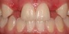
When evaluating the data presented on FPDs, certain conclusions can be made. FPDs have short lifespans after 10 years, with caries and endodontic problems the primary cause of failure.8,23-25 Once a complication occurs, the failure of FPDs often results in the loss of abutment teeth and the need for additional abutment teeth and pontics. Also, FPD retainer crowns/abutments have lower survival rates than single (non-splinted, stand-alone, tooth-borne) crowns.8,18 RCT FPD abutment teeth have significantly lower success rates than non-RCT-abutments or single crowns.19,20 With single crowns, RCT single crowns (Figure 6) have a similar survival rate as non-RCT crowns.19,20
The most notable insight from the presented data is that teeth requiring single full-coverage restorations have satisfactory survival rates.3,8 However, once the crowned tooth is splinted to another tooth, whether it is an abutment or pontic, the survival rate of those teeth decreases significantly.8,18-20 If an abutment tooth also had root canal therapy, the survival rate is further affected.19,20 Consequently, in the author’s experience, FPD therapy can be looked at as a subtractive treatment approach, both from a biological standpoint and also from the number of teeth required to replace the missing teeth.
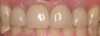
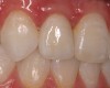
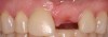
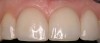
When a patient presents with a missing anterior tooth and heavily treated adjacent teeth, although conceptually an FPD option may seem the most appropriate choice, it can be argued that it would not be the most favorable option. If long-term treatment outcome is a strong motivator for the patient, then a treatment plan that includes individual restored units would be the more appropriate choice. Consequently, the patient would most likely have improved benefit with a single implant-supported restoration and treating the adjacent teeth as separate, single units (Figure 7).
Implant therapy does have its disadvantages. It involves surgical intervention, both with placement and possible adjunct bone and soft tissue augmentation to allow for ideal positioning of the implant body.13,14 Consequently, additional initial costs are often associated with this treatment modality when compared with FPDs.32 It also takes time, considerably more than FPD therapy.
From a treatment planning and case acceptance standpoint, the argument for and against any one of the available tooth replacement options can be made. However, the literature indicates that single-implant dental therapy will provide greater overall advantages when compared to the other treatment modalities. Advantages include:3,13-16,29
- Biologically conservative: healthy teeth are preserved/conservation of tooth structure.
- Higher survival rates compared with FPDs.
- Preservation of alveolar bone and soft-tissue profile.
- Improved oral hygiene.
- Patient satisfaction.
- Less cost in the future to correct problems associated with traditional treatment options (ie, secondary caries, root canal therapy, etc.). If one is to consider revision considerations of an implant crown versus a conventional 3-unit bridge: FPD revisions are required on average every 10 years, and, often, root canal therapy and additional abutments are required. With implant therapy, if the restorative crown requires revision, additional surgical fees are not usually required. Consequently, the cost to the patient over their lifetime is potentially less for implant therapy than conventional bridge therapy.
- Dental implants decrease the need for additional therapy; they are biologically conservative. They are an additive treatment approach, not subtractive.
CONCLUSION
When comparing all of the options presented, especially implant therapy versus FPD therapy, it can be argued that the benefits of implant therapy outweigh the negatives in most clinical presentations. Also, from a cost-to-benefit perspective, depending on how many years of service the patient expects from his or her dentition, implant therapy will usually exceed FPD therapy.
REFERENCES
1. Meyenberg KH, Imoberdorf KJ. The esthetic challenges of single tooth replacement: a comparison of treatment alternatives. Pract Periodontics Aesthet Dent. 1997;9(7):727-735.
2. Willis MS, Esqueda CW, Schacht RN. Social perceptions of individuals missing upper front teeth. Percept Mot Skills. 2008;106(2):423-435.
3. Torabinejad M, et al. Outcomes of root canal treatment and restoration, implant-supported single crowns, fixed partial dentures, and extraction without replacement: a systematic review. J Prosthet Dent. 2007;98(4):285-311.
4. Wostmann B, Budtz-Jorgensen E, Jepson N, et al. Indications for removable partial dentures: a literature review. Int J Prosthodont. 2005;18(2):139-145.
5. Miyamoto T, et al. Treatment history of teeth in relation to the longevity of the teeth and their restorations: outcomes of teeth treated and maintained for 15 years. J Prosthet Dent. 2007;97(3):150-156.
6. Williams VD, Thayer KE, Denehy GE, Boyer DB. Cast metal, resin-bonded prostheses: a 10-year retrospective study. J Prosthet Dent. 1989;61:436-441.
7. Wyatt CC. Resin-bonded fixed partial dentures: what’s new? J Can Dent Assoc. 2007;73(10):933-938.
8. Goodacre CJ, Bernal G, Rungcharassaeng K, Kan JY. Clinical complications in fixed prosthodontics. J Prosthet Dent. 2003;90(1):31-41.
9. Albrektsson T, Wennerber A. The impact of oral implants - past and future, 1966-2042. J Can Dent Assoc. 2005;71(5):327.
10. Lowe RA. Predictable fixed prosthodontics: technique is the key to success. Compend Contin Educ Dent. 2002;23(3 Suppl 1):4-12.
11. Cronin RJ, Cagna DR. An update on fixed prosthodontics. J Am Dent Assoc. 1997;128(4):425-436.
12. Raigrodski AJ. Contemporary materials and technologies for all-ceramic fixed partial dentures: a review of the literature. J Prosthet Dent. 2004;557-562.
13. Buser D, Martin W,Belser UC. Optimizing esthetics for implant restorations in the anterior maxilla: Anatomic and surgical considerations. Int J Oral Maxillofac Implants. 2004;19:43-61.
14. Carrion JB, Barbosa IR. Single implant-supported restorations in the anterior maxilla. Int J Periodontics Restorative Dent. 2005;25(2):149-155.
15. Leblebicioglu B, Rawal S,Mariotti A. A review of the functional and esthetic requirements for dental implants. J Am Dent Assoc. 2007;138(3):321-329.
16. Den Hartog L, Slater JJ, Vissink A, et al. Treatment outcome of immediate, early and conventional single-tooth implants in the aesthetic zone: a systematic review to survival, bone level, soft-tissue, aesthetics and patient satisfaction. J Clin Periodontol. 2008;35(12):1073-1086.
17. Zitzmann NU, Margolin MD, Filippi A, et al. Patient assessment and diagnosis in implant treatment. Aust Dent J. 2008;53(Suppl 1):S3-S10.
18. Cheung GS, Lai SC, Ng PR. Fate of vital pulps beneath a metal-ceramic crown or a bridge retainer. Int Endod J. 2005;38(8):521-530.
19. De Backer H, Van Mack G, Decock V, Vanden Berghe L. Long-term survival of complete crowns, fixed dental prostheses, and cantilever fixed dental prostheses with posts and cores on root canal-treated teeth. Int J Prosthodont. 2007;20(3):229-234.
20. Tan K, Chan Es, Sim CP, et al. A 5-year retrospective study of fixed partial dentures: success, survival, and incidence of biological and technical complications. Singapore Dent J. 2006;28(1):40-46.
21. De Backer H, Van Mack G, De Moor N, Vanden Berghe L. Long-term results of short-span versus long-span fixed partial dental prostheses: an up to 20-year retrospective study. Int J Prosthodont. 2008;21(1):75-85.
22. Pjetursson BE, Tan K, Lang NP, et al. A systematic review of the survival and complication rates of fixed partial dentures (FPDs) after an observation period of at least 5 years. Clin Oral Implants Res. 2004;15(6):667-676.
23. Glantz PO, Nilner K. Patient age and long term survival of fixed prosthodontics. Gerodontology. 1993;10:33-39.
24. Yi SW, Ericsson I, Carlsson GE, Wennstrom JL. Long-term follow-up of cross-arch fixed partial dentures with advanced periodontal destruction. Evaluation of the supporting tissues. Acta Odont Scand. 1995;53(4):242-248.
25. Scurria MS, Bader JD, Shugars DA. Meta-analysis of fixed partial denture survival: prostheses and abutments. J Prosthet Dent. 1998;79(4):459-464.
26. Abt E. Growing base on evidence on the survival rates of implant-supported fixed prostheses. Evid Based Dent. 2008;9(2):51-52.
27. Romeo E, Lops D, Margutti E, et al. Long-term survival of oral implants in the treatment of full and partial arches: a 7-year prospective study with the ITI dental implant system. Int J Oral Maxillofac Implants. 2004;19(2):247-259.
28. Krenmair G, Schmidinger S, Waldenberger O. Single-tooth replacement with the Frialit-2 system: a retrospective clinical analysis of 146 implants. Int J Oral Maxillofac Implants. 2002;(1):78-85.
29. Priest G. Single-tooth implants and their role in preserving remaining teeth: a 10-year survival study. Int J Oral Maxillofac Implants. 1999;14(2):181-188.
30. Lindh T, Gunne J, Tillberg A, Molin M. A meta-analysis of implants in partial edentulism. J Clin Oral Impl Res. 1998;9:80-90.
31. Engquist B, Nilson H, Astrand P. Single-tooth replacement by osseointegrated Branemark implants. A retrospective study of 82 implants. Clin Oral Impl Res. 1995;6:238-245.
32. Bouchard P, Renouard F, Bourgeois D, et al. Cost effectiveness modeling of dental implant vs. bridge. Clin Oral Implant Res. 2009;20(6):583-587.
About the Author
Aldo Leopardi, DDS, MS, Prosthodontist, Private Practice, Greenwood Village, Colorado