You must be signed in to read the rest of this article.
Registration on CDEWorld is free. Sign up today!
Forgot your password? Click Here!
Approximately 390,000 new cases of oral cancer are diagnosed worldwide each year,1 with radiation therapy playing a central role in treatment.2 While critical for tumor control, radiation produces significant oral complications including xerostomia, compromised tooth structure, and impaired bonding substrates that challenge conventional restorative treatment approaches. This article reviews the radiation-induced changes that affect restorative prognosis; examines the comparative performance of restorative materials in irradiated patients; and presents a clinical decision framework for material selection based on treatment phase, fluoride compliance, and salivary function.
Radiation-Induced Oral Complications
Radiation therapy triggers a cascade of systemic and oral complications. Systemic effects such as fatigue, weight loss, and immunosuppression impair wound healing and increase infection risk.3 Common oral complications include mucositis, ulcerations, candidiasis, dysphagia, and taste alterations.2 Salivary gland damage results in xerostomia, affecting up to 80% of patients receiving radiation to the head and neck region, and markedly increases caries risk.4 Studies report a 2- to 3-fold higher incidence of caries compared to non-irradiated patients, with radiation-related caries typically manifesting within 3 months of treatment completion due to loss of saliva’s protective functions.5,6 Additionally, radiation-induced fibrosis may cause trismus and reduced tongue mobility, complicating dental care delivery.7
Perhaps most insidious are the structural changes that initially remain subclinical. Studies using scanning electron microscopy reveal widened interprismatic spaces in enamel, increased cracking, and higher prevalence of craze lines, particularly in cervical regions. Chemical analyses demonstrate decreased crystallinity, increased crystal size, and reductions in phosphate and carbonate content, all indicating compromised mineral integrity.8 These alterations substantially affect restorative prognosis even when clinical changes are not yet apparent.
The Critical Role of Timing
Restorations placed before radiation therapy demonstrate preserved hybrid layers and stronger bond strength compared to those placed after radiation exposure. Post-radiation bonding is compromised due to altered enamel and dentin properties, collagen oxidation, and increased matrix metalloproteinase activity that degrades the adhesive interface.9,10 The clinical implication is clear: complete all necessary restorative treatment before radiation therapy whenever possible to maximize restoration longevity.
Comparative Material Performance
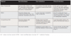
Not all restorative materials respond equally to the radiation-altered oral environment. Understanding material-specific performance characteristics is essential for treatment planning.
Amalgam and Metallic Restorations
Amalgam exhibits structural stability following radiotherapy but demonstrates significant drawbacks. The metallic structure amplifies photon energy, increasing radiation backscatter that can worsen mucositis in adjacent soft tissues.11 Additionally, amalgam offers no fluoride release and requires invasive preparation design, which is not ideal for already-weakened tooth structure. Amalgam should be avoided in both pre- and post-radiotherapy patients.
Resin Composite
Resin composite is mechanically stable after radiotherapy and benefits from minimally invasive preparation design. Unlike glass ionomers, composites polymerize through light-activated reactions independent of water availability, making them less sensitive to reduced salivary flow at placement. Studies demonstrate significantly better marginal adaptation and retention of anatomical form compared with glass ionomer cement in xerostomic patients.12 Clinical survival rates approach 85% to 90% at 10 years when placed before radiation.13,14
Post-radiation placement requires special consideration. Self-etch adhesive systems demonstrate superior performance compared to total-etch protocols in irradiated dentin.15 Self-etch adhesives utilize milder acidic monomers that partially demineralize while preserving residual hydroxyapatite around collagen fibrils. This reduces dependence on the intact but radiation-damaged collagen scaffold and provides additional chemical bonding between functional monomers and hydroxyapatite.16
Glass Ionomer Cements
Conventional glass ionomer cement (GIC) is generally the least favorable option. While GICs offer fluoride release that aids remineralization, they experience significant compositional breakdown following radiotherapy. The material relies on water-mediated acid-base reactions; in xerostomic patients, reduced salivary flow limits water availability during critical early maturation, compromising initial setting and mechanical properties.17
Additionally, radiation-induced reductions in hydroxyapatite weaken the ionic bonding mechanism of GIC. Restoration hardness declines, allowing microcrack formation.18,19 Surface integrity deteriorates faster, and durability is limited. Clinical survival rates approximate 80% at 6 years, substantially lower than composite.14,20
Resin-modified glass ionomer (RMGI) occupies an intermediate position. The resin component improves early strength and makes RMGI less sensitive to moisture loss than conventional GIC. RMGI maintains hardness and shows higher compressive strength than GIC following radiation.19 However, RMGI generally does not achieve the long-term durability of composite. Its role is primarily for low-stress restorations or patients with poor fluoride compliance where fluoride release provides added protection.
Other Materials
Ceramics and zirconia remain chemically stable with no degradation following radiation but require invasive preparation design, which is not ideal for weakened teeth. Bioactive materials show promise for non-irradiated teeth, providing sustained calcium, phosphate, and fluoride ion release.21 However, evidence in high-risk and radiation-treated patients remains scarce, with virtually no long-term data available for these populations. The existing literature is largely limited to studies in healthy, low-risk patients, leaving a critical gap in our understanding of how bioactive restorations perform under the challenging oral conditions imposed by radiation therapy.
Clinical Decision Framework
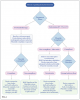
Material selection should be tailored to patient-specific risk factors, particularly fluoride compliance, oral hygiene practices, and salivary function (Figure 1).
Pre-Radiotherapy Phase
Before radiation begins, the primary goal is reducing future caries risk and ensuring optimal dental health. A baseline risk assessment should evaluate caries history, anticipated salivary flow reduction based on radiation field and dose, and patient compliance. Preventive strategies include professional fluoride varnish applications, high-concentration fluoride toothpaste (5,000 ppm), custom fluoride trays with neutral sodium fluoride gel, and patient-centered oral hygiene instructions.22
All active caries should be addressed before radiation, as the oral environment will become more cariogenic. Resin composite is the material of choice, offering superior long-term marginal adaptation and integrity. For patients unlikely to comply with fluoride protocols, RMGI may be selected for its fluoride-releasing properties, though patients should understand the reduced long-term prognosis compared to resin composite.
Post-Radiotherapy Phase
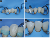
Post-radiation management is more complex due to the permanently altered environment. Radiation-related caries should be addressed promptly. Resin composite remains preferred when adequate isolation can be achieved, using self-etch adhesive systems to maximize bond durability.23 Figure 2 illustrates a clinical case of post-radiation restorative management in a head and neck cancer patient. In cases where isolation is severely compromised or patient compliance is poor, RMGI may serve as an interim or definitive restoration.
Oral Hygiene and Fluoride Protocols
Implementing a strict oral hygiene protocol and fluoride therapy remains paramount for patients undergoing or having completed radiation therapy, as they face lifelong caries risk. Evidence-based protocols include professional fluoride varnish applications every 3 months minimum, daily use of high-concentration fluoride toothpaste, and custom fluoride trays with 1.1% neutral sodium fluoride gel for 5 minutes daily.22,24 When composite restorations are used in conjunction with fluoride varnish for xerostomic patients, outcomes are optimized.14
Conclusion
Resin composite with self-etch adhesive systems represents the gold standard for both pre- and post-radiation restorations, offering superior survival rates compared to glass ionomer alternatives. The single most important clinical decision is completing all restorative treatment before radiation therapy whenever possible, as bond strength and longevity are significantly compromised when procedures are performed on irradiated tooth structure.
Acknowledgments
The authors thank Aljomar José Vechiato Filho, DDS, MSc, PhD; Dental Oncology Service, Instituto do Câncer do Estado de São Paulo, ICESP-FMUSP; and the Department of Periodontology, Guarulhos University, São Paulo, Brazil, for providing the clinical case images used in this manuscript, and the faculty of RSDM 5511 at the Dental College of Georgia, Augusta University, for their continued educational support.
Conflicts of Interest
The authors declare no conflicts of interest.
About the Authors
Elizabeth Winders, DMD Candidate
Dental College of Georgia
Augusta University Augusta, Georgia
Tomi Adeogun, DMD Candidate
Dental College of Georgia
Augusta University
Augusta, Georgia
Daniel Lee, DMD Candidate
Dental College of Georgia
Augusta University
Augusta, Georgia
Mildred Nyaniba Bonful, DMD Candidate
Dental College of Georgia
Augusta University Augusta, Georgia
Rafael Rocha Pacheco, DDS, MSc, PhD
Associate Professor and Associate Dean for Digital Technologies
Dental College of Georgia
Augusta University
Augusta, Georgia
Gabriela de Alencar Pinto Magalhaes, DDS, MSc, PhD
Assistant Professor
Department of Restorative Sciences
Dental College of Georgia
Augusta University
Augusta, Georgia
References
1. Bray F, Laversanne M, Sung H, et al. Global cancer statistics 2022: GLOBOCAN estimates of incidence and mortality worldwide for 36 cancers in 185 countries. CA Cancer J Clin. 2024;74(3):229-263.
2. Shrateh ON, Habib A, Ur Rehman Shamsi H, et al. Treatment options for radiation-induced xerostomia in patients with head and neck cancer: a systematic review and meta-analysis. Br J Oral Maxillofac Surg. 2026;64(1):24-32.
3. Russi EG, Raber-Durlacher JE, Sonis ST. Local and systemic pathogenesis and consequences of regimen-induced inflammatory responses in patients with head and neck cancer receiving chemoradiation. Mediators Inflamm. 2014;2014:518261.
4. Nathan CO, Asarkar AA, Entezami P, et al. Current management of xerostomia in head and neck cancer patients. Am J Otolaryngol. 2023;44(4):103867.
5. Walker MP, Wichman B, Cheng AL, Coster J, Williams KB. Impact of radiotherapy dose on dentition breakdown in head and neck cancer patients. Pract Radiat Oncol. 2011;1(3):142-148.
6. Gupta N, Pal M, Rawat S, et al. Radiation-induced dental caries, prevention and treatment—a systematic review. Natl J Maxillofac Surg. 2015;6(2):160-166.
7. Patel Y, Bahlhorn H, Zafar S, Zwetchkenbaum S, Eisbruch A, Murdoch-Kinch CA. Survey of Michigan dentists and radiation oncologists on oral care of patients undergoing head and neck radiation therapy. J Mich Dent Assoc. 2012;94(7):34-45.
8. Madrid CC, de Pauli Paglioni M, Line SR, et al. Structural analysis of enamel in teeth from head-and-neck cancer patients who underwent radiotherapy. Caries Res. 2017;51(2):119-128.
9. Madrid Troconis CC, Santos-Silva AR, Brandão TB, Lopes MA, de Goes MF. Impact of head and neck radiotherapy on the mechanical behavior of composite resins and adhesive systems: a systematic review. Dent Mater. 2017;33(11):1229-1243.
10. Rodrigues RB, Carvalho AJD, Felipe ESBV, Simamoto-Júnior PC, Novais VR. Impact of radiotherapy in chemical composition and mechanical properties of human cervical dentin: an in vitro study. J Appl Oral Sci. 2025;33:e20240279.
11. Chin DWH, Treister N, Friedland B, et al. Effect of dental restorations and prostheses on radiotherapy dose distribution: a Monte Carlo study. J Appl Clin Med Phys. 2009;10(1):80-89.
12. De Moor RJ, Stassen IG, van ‘t Veldt Y, Torbeyns D, Hommez GM. Two-year clinical performance of glass ionomer and resin composite restorations in xerostomic head- and neck-irradiated cancer patients. Clin Oral Investig. 2011;15(1):31-38.
13. Fan S, Diaz L, Sáenz-Ravello G, Valmaseda-Castellon E, Al-Nawas B, Schiegnitz E. Comprehensive update on implants in patients with head and neck cancer (2021-2024): systematic review and meta-analysis of the impact of radiotherapy and chemotherapy on implant survival. Clin Oral Implants Res. 2025;36(9):1035-1052.
14. Palmier NR, Madrid Troconis CC, Normando AGC, et al. Impact of head and neck radiotherapy on the longevity of dental adhesive restorations: a systematic review and meta-analysis. J Prosthet Dent. 2022;128(5):886-896.
15. Bernard C, Villat C, Abouelleil H, Gustin MP, Grosgogeat B. Tensile bond strengths of two adhesives on irradiated and nonirradiated human dentin. Biomed Res Int. 2015;2015:798972.
16. Yoshida Y, Nagakane K, Fukuda R, et al. Comparative study on adhesive performance of functional monomers. J Dent Res. 2004;83(6):454-458.
17. Eggmann F, Hwang JD, Ayub JM, Mante FK. Impact of irradiation on the adhesive performance of resin-based dental biomaterials: a systematic review of laboratory studies. Materials (Basel). 2023;16(7):2580.
18 Atalay C, Yazici AR. Effect of radiotherapy on the surface roughness and microhardness of contemporary bioactive restorative materials. Support Care Cancer. 2024;32(5):295.
19. Sivavong P, Sanprasert C, Leekhaphan P, et al. Effect of ionizing radiation on the mechanical properties of current fluoride-releasing materials. BDJ Open. 2024;10(1):10.
20. Dezanetti JMP, Nascimento BL, Orsi JSR, Souza EM. Effectiveness of glass ionomer cements in the restorative treatment of radiation-related caries—a systematic review. Support Care Cancer. 2022;30(11):8667-8678.
21. de Carvalho LF, Gimenes ESM, Barboza ADS, et al. Effectiveness of bioactive resin materials in preventing secondary caries and retention loss in direct posterior restorations: a systematic review and meta-analysis. J Dent. 2025;152:105460.
22. Weyant RJ, Tracy SL, Anselmo TT, et al. Topical fluoride for caries prevention: executive summary of the updated clinical recommendations and supporting systematic review. J Am Dent Assoc. 2013;144(11):1279-1291.
23. Arid J, Palma-Dibb RG, de Oliveira HF, et al. Radiotherapy impairs adhesive bonding in permanent teeth. Support Care Cancer. 2020;28(1):239-247.
24. Marinho VC, Worthington HV, Walsh T, Clarkson JE. Fluoride varnishes for preventing dental caries in children and adolescents. Cochrane Database Syst Rev. 2013;(7):CD002279.