You must be signed in to read the rest of this article.
Registration on CDEWorld is free. Sign up today!
Forgot your password? Click Here!
Implant success can be measured by a variety of factors, including long-term bone and soft-tissue maintenance, high esthetics, lack of prosthetic issues, and implant strength with resistance of the implant to fracture. Numerous studies have suggested that platform-shifting may be a useful approach to help stabilize bone and soft tissues.1-4It is imperative that surgeons placing platform-shifted implants review such studies to understand what is necessary to achieve the preservation of crestal bone and maintenance of soft tissue, which are critical for long-term implant stability and survival.
Platform-shifting is a technique utilizing an implant that enables the placement of an abutment or crown that is narrower than the implant platform. While platform-shifting has been in use for approximately 25 years, recently the importance of vertical soft tissue coronal to the implant's platform shift has been an area of increased clinical focus, and the three-dimensional
positioning of the "implant platform depth" (IPD) has been found to be one of the key determinants of implant platform position and long-term implant success.1 An understanding of the anatomical landmarks is key in determining the optimal IPD. This article discusses the measurement of the IPD in base-
level abutment restorations utilizing platform-shifted implants, with an emphasis on anatomical landmarks for improving long-term crestal bone preservation.
Crestal Bone Preservation and Soft-Tissue Maintenance
Long-term dental implant success is based on many key biological factors, including the preservation of crestal bone and maintenance of the soft tissues. Varying degrees of surface roughness have been shown to contribute to improved implant and abutment integration5; there is evidence that having a rough implant surface subcrestal can help prevent bone loss and that a smooth surface can cause more bone loss.5 In a recent study, the use of an anodization process was found to allow for the soft tissues to adhere to the implant and to create an area of soft-tissue attachment.5 It has been demonstrated that subcrestal placement of a roughened surface is advantageous to a smooth surface.5
Crestal Bone Loss
The amount of crestal bone present around a dental implant is one of the determinants of long-term implant success,6 whereas bone loss around an implant increases the likelihood of implant failure.6,7 Bone loss around an implant can advance further bone loss due to the lack of seal around the gingival biologic interface.6,7
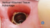
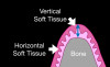
The crestal biologic interface, which includes vertical and horizontal bone and keratinized vertical soft tissue, represents an interface of the implant to the body (Figure 1). It is therefore essential to have a well-defined crestal biologic interface over the site at which the dental implant communicates with the oral cavity. The crestal biologic interface should have a minimum of 3 mm to 4 mm of vertical soft tissue supracrestally with a keratinized outer layer.8Once the implant has healed, it is desirable to have probing depths around dental implants that are 2.5 mm to 4 mm.
In general, probing evaluations may be greater around implants than teeth.8 The vertical tissue above the platform shift (ie, the IPD) has been described by Linkevičius et al as one of the key determinants of maintaining crestal bone.9 Currently it is proposed that there should in fact be at least 4 mm of vertical soft tissue over the IPD.9 The 4 mm is required to establish vertical tissue above the IPD followed by the emergence shape initiating from the abutment platform depth (APD). Knowing where to place the IPD as well as how the abutment interfaces with the connective tissue is critical to long-term implant success.
It is believed that if crestal bone loss occurs, the biological interface represents a woundlike response to the body. Sousa et al postulated that this interface must be a layer of dense, collagen-
rich connective tissue having a keratinized epithelium lining with a well-defined lamina propria firmly attached to the periosteum of the bone.10 They reported that implants with healing abutments wider than 45° had more bone loss than those that were narrower than 45°. Thus, the vertical shape of the abutment leaving the implant has a better chance of crestal bone maintenance by leaving the implant in a vertical orientation and then shaping the emergence from the abutment platform level (APL). The tissue around the APL has been previously discussed in a randomized study of the one-abutment one-time protocol. Rios-Santos et al reported that the vertical height of the abutment, and not the one-abutment one-time protocol, was shown to influence the bone maintenance and therefore the maintenance of the soft tissues11; thus the researchers concluded that factors protecting the bone were more highly associated with the vertical shape of the abutment leaving the implant than how many times an abutment was placed and removed.
Crestal bone loss can have many etiologies. One cause of bone loss around implants is collectively called peri-implantitis, the most common complication in implant dentistry. The remodeling of the bone and soft tissue caused by peri-implantitis is a highly undesirable outcome for both the patient and the dental team. Peri-implantitis causes an inflammatory process in soft tissues12in which neutrophils are drawn to the site of bacterial infection, leading to osteoclastic activity that often results in bone loss at the interface between the implant and the soft-tissue seal around the implant.13 The most frequently cultured peri-implant-related bacteria are the gram-negative anaerobes, such as Prevotella inter-
media, Porphyromonas gingivalis, Aggregatibacter actinomycetemcomitans, and Bacteroides forsythus. From the standpoint of the overall health of the patient, the importance of preventing chronic, long-term effects of infection at the peri-implant site cannot be overstated.
Goodacre et al noted that biological failures are the most common implant-related complications that occur after implants are loaded. Implant failure primarily occurs within 18 months of initial loading.14
In addition, crestal bone loss can result from pressure from the bolus of food hitting the soft tissues, which can push the soft tissues away from the soft-tissue implant interface, causing an opening for the bacteria to enter the sulcus and possibly past the lamina propria to the bone.10 Heavy occlusal forces can also be magnified by the cantilevering of the prosthetics and the position of the implant in the arch, the opposing arch, the masticatory dynamics, and parafunctional habits.10Other forces on the implant interface include excessive water jetting, heavy toothbrush forces, and even playing certain musical instruments such as woodwinds.10 Pre-existing periodontal disease can lead to
crestal bone loss in adjacent teeth that can affect the implant
interface.10 Smoking and diabetes have also been associated with crestal bone loss around dental implants.15
Peri-implant Soft Tissue
Ample soft tissue is necessary for implant success as well, as this helps in the esthetic results of the treatment.16 The anatomical importance of soft tissues in implantology is well-established.17,18In the seminal study by Abrahamson et al using a dog model,19researchers described the vertical soft tissue of the crestal biologic interface, as they demonstrated the zone of attachment of the junctional epithelium over the implant platform and reported that the soft tissues around the implant outlined the connective tissue and junctional epithelium around the dental implant. Initially measured on platform-matched implant systems, the connective tissue was found to be on average 1 mm to 1.5 mm, while the junctional epithelium was reported to measure approximately 1.8 mm.19This finding was significant because the tissues reportedly performed better, with less bone loss, when the tissues were dense, collagen-rich connective tissue having a keratinizing epithelium lining with a lamina propria firmly attached to the
periosteum of the bone. When researchers repeated the studies on the platform-switched implant systems, they found that the implant had less bone loss.20 It has also been found that minimal tissue coverage can lead to an acceleration of crestal bone loss.20
Platform-Shifting
One critical method that has helped improve bone and soft-tissue maintenance around the implants is "platform-shifting" (or, "platform-switching").1,4 Platform-shifting is the use of an abutment that is smaller in diameter than the implant platform to which it is connected, whereas platform-matching refers to matching the implant diameter with the diameter of the abutment.
Recent studies have compared bone loss with platform-
matching versus platform-shifting.1,2,4 In a meta-analysis of 10 studies (1,239 implants), Atieh et al demonstrated that platform-
shifted implants are associated with significantly less bone loss than platform-matched implants.1,4
Improvements in the stability of the gingival complex have been seen with platform-shifted implants, with ample soft tissue associated with stability in the crestal bone.20 In addition, platform-shifting appears to provide a "biological gasket" or seal that inhibits bacteria from entering the abutment-implant interface (the connection between the implant fixture and its restorative abutment), which further prevents bone loss.1,4 The current author postulates that the thickness of the vertical tissue appears to provide a minimal thickness required to meet the forces exerted on the complex during the mastication of food.
Although platform-shifting has been in use for approximately 25 years, in recent years the importance of vertical soft-tissue
above the platform shift has become an area of increased clinical focus.9As explained by Velijanovski et al, vertical soft-tissue thickness is an etiologic factor in crestal bone loss.21 Thus, it is the depth of the platform shift that is one of the key determinants of the implant platform position with these implants. The position of the platform shift also appears to play a role in the maintenance of implant crestal bone.4 It is essential to make certain that the platform shift is not too shallow, as platform shift that is too close to the gingival surface has been shown to cause an increase in crestal bone loss.9,21
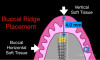
When we look at the relevance of the platform shift, the step in the platform appears to maintain the thickness and seal of the gingival biologic interface. There now have been modifications to the round platform shift to a trioval shape. The trioval shape still has a 4-mm interface on the smaller implants that helps to provide the soft-tissue seal. The new design has also utilized a base-level abutment to have a vertical interface from the implant of 8°.
Trioval and Conical Connections
Implant platforms that feature a platform-shift include external hexagon implant-abutment connections, internal hexagon connections, trioval connections, and conical connections.22 Both trioval and conical connection platforms have platform-
shifting capabilities and thus may help preserve crestal bone.23 With a trioval conical connection (TCC), the connection features a unique trioval shape, designed to be self-centering to lock the abutment in place and to tightly connect with the abutment.24
The TCC has a platform shift that shows the reduction in platform diameter from the implant diameter, which allows for a connective tissue seal around the implant-bone interface. As discussed earlier, an active defense response at the implant interface can occur when an interface is under attack by oral bacteria, which can be detrimental to bone around a dental implant.13The TCC and conical connection implants allow for the lamina propria to seal the connective tissues at the level of what the body interprets as a mechanical injury.12 The junctional epithelium then provides the vertical tissue thickness to protect the bone from osteo-
clastic activity that would otherwise lead to crestal bone loss.
Emergence Transition
The emergence transition of the abutment transition is an obvious necessity following the base-level abutment transition to the tissue level (Figure 2). If the base abutment is at 25°, then the emergence transition must occur to have a transition to the zenith on the buccal and lingual soft tissue. The desired wineglass shape of the transition from the vertical component of the base abutment creates an attachment of the junctional epithelium. Interproximal contact points are also crucial in maintaining and promoting papilla formation. It is usually necessary to place an "anatomic healing abutment" or an immediate temporary to establish both esthetics and the protective vertical soft tissue needed for maintaining crestal bone.
Implant Platform Depth
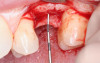
The position of the IPD has become a focus for achieving long-term implant success. The IPD has a major influence on how the body will respond to the everyday needs of the implant system. In particular, the IPD has an influence on the bacterial seal, the forces of the food bolus hitting the soft tissue surrounding the crown, and the bone forces distributed through mastication.9The optimal depth of implant placement should thus be clearly defined prior to placing the implant. To define the depth of the IPD, it is essential to choose a reference point of an attached vertical tissue that will require some future healing. The soft-tissue healed zenith has been proposed as the point of reference for the vertical tissue measure9 (Figure 3). It is important to note that this zenith can be on any aspect of the proposed implant site. For example, the point of reference on an immediate implant placement may have recession that would have to be calculated into the vertical soft-tissue height.
Where Should the IPD Be Measured?
The IPD helps determine the depth, angle, and position of the implant. In the placement of a dental implant, the IPD can generally be measured from the relative position of either the bone level, the cementoenamel junction (CEJ) of adjacent teeth, or the soft-tissue level. However, determining the IPD is complicated by the fact that the point of measure is started on a 3D surface that is often not structurally flat. To achieve the minimal thickness of tissue, the author proposes that the depth should be measured from the minimal position of the free gingival margin relative to the soft-tissue needs of the implant. Both
platform-matched and platform-shifted implants begin with placements to the level of the bone.
Bone-Level Implants
Many implants are currently labeled and presented by manufacturers as "bone-level" solutions; surgeons placing the implants are thus required to position the implants relative to the bone. Clinicians may even choose to place the implant 1 mm sub-
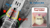
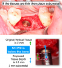
crestal, but this is a placement relative to the level of the implant in the bone. It is interesting to question the bone position as the point chosen for implant placement, as often the bone is not level and is very scalloped. Figure 4 shows the original vertical tissue to be at 1.8 mm.
CEJ-Level Implants
Other practitioners choose a relative point for implant placement based on the CEJ of the adjacent teeth. Although the CEJ may be a clearly defined point, it cannot be directly related to the soft tissue above the dental implant platform. For example, in a tooth with an altered passive eruption, the CEJ is at bone level, leading to a very short tooth and subsequent shallow implant placement.
Zenith-Based Implant Placement
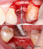
More recently, the depth of the implant has been described as a position of the IPD relative to the soft tissue9 (Figure 5). Linkevičius reported that vertical soft tissue is a key determinant of crestal bone maintenance.9 In the immediate placement of implants, it is important to note that tissues are usually scalloped; thus, the vertical tissue depth requires further description.
In the gingival zenith-based approach to implant placement, the position of the vertical soft tissue in relation to the gingival zenith is considered. As explained by Pawar et al, "The zenith is defined as the most apical point of the gingival marginal scallop. This important landmark [has been] described as having a specific spatial orientation in the apico-coronal and mesiodistal directions."25
While the gingival zenith is often an esthetic position that defines an ideal soft-tissue architecture, it can also be defined by its relative position to buccal and lingual bone loss and can define the thickness of tissue required to seal a dental implant. In a report published in 2015, Linkevičius et al described the tissue as the key to "zero bone loss concepts."9Furthermore, he discussed the relative position to be 2.5 mm of tissue, which is the required amount of tissue to minimize crestal bone loss. More
recently, it has been proposed that the depth of the IPD should be
4 mm from the zenith of the vertical soft tissues (Figure 6).26 It has been demonstrated that a vertical tissue angle helps minimize crestal bone loss.27 The TCC implant has a vertical abutment
system of 25°. The abutments have been designed to be very
vertical on the implant initial transition and then flare in the
junctional epithelium.
IPD for Base-Level Abutment Restoration With the Trioval Conical Connection
To determine the depth of a platform-shifted dental implant with the TCC and conical connections, it is desirable to use the TCC base abutment to establish the shape of the vertical tissues above the IPD to have a minimum of 4 mm of vertical tissue over the platform shift. If the vertical tissue is thin, then the implant could be placed subcrestal or the tissue could be augmented. To determine the depth of a platform-shifted dental implant with the TCC and conical connections:
1. Measure the available vertical tissue height (analog and/or digital measurement).
2. Mark and verify the proposed zenith; check the digital wax.
3. Mark the zenith reference; 90° from proposed zenith.
4. Calculate the IPD to be 4 mm below the zenith reference.
Subcrestal Placement
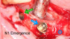
Subcrestal placement of platform-shifted implants can prevent the loss of crestal bone in sites having a vertical soft-tissue thickness of less than 3 mm (Figure 7).21 When the implant is placed subcrestal, the bone often requires modification to allow for abutment seating. It is important to use a surgical bone mill following the placement of a subcrestal implant to allow for the base abutment to be seated to enhance the vertical soft tissue. The conical connection bone mill shown in Figure 8 demonstrates the position of the bone before and after the shaping procedure with the bone mill. This allows the vertical soft tissues to be increased and secure the site (Figure 9).9
Conclusion
The preservation of crestal bone and maintenance of the peri-
implant soft tissues are of paramount importance for long-term dental implant stability and survival. Platform-shifting with combined vertical soft tissue above the platform shift is one method that has helped improve bone and soft-tissue maintenance around the implants. It is critical to ensure that the platform shift at the IPD level is also at an abutment angle that is less than 45° leaving the implant. Using a base-level abutment at a level of
1.75 mm with emergence at the APL will establish a more protective barrier that resides in the junctional epithelium to meet the aforementioned criteria for success. The final emergence profile of the transmucosal abutment establishes the emergence profile required for esthetics and soft-tissue seal. Furthermore,
crestal bone loss can occur as a result of osteoclast activity to fight bacteria at the abutment-implant interface; trioval and conical connection platforms, which have platform-shifting capabilities, foster a connective tissue seal around this interface. In addition to contributing to implant success, from the standpoint of maintaining the overall health and well-being of the patient, the importance of preventing infection at the peri-implant site and its potential chronic, long-term complications cannot be overstated.
The concept of "zero bone loss" and how it is one of the most important bodily responses to dental implants is an important topic in implantology and the basis of a valuable approach to long-term implant success and survival. In his book Zero Bone Loss Concepts, Linkevičius outlines many of the key factors that contribute to peri-implant bone stability.26 What is clear is that IPD is an important factor in achieving long-term implant success. Minimizing bone loss around dental implants has a direct relationship to the inflammatory response and can be achieved in part by placement of the implant in the ideal position for the particular implant system being utilized. Ultimately, it is the response of the body to the implant design and position that will determine the long-term success of the surrounding bone and soft tissues.
References
1. Atieh MA, Ibrahim HM, Atieh AH. Platform switching for marginal bone preservation around dental implants: a systematic review and meta-analysis. J Periodontol.2010;81(10):1350-1366.
2. Serrano-Sánchez P, Calvo-Guirado JL, Manzanera-Pastor E, Lorrio-Castro C, Bretones-López P, Pérez-Llanes JA. The influence of platform switching in dental. A literature review. Med Oral Patol Cir Bucal. 2011;16(3):e400-e405.
3. Lazzara RJ, Porter SS. Platform switching: a new concept in implant dentistry for controlling postrestorative crestal bone levels. Int J Periodontics Restorative Dent.2006;26(1):9-17.
4. Gupta S, Sabharwal R, Nazeer J, Taneja L, Choudhury BK, Sahu S. Platform switching technique and crestal bone loss around the dental implants: a systematic review. Ann Afr Med. 2019;18(1):1-6.
5. Milleret V, Lienemann PS, Gasser A, Bauer S, Ehrbar M, Wenner-
berg A. Rational design and in vitro characterization of novel dental implant and abutment surfaces for balancing clinical and biological needs. Clin Implant Dent Relat Res. 2019;21 Suppl:15-24.
6. Uppala S, Parihar AS, Modipalle V, et al. Crestal bone loss around dental implants after implantation of Tricalcium phosphate and platelet-rich plasma: a comparative study. J Family Med Prim Care. 2020;9(1):229-234.
7. Desai MH, Patil VA. Platform switching: a panacea for bone loss?? J Indian Soc Periodontol. 2013;17(5):681-683.
8. Greenstein G, Cavallaro J, Tarnow D. Dental implantology: numbers clinicians need to know. Compend Contin Educ Dent. 2019;40(5):e1-e26.
9. Linkevičius T, Puisys A, Steigmann M, Vindasiute E, Linkeviciene L. Influence of vertical soft tissue thickness on crestal bone changes around implants with platform switching: a comparative clinical study. Clin Implant Dent Relat Res.2015;17(6):1228-1236.
10. Sousa V, Mardas N, Farias B, et al. A systematic review of
implant outcomes in treated periodontitis patients. Clin Oral
Implants Res. 2016;27(7):787-844.
11. Ríos-Santos JV, Tello-González G, Lázaro-Calvo P, et al. One abutment one time: a multicenter, prospective, controlled, random-
ized study. Int J Environ Res Public Health. 2020;17(24):9453.
12. Assery NM, Jurado CA, Assery MK, Afrashtehfar KI. Peri-
implantitis and systemic inflammation: a critical update. Saudi Dent J. 2023;35(5):443-450.
13. Ericksson I, Persson LG, Berglundh T, Marinello CP, Lindhe J, Klinge B. Different types of inflammatory reactions in peri-implant soft tissue. J Clin Periodontol. 1995;22(3):255-261.
14. Goodacre CJ, Bernal G, Rungcharassaeng K, Kan JY. Clinical complications with implants and implant prostheses. J Prosthet Dent.2003;90(2):121-132.
15. Klokkevold PR, Han TJ. How do smoking, diabetes, and periodontitis affect outcomes of implant treatment? Int J Oral Maxillofac Implants.2007;22 Suppl:173-202. [published correction appears in Int J Oral Maxillofac Implants. 2008;23(1):56].
16. Jose EP, Paul P, Reche A. Soft tissue management around the
dental implant: a comprehensive review. Cureus.2023;15(10):
ee48042.
17. Linkevičius T, Apse P, Grybauskas S, Puisys A. Reaction of crestal bone around implants depending on mucosal tissue thickness. A 1-year prospective clinical study. Stomatologija.2009;11(3):83-91.
18. Di Gianfilippo R, Valente NA, Toti P, Wang HL, Barone A. Influence of implant mucosal thickness on early bone loss: a systematic review with meta-analysis. J Periodontal Implant Sci. 2020;50(4):209-225.
19. Abrahamson I, Berglundh T, Lindhe J. The mucosal barrier
following abutment dis/reconnection. J Clin Periodontol.1997;
24(8):568-572.
20. Farronato D, Manfredini M, Farronato M, Pasini PM, Orsina AA, Lops D. Behavior of soft tissue around platform-switched implants and non-platform-switched implants: a comparative three-year clinical study. J Clin Med. 2021;10(13):2955.
21. Veljanovski D, Atanasovska-Stojanovska A, Pivkova-Veljanovska A, Mijiritsky E, Bollen C. The vertical soft tissue thickness and subcrestal implant placement as factors for peri-implant crestal bone stability. J Med Sci. 2021;9(D):257-263.
22. Strietzel FP, Neumann K, Hertel M. Impact of platform switching on marginal peri-implant bone-level changes. A systematic review and meta-analysis. Clin Oral Implants Res. 2015;26(3):
342-358.
23. Gamborena I, Sasaki Y, Blatz MB. Transmucosal abutments in the esthetic zone: surgical and prosthetic considerations. J Esthet Restor Dent. 2023;35:148-157.
24. Fabbri G, Staas T, Urban I. A retrospective observational study assessing the clinical outcomes of a novel implant system with low-speed site preparation protocol and tri-oval implant geometry.
J Clin Med. 2022;11(16):4859.
25. Pawar B, Mishra P, Banga P, Marawar PP. Gingival zenith and
its role in redefining esthetics: a clinical study. J Indian Soc Perio-
dontol. 2011;15(2):135-138.
26. MacLean S. Successful implant planning and surgery: an eight-step digital protocol. Compend Contin Educ Dent. 2020;41(7):e6-e7.
27. Linkevičius T. Zero Bone Loss Concepts. Batavia, IL: Quintessence Publishing; 2019.