You must be signed in to read the rest of this article.
Registration on CDEWorld is free. Sign up today!
Forgot your password? Click Here!
Adhesive dentistry impacts nearly every aspect of restorative procedures. Whether the clinician is performing routine direct restorations, cosmetic procedures, or placing indirect fixed prosthetics, proper bonding technique is essential to ensuring successful long-term outcomes.
In the early days of restorative dentistry, materials for restorations such as amalgam demanded greater attention to the mechanical principles involved with preparation than to the actual placement of the materials. Although amalgam restorations are not esthetic, they are forgiving from a placement technique perspective, provided that the preparations are done within accepted clinical parameters, as there is little need for concern regarding control of the operative field during placement of these restorations. From a procedural standpoint, the introduction of composite materials has dramatically changed restorative dentistry. The rules and guidelines for preparation have changed for the better with the use of these newer mat- erials, allowing preparations to be more conservative and sacrificing less healthy and viable tooth structure. However, some aspects of adhesive dentistry have made composite materials much more challenging to work with than traditional restorative materials, and the focus of restorative dentistry has changed from mechanical principles to chemical principles. With composite restorations, the process of placing bonded restorations has proved to be an unforgiving one. While every attempt should always be made to place amalgam in a dry preparation, the presence of moisture does not consign that restoration to failure. With composite materials, however, control of the operative field has become a critically important factor. Although "the most important step is the one you are currently performing" remains a valuable maxim, a substantial number of dental practitioners would assert that ensuring control of the field is the most critical objective of any adhesive procedure that involves composite restorations.
The greatest enemy of adhesive dentistry is contamination of the operative field. Among the many contaminants are oil, water, blood, saliva, and crevicular fluid, any of which can potentially lead to failure of the restoration over the long term. In addition, the possibility of under-curing due to poor curing light intensity or curing at too great a distance from target is another factor that must be taken into consideration during adhesive procedures. To prevent or minimize these potential pitfalls, techniques and processes that address these challenges must be undertaken properly to ensure long-term success for bonded restorations.
POSTOPERATIVE SENSITIVITY
One of the most frustrating aspects of adhesive dentistry is postoperative sensitivity from a recently placed restoration. The first step for many clinicians is to adjust the occlusion and then hope that hyperocclusion was the culprit.1,2 Because often the duration of reversible pulpitis is brief,3 this adjustment in combination with the "tincture of time" may be sufficient to alleviate the symptoms.
However, in other cases, postoperative sensitivity may continue. In such cases, troubleshooting can become quite difficult, because the problem of postoperative sensitivity is a multifaceted one and the true cause may be greatly influenced by the restorative technique used by the clinician.4 Some sensitivity may be caused by the introduction of contaminants into the operative field, which results in microleakage that leads to sensitivity,5,6while other cases may result from under-curing.7
Voids
Voids on the pulpal floor can be a common cause of pain during mastication.8 During the placement of the restorative material, occasionally the material can stay lightly attached to the instrument as the instrument is removed from the preparation. This "tug back" can cause the material to lose contact with the pulpal floor and create a small void above the ends of the cut dentinal tubules. Later, with the pressure caused by mastication, the hardened composite presses down on this void and drives the air in the void into the dentinal tubules, creating a pressure differential. This pressure differential causes the fluid in the dentinal tubules to move, which in turn causes the nerves in the pulp to fire. When the pressure is not applied, the pain is not present.
Some clinicians use a layer of flowable composite or a layer of glass ionomer along the dentin to help prevent voids.9While useful and often effective in preventing voids, this technique does increase the number of steps and the procedural time overall, which in turn affects the ability to maintain control over the operative field. Clinicians utilizing this technique should therefore be mindful of the potential for contamination of the operative field.
PREVENTING CONTAMINATION OF THE OPERATIVE FIELD
Oil, gingival crevicular fluid, and moisture from water and saliva are common contaminants that jeopardize control of the operative field. Fortunately, several modalities, including isolation methods, are available to clinicians to help them prevent operative field contamination.
Oil
There are two types of air compressors used in dental offices: "oiled" and "oil-less." The oiled type, as the name implies, uses oil as a lubricant. Some of this oil can find its way into the compressed air delivered to the operatory and can be expressed through the chip air ports of the handpiece. Oil contamination of the preparation causes greatly reduced bond strengths,10,11and these weakened bonds create sensitivity whenever pressure is applied to the restoration. Decreased bond strength also allows the restorative material to be pulled ever so slightly away from the dentin, which will likewise lead to sensitivity.
Reduced bond strength can also result in marginal failure and/or sensitivity.11 With the flexing of the tooth, the weakened bond allows the tooth to microscopically pull away from the restorative material, causing marginal integrity to be lost. Loss of marginal integrity opens up a microscopic gap between the tooth and the restoration that creates a dwelling place for bacteria, and breakdown of the enamel and dentin soon follows.5
Air compressors are a potential source of oil contamination.12 During installation of an oiled compressor, filters are put on the system at the point of exit of the compressed air. As the air passes through these filters, a large amount of the oil is removed and remains in the filtration medium. While this is a worthwhile measure for preventing oil contamination in the operatory, the filters must be evaluated and replaced on a regular basis or they will become saturated and oil will enter the delivery line. It is therefore important that these filters be checked regularly and that they be replaced when indicated.
Many dental handpiece manufacturers also recommend oil to keep the parts moving freely. Therefore, even in an opera- tory where the delivered air is pristine, oil contamination of the preparation can still be a potential problem. In order to prevent the handpiece from spraying oil into the preparation, it is imperative to use only the amount of oil recommended in the handpiece instructions for use (IFU). It is also advisable for the clinician or auxiliary to run the handpiece for 30 seconds to help remove any residual oil that would otherwise remain in the device.
The "oil-less" compressor types are preferred because of their lack of contamination potential. They are also a quieter option, which helps decrease stress for both the patient and the clinician. However, they are more expensive, which must be factored in when budgeting for the system.
Gingival Crevicular Fluid
Controlling the field along the gingival margin, where it can be difficult to obtain appropriate isolation,13,14 is critical when performing restorations in the esthetic zone. Crevicular fluid at the gingival margin is frequently hard to detect without high magnification and bright auxiliary lighting, but it can be the bane of cosmetic restorations.
In the author's experience, frequently at 12 to 18 months postoperatively a slight brownish stain will be noticed at the gingival margin. Left untreated, this stain will continue to expand along the margin in a gingival/incisal direction until finally it becomes noticeable to the naked eye.
The use of an aluminum chloride hemostatic agent is an effective means of preventing this breakdown. In general medicine, these agents are used to control excessive sweating, while in dentistry, they are highly effective in eliminating the seepage of crevicular fluid along the gingival margin15 and do not create any staining when used.
Of course, efficient use of time is also necessary to provide the best results, as any chemical applied to the tissue begins to break down the moment it is used. Decreasing cure times greatly improves long-term clinical and esthetic success.
Rubber Dam
Use of a rubber dam is possibly the most common method for isolating the operative field to prevent moisture contamination, as it has widespread availability and is well-recognized throughout the profession of dentistry. Rubber dams are inexpensive and require minimal training for placement by an auxiliary. The most common difficulty encountered when using a rubber dam appears to be not with the use of the clamp, but with the placement of the frame.16
For this reason, it is tremendously beneficial to use a rubber dam that has a flexible plastic frame already attached to the dam. These dams are much easier and more efficient to place than those that require placement of the frame; after the clamp is placed, the punched and framed dam is simply slipped over the clamp. Because the frames for these products are flexible, once the framed dam is placed over the clamp, the dam opens to provide isolation and a clear view of the operative field, with no other effort required by the team.
The other potential problem is leakage of the dam, which allows contaminants to seep around the clamped tooth.17,18 Dental caulking and putty materials are available that can be placed around the tooth and clamp to work as a seal to prevent liquids from seeping.19
Isolation and Vacuum Products
Other options to help the clinician maintain control over the operative field include isolation and vacuum devices (often single-unit products) that can provide multiple functions, ie, retraction, opening of the oral cavity, light, and most importantly, evacuation.
These devices are made of soft latex-free polymer in a "U" shape and connect to the high-speed suction hose. The bottom of the "U" is formed into a bite block that allows the patient to close their mouth comfortably while also providing for opening of the oral cavity to provide access and visualization. The legs of the "U" have soft flanges that extend upwards and downwards to allow retraction of the tongue and buccal mucosa. These legs, in addition to providing retraction, are also hollow and function as a connection to the high-speed suction. This yields an operative field that is kept consistently dry. Mouthpieces with a built-in light, which may include orange filters for bonding procedures, provide incredibly bright auxiliary lighting.20
EFFICIENCY FOR MAINTAINING CONTROL OF THE OPERATIVE FIELD
Although the use of various agents, devices, and processes can be valuable in helping provide a much drier operative field, no product will work in every case nor for an extended time. For this reason, efficiency in restorative procedures is critical to successful outcomes.
Regardless of whether a direct or indirect restoration is being placed, once the process of bonding the restoration has begun, the clinical team is engaged in a game of "beat the clock" against contamination. The more time needed to complete a restoration, the greater the probability of contamination. Therefore, minimizing the number of steps and the amount of time needed to complete the procedure is critical.
Curing
One of the best ways to increase the clinical efficiency of any bonded procedure is to decrease the amount of time spent curing. However, curing times can be decreased only by utilizing a curing device with a known high output that is consistent throughout the lifetime of the device.
Light-emitting diode (LED) lights are currently the most common piece of hardware in dental curing devices. LED lights are usually bright, durable, and affordable. However, there are factors that can affect the intensity of their output throughout their lifecycle. Battery capacity is one such determinant of longevity of output intensity. As batteries age, their ability to hold a charge lessens, and this can affect how much energy is available to power the LED array. Power output may also wane on a daily basis; although a curing device charged overnight begins the clinical day with a full charge, throughout the day the amount of remaining battery power decreases, causing some devices to lose curing intensity. This can greatly increase frustration for the clinician when troubleshooting problems such as patient complaints of sensitivity.
In addition, over time the glass or plastic construction of the LEDs can discolor or lose some transparency. As this happens, the amount of photons available for the curing process declines and curing efficiency decreases.21
Laser Curing
One innovative tool that has dramatically improved procedural outcomes is the handheld curing laser. In this age of rapidly increasing technologic advances, the term "disruptor" has come into common usage to describe any innovation that produces radical change in a market or a procedure. In that sense, the handheld curing laser is an apt example of a disruptive innovation in restorative dentistry.
Light-cured composites contain a chemical, the photoinitiator, that causes rapid physical change to the material when it is exposed to a specific wavelength of light. Although both the current generation of LED curing lights and the handheld curing laser produce this wavelength, laser light has certain characteristics that make it preferable to standard LED light.
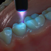
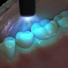
The first and perhaps most important difference is that laser light is collimated (Figure 1). With a non-laser light source, regardless of whether the light is an LED curing light or any other non-laser light, the light begins to diverge the moment it exits the light-curing device (Figure 2). This phenomenon is the reason that the rooms in a house are well-lit at night: photons generated by the light provide circumferential divergence, by which the photos exit and travel in every direction. However, although generalized, even light distribution is desirable when using lighting to illuminate a room, a vastly different requirement is called for with high-speed photopolymerization. In curing devices, beam divergence is synonymous with waste, as it does not contribute to the process of photopolymerization of the composite material. In other words, even if an LED device has an incredibly high mw/cm2 output, if a large portion of the photons miss the target, the intensity of its output is of no use.
Two of the critical properties of laser light are its monochromaticity and its collimation.22Monochromatic light refers to light beams in which every photon is the same wavelength (ie, color, in this case), while with collimated light, all of the photons exit the device and travel in the same direction, rather than disperse.
With collimation, every photon is directed at the target, allowing the laser to cure incredibly quickly and from notably further away from the target, if necessary. According to the manu- facturer, the collimation and monochromatic properties of the handheld curing laser also allow for a 1-second curing cycle, although two to three successive cycles may be necessary to achieve complete cure.23 If a treatment plan involves a 20-second curing cycle, a substantial amount of time can thus be saved by using a laser. This translates to significantly less time spent attempting to control the operative field, which in turn will result in less contamination and better clinical outcomes, with the adjunctive benefit of less stress for the clinical team.
This focused intensity also creates better depth of cure in restorations. For example, for the proximal box in Class 2 restorations, the target is the floor of the box. The proximal box is dictated by the size of the tooth, as well as by the amount of tooth structure that needs to be removed because of the presence of caries. In a situation presented clinically with a deep and wide proximal box, beam intensity is absolutely critical for the long-term success of the restoration.
Manufacturers test the intensity of their lights with the device sitting directly on top of the radiometer. However, for every 1 mm that the light source is moved away from the target, a precipitous decrease in intensity occurs (approximately 10% for every millimeter of distance from the target).24 A proximal box can easily have a depth of 5 mm, which would translate to a light output of approximately 50%, converting a 1,000 mw/cm2output to an output of approximately 500 mw/cm2 at the floor of the proximal box. For a proximal box of even greater depth due to the large size of the tooth or gingival recession, the output received at the floor of the box could theoretically be much less, thus creating even greater potential for postoperative complications as well as decreased longevity of the restoration. Curing this area with a laser would result in a minimal decrease in intensity owing to the collimation of the beam.
Many clinicians, knowing a composite restoration cannot be over-cured, will perform multiple cure cycles from different directions. Although this is an acceptable method to ensure consistency of curing, the practitioner should be aware that each curing cycle also generates heat. Over-curing has the potential to turn the tooth into a heat sink as the curing cycles build up the temperature. This is especially true in the case of smaller teeth; thus, a molar undergoing a small Class 2 restoration will not build up as much heat as a lateral incisor that has undergone a 3/4 crown preparation. Because an increase of 5°C in a tooth can cause pulpal inflammation, it is important for the practitioner to be mindful of the heat generation that curing causes.
A curing laser can create a depth of cure of 4 to 8 mm, depending on the shade of the composite used. Lighter composites allow deeper penetration of the beam, while darker shades permit less depth of cure. The effectiveness of curing can also be greatly affected by the technique employed by the individual operator who determines light placement. Because of the importance of protecting one's eyes from curing lights, many operators, whether the curing is performed by the clinician or the dental assistant, choose to turn their head away from the light source instead of using protective glasses. In doing so, frequently the operator's hand and arm will move as the head is turning, causing the curing exposure to go off target. Because of the beam divergence of traditional LED curing devices, some photons will strike the restoration, leading to polymerization of the material that is closest to the light source. The end result is less than desirable. The material will appear to be cured when engaged by an explorer, but material deeper inside the preparation is much less photopolymerized. When the patient is no longer anesthetized and occludes on the restoration, the less polymerized mat- terial flexes, causing postoperative sensitivity. Although the handheld curing laser also requires the use of eye protection, because it entails only 1-second curing cycles, the amount of time that eye protection needs to be worn, though critical, is incredibly short.
CONCLUSION
The greatest enemy of adhesive dentistry is contamination of the operative field. With the growing popularity of composite restorations, the adoption of procedures that require bonding continues to increase. However, placement of composite restorations can present challenges not posed by the use of traditional mat- erials such as amalgam, and the long-term success of bonded restorations may be threatened by factors that interfere with control over the operative field, including inefficient use of procedural time, poor curing light intensity, and the presence of oral contaminants. These clinical factors also increase the likelihood of postoperative sensitivity. Minimizing the procedural time necessary is the most direct and simplest way to establish and maintain control of the operative field, as shortening the duration of the procedure helps reduce the opportunities for oral contamination of the field and for light-curing devices to experience diminished power output. The oral environment can be an unforgiving place to perform restorative preparations and place composite restorations. Small sacrifices in control of the operative field can result in less-than-optimal experiences and results.
The use of a handheld curing laser greatly shortens the curing cycle and therefore allows the dental clinician and team to operate at peak efficiency while also achieving superior clinical results. By minimizing the time spent curing adhesive materials, the clinician can accomplish adhesive procedures in less time, with less stress, and be assured that the final restorations will achieve high-quality esthetics and optimal longevity.
About the Author
John C. Flucke, DDS
Chief Dental Editor and Technology Editor
Dental Products Report
Private Practice
Lee's Summit, Missouri
Queries to the author regarding this course may be submitted to authorqueries@broadcastmed.com
References
1. Berkowitz GS, Horowitz AJ, Curro FA, et al. Postoperative hypersensitivity in class I resin-based composite restorations in general practice: interim results. Compend Contin Educ Dent. 2009;30(6):356-363.
2. Stevenson RG III. Best practices: restorative complications. In: Termele DA, ed. Avoiding and Treating Dental Complications: Best Practices in Dentistry. 1st ed. John Wiley & Sons; 2016:1-28.
3. Chen E, Abbott PV. Dental pulp testing: a review. Int J Dent. 2009;365785.
4. Rajnekar R, Mankar N, Nikhade P, Chandak M, Ikhar A, Burde K. Clinical efficacy of two different desensitizers in reducing postoperative sensitivity following composite restorations. Cureus. 2022;14(6):325977.
5. Mathivandani V, Kommi V, Arivarasu L, Keerthi Sasanka L. Marginal integrity around bonded fixed prosthesis after instrumentation with subgingival scalers and curettage - a review. Eur J Mol Clin Med. 2020;7(1):3175-3181.
6. Kumar P, Shenoy A, Joshi S. The effect of various surface contaminants on the microleakage of two different generation bonding agents: a stereomicroscopic study. J Conserv Dent.2012;15(3):265-269.
7. Nassar H, El-Shamy H. Bonding system choice practices among senior dental students. J Int Soc Prev Community Dent.2017;7(Suppl 3):S143-S148.
8. Bhatti UA. The phenomenon of postoperative sensitivity and composite restorations - a review. J Pak Dent Assoc. 2019;28(1):33-40.
9. Chuang SF, Liu JK, Chao CC, Liao FP, Chen YH. Effects of flowable composite lining and operator experience on microleakage and internal voids in class II composite restorations. J Prosthet Dent. 2001;85(2):177-183.
10. Bona Matos A, Cerqueira Oliveira D, Nilo Vieira S, Garone- Netto N, Powers JM. Influence of oil contamination on in vitro bond strength of bonding to dental substrates. Am J Dent.2008;21(2):101-104.
11. Vukelja J, Sever EK, Sever I, Mrmek SJ, Tarle Z. Effect of conventional adhesive application or co-curing technique on dentin bond strength. Materials (Basel). 2021;14(24):7664.
12. Borsatto MC, Thomaz MY, Giamatei Contente MMM, et al. Bonding agent underneath sealant: shear bond strength to oil-contaminated enamel. Braz Dent J. 2010;21(1):50-54.
13. Koppolu M, Gogala D, Mathew VB, Thangala V, Deepthi M, Sasidhar N. Effect of saliva and blood contamination on the bond strength of self-etching adhesive system - an in vitro study. J Conserv Dent. 2012;15(3):270-273.
14. Hoorizad MY, Heshmat H, Hosseini TA, Kazemi SS, Tabatabaei SF. Effect of hemostatic agent on microshear bond strength of total etch and self-etch adhesive systems. Dent Res J (Isfahan). 2019;16(6):361-365.
15. Anil A, Sekhar A, Thomas MS, Ginjupalli K. Haemostatic agents on the shear bond strength of self-adhesive resin. J Clin Exp Dent. 2015;7(3):e356-360.
16.Ahlers MO. A new rubber dam frame design-easier to use with a more secure fit. Quintessence Int. 2003;34(3):203-210.
17. Cochrane Oral Health Group, Nagraj SK, Eachempati P, Paisi M, Nasser M, Sivaramakrishnan, Verbeek JH. Interventions to reduce contaminated aerosols produced during dental procedures for preventing infectious diseases. Cochrane Database Syst Rev. 2020;(7):CD013686.
18. Khoroush M, Keshani F, Esmaeili M, Shirazi MH. Marginal integrity of cervical restorations with caries-affected dentinal walls: effect of contamination with hemostatic agents. J Dent (Tehran). 2018;15(4):214-221.
19. Darcey J, Jawad S, Taylor C, et al. Modern endodontics principles part 4: irrigation. Dent Update. 2016;43(1):20-22, 25-26, 28-30.
20. Hayward R, Osman Y, Grobler SR. A clinical study of the effectiveness of a light emitting diode system on tooth bleaching. Open Dent J. 2012;6:143-147.
21. Jadhav S, Hegde V, Aher G, Fajandar N. Influence of light curing units on failure of direct composite restorations. J Conserv Dent. 2011;14(3):225-227.
22. Patil UA, Dhami LD. Overview of lasers. Indian J Plast Surg. 2008;41(Suppl):S101-S113.
23. Christensen GJ. Curing lights: laser diode versus other light sources. Clinicians Report. July 2022;15(7):1-4.
24. Monet Curing Laser Upgrades to 1-Second Cure. AEGIS Dental Network news. https://www.aegisdentalnetwork.com/news/2021/11/8/monet-curing-laser-upgrades-to-1-second-cure. Posted November 8, 2021. Accessed September 12, 2022.