You must be signed in to read the rest of this article.
Registration on CDEWorld is free. Sign up today!
Forgot your password? Click Here!
Not long ago, the concept of digital dentistry was equated only with producing same-day ceramic restorations with a chairside CAD/CAM system. It was also thought that digital dentistry required fundamentally changing the daily workflows of the dental practice and, for all intents and purposes, essentially starting an in-office dental laboratory. If the clinician was unable to complete the entire workflow-from data acquisition, design, manufacturing, post-processing, to delivery-within a single appointment, he or she would be seen as falling short of accomplishing the purported function of the system. Additionally, an unexpected error during any part of the workflow, whether in scanning, design, milling, or post-processing, was believed to derail the clinician's treatment goal for that patient visit, and having a backup system with analog impression material was therefore considered not only prudent but necessary. In the author's experience as an early adopter of chairside CAD/CAM, managing all of these variables in real time in a busy practice sometimes proved to be stressful. When the management of the patient and the technology was predictable, the system was beneficial, impressive to the patient, efficient, and profitable. However, when complications arose, the opposite outcomes often resulted.
Introducing and adopting any new technique, technology, or workflow necessarily involves a period of adjustment and growth for the clinician. On the journey from adoption to mastery, the clinician's skills and understanding must develop by degrees through the "crawl, walk, run" phases-until, ultimately, he or she will learn to "fly." With early same-day chairside CAD/CAM systems, however, the clinician was essentially required to operate the system at the "flying" level on day one in order to successfully produce a final restoration. Also, these early CAD/CAM systems were closed systems, in which the system does not allow the use of its digital data with other manu-
facturers' software or equipment and thus does not permit the clinician to have choices regarding the software or hardware to be used.
Over the last 5 to 10 years, the landscape and narrative of digital technology in dentistry has essentially been rewritten. Closed systems have given way to open systems, providing clinicians with unprecedented choice, freedom, and flexibility in terms of equipment and software choices.1 Open systems allow clinicians the freedom to choose from among several different intraoral scanners, CAD design software programs, 3D printers, and mills. Today, dentists do not have to master the entire digital workflow to be able to utilize digital techniques on day one. The advent of open systems has offered dentists not only freedom and choice with regard to what equipment they choose, but also flexibility with regard to the rate at which they adopt new techniques. In addition, CAD/CAM workflows for same-day restorations are not the only digital workflows available in dentistry today. Data alignment and comparison using companion digital software offer many additional workflows and capabilities, including nonrestorative workflows such as digital smile design, implant planning, orthodontics, and patient monitoring.2
PATHWAYS TO ADOPTING DIGITAL TECHNIQUES OVER TIME
Using the "crawl, walk, run, fly" analogy, the "crawling" phase in digital dentistry includes understanding the basic operations of an intraoral scanner and using it for simple diagnostic scans. Soon after becoming familiarized with these basic operations, the clinician may proceed to the "walking" phase of routine restorative dentistry and become proficient in producing one to two units of indirect restorations, as well as implant restorations and clear aligners. As the clinician's confidence builds, the "running" phase begins, in which the practitioner learns to leverage data alignment and digital photography for digital smile design, implant planning, larger restorative cases, and removable prosthesis cases.3 When the clinician eventually enters the "flying" phase, he or she will have gained mastery in managing in-house design, printing, milling, and complex cases such as full-arch rehabilitation. Today we know that this process can easily take several years. Open systems allow individual dental practitioners to follow whatever path and pace is appropriate for them.
SELECTING AN INTRAORAL SCANNER
Formerly, the marketplace for intraoral scanners was occupied by only one or two products. Today, there are at least a dozen systems vying for market share globally-including not only those produced by the largest dental technology manufacturers, but also those launched by newcomer and startup companies. Choosing the "right" intraoral scanner in what was once a narrow and focused market can be a daunting proposition. In this situation, dental practitioners should begin by testing, handling, and using as many different systems as possible to find the ideal system for their own individual needs, practice situation, and physical space. The range of precision and the image trueness achieved by the scanner are other important factors for the clin-
ician to consider when evaluating scanners.9 Whereas 10 years ago the only relevant question was if the intraoral scanner could reliably replace physical impression material, today's intraoral scanner also serves as a diagnostic as well as a restorative tool.4Attitudes and understanding about intraoral scanners are quickly changing as these digital tools adopt ancillary features. Color scanning, intraoral cameras, shade mapping, caries detection, transillumination, and delivery systems are all variables to consider when choosing an intraoral scanner.5 Combining utilities such as shade mapping into the intraoral scanner can potentially eliminate the need for additional peripherals such as dedicated shade measuring devices.6,7
BUILDING TRUST THROUGH DIGITAL DENTISTRY
The role of digital technology in dentistry is evolving. In the past, technology was used primarily as a means to an end to deliver definitive treatment. Today, it can be used at the very beginning of the patient relationship to provide transparencyin the patient examination. The modern dental patient of the 2020s is different from patients of previous generations in that he or she is generally better informed and educated about healthcare and dental care. The advent of smartphones and web-based information empowers today's dental patient to expect more from healthcare providers. Modern patients cannot simply be informed of a finding and told what the treatment plan should be. In the author's experience, patients need to be included in the process of discovery, diagnosis, and treatment planning. Trust is the most important pillar for healthcare providers and is essential to maintaining a successful relationship with the patient. Using the intraoral scanner as a communication tool to provide transparency helps earn the patient's trust earlier in the process, by empowering the patient to understand their condition and make better-informed healthcare decisions for themselves.
THE ROLE OF INTRAORAL SCANNING THROUGH EACH PHASE OF THE TREATMENT PROCESS MAP
Similar to an engineering process map, every task performed in the dental office can be seen as having an input, a process, and an output. Even the process of a new patient moving through different phases of treatment can be thought of in terms of process mapping. In this context, the new patient himself or herself can be considered the "input." The process is the specific algorithm and steps that the dentist follows during patient care, and the "output" is a healthy patient who remains in the practice over the long term in the recall program.
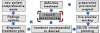
If intraoral scanning were simply a replacement for impression material, it would be used only in the definitive treatment phase, whereas intraoral scanning and digital software currently can be used in all phases of the process map. When any patient requires intervention or treatment, a standardized process can be followed consistently for every case. From the new patient data collection visit, a list of findings and options is developed. These findings and options are used to determine a definitive diagnosis and treatment plan. Next, the patient undergoes a 2-dimensional/3-dimensional (2D/3D) virtual planning phase, followed by a live preview or mock-up phase, and eventually, the definitive treatment phase. The intraoral scanner and associated digital software are used during every phase, aiding the clinician and engaging the patient throughout the process (Figure 1).
USING INTRAORAL SCANNERS TO AID IN DIAGNOSIS
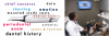
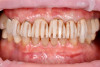
Ensuring that intraoral scanning and the transparency it provides is a prominent feature of the comprehensive new patient visit not only helps to engage the patient, but also aids in the gathering of vital information. The intraoral scan should be displayed in real time in front of the patient for him or her to see. At the same time, real-time observation of the intraoral scan can help the dentist discover the chief concern, obtain mounted study scans, chart restorations and missing teeth, and identify structural problems in the teeth and the soft tissues (Figure 2).
After the scans are used to engage and communicate these findings with the patient and a treatment plan has been agreed upon, the intraoral scanner will be utilized in the restorative phase of treatment, as well. Today's intraoral scanners can be used for crown and bridge restorations, implant restorations, and removable prosthetics, among other restorative procedures.
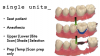
INTRAORAL SCANNING FOR STREAMLINING SINGLE-UNIT RESTORATIONS
Every general dental practice commonly provides single-unit restorations. The intraoral scanner is a useful tool for helping ensure that these procedures are performed accurately.8 Whereas analog techniques necessitated that the final impressions be made at the end of the visit after tooth preparation, intraoral scanning allows the dentist to obtain all the necessary data except for the preparation itself before treatment begins.
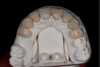
A common digital workflow begins with capturing the preparation arch, opposing arch, and bite registrations immediately after delivering local anesthesia.10Time is managed more efficiently using this technique, and the correct bite registration can be obtained before the patient has been holding their mouth open for an extended period and the local anesthesia has altered their perception of pressure and their movements. The tooth that will be prepared is then digitally erased from the initial scan (Figure 3) and recaptured following tooth preparation, at the end of the appointment. The tooth preparation can be captured with a retractor present to maintain a clean field isolated from saliva and other structures in the mouth.11
With this efficient single-unit restoration workflow, a model-
less technique can be employed when monolithic materials are used, significantly improving return times from the laboratory. If layered restorations are preferred, 3D printed models can be made with suitable accuracy.12 The marginal fit of single-unit restorations made digitally is statistically similar to those manufactured analog.13-17 Chairside adjustments can be minimized, and the patient can receive their final restoration while limiting the time that a provisional restoration is used. In the author's experience, the time and cost savings realized by eliminating impression materials as well as packing, shipping, and tracking of impressions, and the reduced delivery times effectively offset the purchase price of the intraoral scanner within a short period of time.
USING INTRAORAL SCANNING FOR DATA COLLECTION AND MANAGEMENT
Beyond restorative dentistry, the intraoral scanner is an essential data collection and management device. Performing scans for every new patient as well as for existing patients at regular intervals builds a digital library for every patient in the dental practice. Building these patient-specific "digital libraries" as soon as possible helps create opportunities to have more substantive discussions with patients, as well as to deliver the most appropriate care. Using the built-in restorative analysis tools included with every major intraoral scanning system allows the clinician to visually engage the patient, helping communicate findings that are traditionally difficult, such as occlusal pathology. Combining analog articulating ribbon and intraoral scanning helps the practitioner show patients findings such as malocclusions, tooth contacts, and eccentric balancing contacts. As mentioned earlier, displaying these findings via visual aids on a monitor can significantly improve the quality of dentist-patient communications.
Several intraoral scanners also offer built-in data alignment software to allow the clinician to align and compare scans taken over time. This is quickly becoming an essential feature of intraoral scanners in dentistry. As the clinician builds patient-
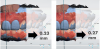
specific digital libraries, it becomes possible to make objective measurements and comparisons of changes in the patient's mouth over time. Discussions with the patient during recall visits regarding tooth position, tooth wear, and tissue levels can be greatly enhanced by using 2D cross-sectional tools in the software and obtaining specific measurements. Using these tools, the practitioner is able to make critical observations at a level previously impossible in clinical dentistry; even changes down to one-tenth of a millimeter can be observed and measured with systems available today (Figure 4).
Intraoral scanning is also useful for the long-term management of patients. Obtaining full-arch scans that provide as much information as possible can reap rewards in the future, regardless of the procedure that is performed at that later date, by improving the quality of the digital libraries. For example, a patient who presents with a palatal ulceration can be evaluated by comparing previous full-arch scans of the area in question, thus helping to clarify the timeline of the current complaint (Figure 5).
DIGITAL SMILE DESIGN
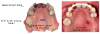
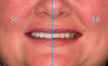
Digital smile design is another important workflow for utilizing digital techniques in the modern dental practice. Digital smile design essentially involves aligning smiling and retracted digital facial portraits (Figure 6) and overlaying proposed tooth shapes over the patient's existing dentition to virtually plan the final treatment goals.18 Many applications are available today to accomplish this. A 2D wireframe architectural drawing is used to help communicate the vision of treatment with both the patient and treatment team, including laboratory partners. Today's digital smile design software applications also allow addition of color and texture to the wireframe drawing to provide the patient with a potential "vision" of the treatment outcomes. In the author's experience, this is especially effective if the process is carried out in real time, with the patient in a non-clinical environment. Enabling the patient to move a digital slide back and forth between images of their preoperative condition and their planned outcome allows them to visualize exactly what the clinician is planning before any potentially irreversible treatment has begun. Showing a "50-50" screenshot can also be useful in communicating the starting point and potential end point, using only photography and mouse clicks (Figure 7).
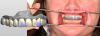
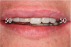
Once the patient has agreed to the treatment plan with the aid of the digital smile design process, the 2D planning phase of the process map begins, and the "output" of the smile design is simply the 2D wireframe drawing. The image is sent to the dental laboratory technician (or used by the treating clinician in some cases) to build a digital wax-up. The images are aligned to the 3D intraoral scan, essentially acting as a virtual facebow aligning the digital model with the maxilla. The wireframe drawing of the proposed outcome is carefully considered and interpreted during the placement, morphing, and refining of the 3D virtual teeth that correspond to the shapes planned in the smile design (Figure 8). Whereas early digital techniques generally used generic tooth shapes, today's systems have multiple libraries including standard shapes, as well as custom libraries digitized from expert laboratory technicians or based on actual patients. These libraries can in turn be selected in the dental CAD design software to faithfully transfer the plan into three dimensions. The output of the digital wax-up is often an STL model of the wax-up. This wax-up model can be printed with a desktop 3D printer and used to create a standard putty matrix. The patient is then invited back for the 3D preview phase, at which point a bis-acryl mock-up can be applied to additive cases to transfer the changes made in the CAD software. The bis-acryl mock-up is applied without any bonding or adhesive steps, to allow easy removal. This step is essential to ensure that the design made virtually will in fact work both esthetically and functionally prior to definitive tooth preparation. It also offers the clinician an opportunity to perform a "dress rehearsal" of the case before the preparation visit. Next, one half of the mock-up is removed and a photograph is taken to provide one last "50-50" view for the patient's approval (Figure 9).
On preparation day, the putty matrix is utilized once again to apply the mock-up prior to beginning tooth preparation. This allows the most conservative reduction of tooth structure by guiding the preparation to follow the planned design instead of the current condition of the teeth (Figure 10). Areas that are additive to the preoperative condition of the teeth can be prepared less while proper material thicknesses are maintained. When the preparations are completed, the putty matrix is utilized once again to produce the provisional restorations. The scanning technique in these larger restorative cases is identical to that used with the basic single-unit protocol. The preparation arch, opposing arch, and bilateral bite scans are obtained prior to beginning the preparation. The preparation arch prescan is captured with the bis-acryl mock-up in place to capture the final tooth contours in the scanning process. The teeth planned for preparation are digitally erased from the prescan and recaptured after preparation and retraction.
These scans are sent to the laboratory following the preparation visit, but the laboratory is instructed to hold off beginning manufacturing until the patient is seen postoperatively. In the author's protocol, the patient is invited back 1 week after preparation/provisionalization so that the clinician can evaluate the esthetics, occlusion, and function of the provisionals. Any necessary changes are made at this point, before the laboratory begins manufacturing of the final restorations. Intraoral scanning of the adjusted and approved provisionals is performed, and the scan is sent to the laboratory. This scan is then merged and aligned with the preparation scans to help guide the design and manufacturing of the final restorations. Studies have confirmed the efficacy of intraoral scanning in multiple-unit fixed restorative treatment, provided that large edentulous spans are not present.19 When this technique is used, the laboratory is not responsible for interpreting or making any global decisions regarding tooth size, contours, and arrangement; they are responsible only for creating restorations based on the approved provisionals. In larger cases involving ceramic layering, printed models can be used to facilitate building and developing contact areas12,20,21 (Figure 11). This technique can significantly reduce the possibility of costly remakes that can be frustrating to the patient and the clinician alike.
At the try-in visit, the restorations are tried individually and as a group to ensure proper fit. If the patient and clinician are satisfied, the restorations are bonded and delivered in the usual fashion, and the patient is scheduled to return to the office
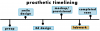
1 week later. At this postoperative visit, any minor changes are made to the occlusion with careful consideration to maximum intercuspation (MIP) and eccentric guidance, including crossover movements. Once the case is finalized and both clinician and patient are pleased with the results, final photographs are taken and a postoperative intraoral scan is obtained. This is an essential scan to include in the patient's digital library. In the event that there is a future problem (eg, breakage), this scan can be used for data comparison. The author's treatment protocol with process mapping includes multiple milestone scans during treatment (Figure 12). The overall treatment protocol is referred to as "prosthetic timelining" and is meant to maximize and leverage available digital technologies for predictable multiple-unit fixed restorative treatments.22
CONCLUSION
The foundation of all digitally driven treatment today depends on the accurate alignment of multiple datasets. Digitizing the patient's dentition as early in the process as possible makes predictable treatment possible. The critical datasets can include facial photography, intraoral scans, facial scans, cone-beam tomo-
graphy, photogrammetry, as well as jaw motion tracking.23,24Lev-
eraging both hardware and software is essential to maximizing the utilization of digital tools in dentistry today. Over the last decade, digital technologies have blossomed and caught up to the needs of the modern clinician. Intraoral scanning began as simply a replacement for physical impression material. Today, it has evolved to be an indispensable tool for communication, diagnosis, treatment, and long-term patient management.
References
1. Rekow ED. Digital dentistry: The new state of the art - is it disruptive or destructive? Dent Mater. 2020;36(1):9-24.
2. Ahmed KE, Whitters J, Ju X, Pierce SG, MacLeod CN, Murray CA. Clinical monitoring of tooth wear progression in patients over a period of one year using CAD/CAM. Int J Prosthodont. 2017;30(2):153-155.
3. Kattadiyil MT, Jekki R, Goodacre CJ, Baba NZ. Comparison of treatment outcomes in digital and conventional complete removable dental prosthesis fabrications in a predoctoral setting.
J Prosthet Dent. 2015;114(6):818-825.
4. Kuhr F, Schmidt A, Rehmann P, Wöstmann B. A new method for assessing the accuracy of full arch impressions in patients. J Dent. 2016; 55:68-74.
5. Dozić A, Kleverlaan CJ, El-Zohairy A, Feilzer AJ, Khashayar G. Performance of five commercially available tooth color-measuring devices. J Prosthodont. 2007;16(2):93-100.
6. Chu SJ, Trushkowsky RD, Paravina RD. Dental color matching instruments and systems. Review of clinical and research aspects. J Dent. 2010;38(Suppl 2): e2-e16.
7. Bidra AS, Taylor TD, Agar JR. Computer-aided technology for fabricating complete dentures: systematic review historical background, current status, and future perspectives. J Prosthet Dent. 2013;109(6):361-366.
8. Patzelt SBM, Lamprinos C, Stampf S, Att W. The time efficiency of intraoral scanners: an in vitro comparative study. J Am Dent Assoc. 2014;145(6):542-551.
9. Lim J-H, Park J-M, Kim M, Heo S-J, Myung J-Y. Comparison of digital intraoral scanner reproducibility and image trueness considering repetitive experience. J Prosthet Dent.2018;119(2):225-232.
10. Wong KY, Esguerra RJ, Chia VAP, Tan YH, Tan KBC. Three-dimensional accuracy of digital static interocclusal registration by three intraoral scanner systems. J Prosthodont. 2018;27(2):120-128.
11. Ting-Shu S, Jian S. Intraoral digital technique: a review. J Prosth-
odont. 2015;24(4):313-321.
12. Patzelt SBM, Bishti S, Stampf S, Att W. Accuracy of computer-aided design/computer-aided manufacturing-generated dental casts based on intraoral scanner data. J Am Dent Assoc. 2014;145(11):1133-1140.
13. Chochlidakis KM, Papaspyridakos P, Geminiani A, Chen C-J, Feng IJ, Ercoli C. Digital versus conventional impressions for fixed prosthodontics: a systematic review and meta-analysis. J Prosthet Dent. 2016;116(2):184-190.
14. Ahlholm P, Sipilä K, Vallittu P, Jakonen M, Kotiranta U. Digital versus conventional impressions in fixed prosthodontics: a review. J Prosthodont. 2016;27(1):35-41.
15. Schaefer O, Decker M, Wittstock F, Kuepper H, Guentsch A. Impact of digital impression techniques on the adaptation of
ceramic partial crowns in vitro. J Dent. 2014;42(6):677-683.
16. Cho S-H, Schaefer O, Thompson GA, Guentsch A. Comparison of accuracy and reproducibility of casts made by digital and conventional methods. J Prosthet Dent. 2015;113(4):310-315.
17. Kale E, Seker E, Yilmaz B, Özcelik TB. Effect of cement space on the marginal fit of CAD-CAM-fabricated monolithic zirconia crowns. J Prosthet Dent. 2016; 116(6):890-895.
18. Coachman C, Calamita MA, Coachman FG, Coachman RG, Sesma N. Facially generated and cephalometric guided 3D digital design for complete mouth implant rehabilitation: a clinical report. J Prosthet Dent. 2017;117(5):577-586.
19. Nedelcu R, Olsson P, Nyström I, Rydén J, Thor A. Accuracy and precision of 3 intraoral scanners and accuracy of conventional impressions: a novel in vivo analysis method. J Dent. 2018;69:110-118.
20. Vögtlin C, Schulz G, Jäger K, Müller B. Comparing the accuracy of master models based on digital intra-oral scanners with conventional plaster casts. Physics in Medicine. 2016;1:20-26.
21. Alharbi N, Wismeijer D, Osman RB. Additive manufacturing techniques in prosthodontics: where do we currently stand? A critical review. Int J Prosthodont. 2017;30(5):474-484.
22. Rajan N. Diagnostic intraoral scanning: technology enhances patient education, diagnosis, and treatment. Dentistry Today. February 1, 2021.
23. Hassan B, Gimenez Gonzalez B, Tahmaseb A, Greven M, Wismeijer D. A digital approach integrating facial scanning in a CAD-CAM workflow for complete-mouth implant-supported rehabilitation of patients with edentulism: a pilot clinical study. J Prosthet Dent. 2017;117(4):486-492.
24. Peñarrocha-Oltra D, Agustín-Panadero R, Pradíes G, Gomar-Vercher S, Peñarrocha-Diago M.Maxillary full-arch immediately loaded implant-supported fixed prosthesis designed and produced by photogrammetry and digital printing: a clinical report.
J Prosthodont. 2017;26(1):75-81.