You must be signed in to read the rest of this article.
Registration on CDEWorld is free. Sign up today!
Forgot your password? Click Here!
Although facially generated treatment planning of the worn dentition includes an evaluation of the esthetics, function, structure, and biology,1 this discussion will concentrate on the evaluation of esthetics and function. Before beginning to evaluate a smile, it is essential that the restorative dentist develop a concept of what a beautiful and natural smile looks like so that he or she can assess the ways in which the patient deviates from the ideal. Beyond the agreed upon standards, each dentist will have his or her own concept of beauty, and this is where the artistic and creative aspect of treatment planning enters. Having a beautiful smile is desirable, but if the upper and lower anterior teeth lack proper function, the patient will not be pleased with the restoration.2 For the upper anterior teeth, the labial surface is the esthetic component and the lingual surface is where the function with the lower incisors takes place. Once restored, the shape of the lower incisal edges helps provide proper contact against the usually concave lingual surface of the upper incisors. This lingual surface also must work in harmony with the temporomandibular joints. The information provided by cone-beam computed tomography (CBCT) makes this diagnostic tool a must for confirming joint status.3 When the goal is comprehensive patient diagnosis and treatment, the interdisciplinary approach can only provide that level of care if the most comprehensive imaging studies are available to allow the doctors to visualize all of the areas of concern. A 2-dimensional image, such as a panoramic radiograph, can act as a screening tool for temporomandibular joint status4; however, small degenerative resorptive lesions on the head of the condyle may be overlooked when using 2D methods, making it difficult to get complete information regarding the condylar condition and position in the fossa.5
Facial Esthetics
The starting point for treatment planning the restoration of anterior teeth is the establishment of tooth position relative to facial structure.6 Lips and the supporting jaw structure frame the teeth. The resulting smile is one of the main defining components of facial beauty. Depending on the observer, perceived beauty may be based on differing values, but the basic elements remain the same for every patient. Excessive gingival display often accompanies worn upper anterior teeth as a result of dental extrusion, and loss of vertical dimension can allow counterclockwise rotation of the mandible as the teeth shift to adjust to the changes caused by wear. Applying the accepted standards, intraoral photographs should be used in conjunction with x-ray images to determine if the upper anterior teeth are in the optimal location for restoration with the best esthetics. Studies have been conducted to determine the feelings of both lay people and dentists regarding which elements make a smile more beautiful and which elements detract from it.7
Cephalometric Analysis
At first, the idea of the orthodontist and restorative dentist reviewing the cephalometric numbers together may seem unusual; however, the time spent will prove valuable.8 The measurements will dictate the optimal position of the upper and lower incisors in order to provide the best appearance and function. Cephalometric measurements allow the dentist to quantitatively evaluate facial and dental structures. They give objective clues to help practitioners understand what is responsible for the subjective symptoms that they see. An extensive examination can be performed; however, a basic assessment is sufficient to yield valuable information that pertains directly to the anterior teeth being evaluated for restoration. Although each patient will deviate from the ideal numbers, they can still provide useful guidance for treatment. The orthodontist has the capability to alter the axial inclination and vertical position of the anterior teeth to improve these relationships. It is not necessary for the restorative dentist to perform a complete cephalometric analysis, as the orthodontist interdisciplinary team member will gladly share this diagnostic data. Some basic cephalometric measurements include the following:
• ANB. This measurement describes the relationship between the upper and lower jaws and can be useful in understanding both the esthetic and functional concerns when evaluating anterior teeth. Ideally, the maxilla is positioned forward of the mandible by 2°. The greater the deviation from the norm, the greater the compromise and camouflage needed to restore with proper esthetics and function. For example, if the ANB is 5° or 3° greater than the ideal, in order to maintain functional contact, the upper incisors will tend to be more retroclined and the lower incisors more proclined. Some more extreme cases may require orthognathic surgery in conjunction with orthodontics in order to achieve desirable esthetics and function.
• 1-NA. The ideal measurement of the axial inclination of the upper incisors relative to the nasion-A point line is 22°. An axial inclination that is upright and retroclined is more likely to accompany a Class II skeletal malocclusion relationship with a retrognathic mandible, whereas a more flared inclination of the upper incisors is more often associated with Class III patients with a prognathic mandible or maxillary skeletal deficiency. Flared and protruding upper incisors are also often found in patients with lower lip entrapment, resulting in a Class II relationship with excessive overbite and overjet. The lateral head film or lateral view of the CBCT study can be helpful in assessing the length of the upper incisor with respect to the resting lip. These measurements can provide valuable information, even if the patient does not require orthodontic treatment. Without these measurements, the restorative dentist may be left to estimate the dental position by eye.
• 1-NB. This measurement represents the axial inclination of the lower incisors to the nasion-B point line. Although the ideal is 25°, this inclination will increase with an increased ANB measurement. In order to achieve an ideal upper incisor inclination, it may be necessary to accept a greater-than-ideal proclination of the lower incisors.
• Interincisal angle.The ideal inside angular relationship of the upper to the lower incisor axial inclination is 130°. This measurement is one of the more informative in orthodontic treatment planning as it relates to preparation of anterior guidance and upper incisor inclination influencing the smile. For example, patients who have a large interincisal angle are more likely to have upright and retroclined upper incisors and be prone to deep anterior vertical overbite relapse due to the difficulty in achieving a lingual stop on the upper incisors.
• Occlusal plane to lip line. This relationship can be very helpful in evaluating resting lip position. The soft tissue outline can be seen on the lateral head film.
Although there are many other measurements that could be considered, the ones presented here offer a good place to start.
Wear and Extrusion
Extrusion of the anterior teeth occurs as the teeth wear and accelerates as the upper lingual functional surface dentin is exposed.9,10 Because dentin is softer than enamel and usually only the enamel perimeter of the mandibular incisal edges remains, resistance to continued wear is reduced. In addition, lost dentin often results in a depression in the middle of the incisal edge, which causes a further reduction in tooth structure. The teeth remain in contact through extrusion during this process, which can result in supraeruption. A lack of vertical clearance can make the restoration of anterior teeth more difficult, and these procedures are often compromised when more aggressive treatment is performed to provide some vertical clearance for the restoration. As the wear continues untreated, the mandible can hinge forward in a counterclockwise rotation, resulting in a worsening of the anterior dental malocclusion. In addition, attrition can allow lingual collapse of the lower anterior teeth (due to their V-shaped anatomy), accompanied by loss of arch perimeter and lower incisor root proximity. As the incisors continue to shorten with wear, the incisal edge becomes smaller from the mesial to distal surfaces and larger from the facial to lingual surfaces. The need for esthetic compromise may be increased due to dental malalignment, malocclusion, and/or uneven or excessive soft-tissue presentation when smiling.
Anterior Dental Function/Envelope Of Function
Poor axial inclination of the upper and lower incisors is often associated with aggressive attrition of the anterior teeth. This includes an excessive interincisal angle of the upper and lower incisor axial relationship. A restricted envelope of function often accompanies an open interincisal angle. It is essential for the lower incisors to be able to move forward as the mandible opens, which is achieved by establishing freedom in centric (ie, long centric) occlusion. The envelope of motion describes the condylar range of motion, and the envelope of function is the anterior functional dental relationship inside this larger envelope.11 As the lower incisors move along the lingual surface of the upper incisors, the vector of force changes from vertical (at full closure) to horizontal (as the mandible opens). There is a dramatic 35% increase in the force generated for each 10° change from vertical to horizontal in the force vector.12 Without a clear understanding of anterior dental function and planning, the restoration can fail. Even if the patient has limited need for orthodontic treatment, working as an interdisciplinary team provides additional input to improve restorative treatment planning.
Case Report
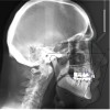
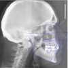
A 76-year-old, retired businessman presented to the office to improve his smile and overall oral health (Figure 1). An esthetic assessment revealed minimal display of the upper anterior teeth and dark discoloration of all teeth (Figure 2). Cephalometric analysis determined that the ANB measurement was 4°, indicating a mild Class II skeletal pattern with upright and retroclined incisors. At 145°, the interincisal angle was excessive, and at 6°, the 1-NA measurement was less than ideal (ie, 22°). If the root angulation of the upper incisors could be shifted toward the palate, it would improve the display and function of the upper anterior teeth with less restrictive anterior guidance (Figure 3).
A more comprehensive examination determined that the patient's anterior teeth bite in an end-to-end relationship with a wide area of contact, which resulted in moderate to locally severe anterior dental wear. Possible crossover bruxing with a flat lower incisor wear table was noted. In the absence of orthodontics, supraeruption of the worn teeth contributes to a poor prognosis for conservative restoration without crown lengthening and gingival reduction. Additional findings included an undersized mesial-distal width of the upper and lower anterior teeth with reduction in the dental arch perimeter; missing teeth Nos. 19 and 30 with distal drift of Nos. 20 and 21, leaving space distal of No. 22; a fixed bridge restoration for tooth No. 19 and an implant fixture in the tooth No. 30 site, both requiring repair; and some concern regarding inadequate oral hygiene.
Treatment with Orthodontic Contributions
Following diagnosis, a treatment plan was selected that involved the use of orthodontics prior to the initiation of restorative therapy. Using this interdisciplinary approach allowed the treatment to be optimized while conserving tooth structure. Benefits to an interdisciplinary approach include the following:
• Minimally invasive preparation. Teeth should be restored using minimally invasive tooth reduction and placement of margins in enamel. Aligning and leveling the cementoenamel junctions of the teeth with orthodontics before restorative preparation minimizes the need for crown reduction and possibly eliminates the need for crown lengthening. In this manner, the treatment no longer has to focus exclusively on the incisal edges. Instead, it can correct the differential extrusion with differential intrusion. For this case, differential intrusion of the anterior teeth was incorporated into removable aligners to level the patient's cementoenamel junctions (Figure 4).
• Smile optimization. When optimizing the smile position of the upper anterior teeth, using cephalometric analysis can provide a more consistent evaluation than a subjective impression of underlying jaw and tooth relationships. Due to the excessive interincisal angle present in this case, the removable aligners were programmed with lingual root torque for the upper anterior teeth (Figure 5 and Figure 6).
• Enhanced dental function. Practitioners should provide anterior guidance that is comfortable and nondestructive. Improving axial tooth inclination with orthodontic treatment allows for less compromise in function. When improperly managed, a lack of anterior guidance can result in peripheral symptoms such as bruxing, muscle spasm, and joint dysfunction. Provisional restorations fabricated from dental materials that are compatible and functionally correct can confirm guidance and elimination of parafunctional habits prior to final restoration (Figure 7).
• Shortest possible treatment time. The orthodontic component of the treatment time is usually the longest portion of the overall treatment time. This can deter patients from committing to comprehensive treatment. In this case, an acceleration auxiliary (AcceleDent®, OrthoAccel Technologies, Inc.) was recommended to shorten the treatment time. The patient wore aligners for 8 months to achieve the desired level of correction, then provisional restorations were placed.
• Increased treatment acceptance. With the addition of removable aligner therapy to the orthodontic treatment armamentarium, many patients who previously refused conventional treatment with bonded brackets may now be willing to proceed (Invisalign, Align Technology, Inc.). In this case, the patient initially agreed to treat the anterior teeth only; however, after seeing the success of the anterior treatment, he ultimately agreed to commit to total restoration. Previously, this patient had been given numerous referrals for orthodontic treatment, but he always declined because the treatment required fixed orthodontic appliances.
• Implant placement.Implant placement simultaneous with orthodontic treatment allows the implant to be restored with a provisional restoration. In addition, early placement allows the orthodontist to use the implant as a source of anchorage and facilitates integration during the finishing stage of orthodontics so that permanent restoration of the implant can coincide with the conclusion of orthodontic treatment (Figure 8).
• Provisional use. When aligners are used to move teeth with provisional restorations in place, a more precise positioning of the teeth is achieved. Provisional use also allows the patient to experience improved function and esthetics before the completion of treatment and provides the restorative dentist with valuable insight into planning the final restoration. For example, placing provisional restorations during orthodontic tooth movement enables the conformation of esthetic components such as color and shape as well as functional correction.
• Splinting.For patients with a restricted envelope of function and possibly some types of temporomandibular joint dysfunction, aligners may be used to achieve a splint-like effect to manage treatment with some potential for occlusal adjustment of the aligner as it proceeds. The occlusal coverage provided by removable aligners also helps to provide protection from continuing attrition during treatment.
Conclusion
How does a restorative dentist go about creating or joining an interdisciplinary treatment team? A dental study club, such as the Seattle Study Club, can provide an effective way to quickly become involved. Associating with other like-minded professionals is a great way to improve diagnosis and treatment planning. It also provides the opportunity to practice referral protocols among members of the potential interdisciplinary team. Begin by reviewing patient records and arriving at an accurate diagnosis, then discuss treatment possibilities and develop a treatment plan. Remember: there is only one correct diagnosis, but there can be several possible treatment plans.
Orthodontic clear aligner treatment provides restorative dentists with a new group of patients who can be conservatively treated. Many of these patients were previously willing to forego needed restorative treatment because they refuse to wear conventional braces. Therefore, it is incumbent upon interdisciplinary dental team members to familiarize themselves with removable aligner treatment and explain the benefits to patients. Because it is the treating doctor who is responsible for the design of the aligners (not the lab) and the supervision of the treatment, there is a learning curve to the achievement of consistent high-quality results. However, it is no longer reasonable to say that this mode of treatment does not provide a satisfactory outcome, as ample evidence of its success is available.13
Acknowledgement
Prosthodontic dentistry for the case was provided by Marc Alexander DDS, MSc, Summerland, California.
About the Authors
Raymond Kubisch, DDS, MSD
Kubisch & Ferris Orthodontics
Santa Barbara, California
Andrew Ferris, DDS, MS
Kubisch & Ferris Orthodontics
Santa Barbara, California
References
1. Spear F. Interdisciplinary management of worn anterior teeth: facially generated treatment planning. Dentistry Today. 2016;35(5):104-107.
2. Dawson PE. Evaluation, Diagnosis, And Treatment Of Occlusal Problems. 2nd ed. St. Louis, MO: C.V. Mosby Co.; 1989.
3. Larson BE. Cone-beam computed tomography the imaging technique of choice for comprehensive orthodontic assessment. Am J Orthod Dentofacial Orthop. 2012;141(4):402,404, 406.
4. Honey OB, Scarfe WC, Hilgers MJ, et al. Accuracy of cone-beam computed tomography imaging of the temporomandibular joint: comparisons with panoramic radiology and linear tomography. AM J Orthod Dentofacial Orthop. 2007;132(4):429-438.
5. Patel A, Tee BC, Fields H, et al. Evaluation of cone-beam computer tomography in the diagnosis of simulated small osseous defects in the mandibular condyle. Am J Orthod Dentofacial Orthop. 2014;145(2):143-156.
6. Arnett GW, Bergman RT. Facial keys to orthodontic diagnosis and treatment planning--part II. Am J Orthod Dentofacial Orthop. 1993;103(5):395-411.
7. Kokich Jr VO, Kiyak HA, Shaprio PA. Comparing the perception of dentists and lay people to altered dental esthetics. J Esthet Dent. 1999;11(6):311-324.
8. Steiner CC. Cephalometrics for you and me. Am J Orthod. 1953;39(10):729-755.
9. Kois J. Functional Occlusion. Kois Center Website. https://www.koiscenter.com/courses/153-functional-occlusion/. Accessed February 7, 2018.
10. Dinning R, Kubisch R. Interdisciplinary restoration of Class II, Division 2 malocclusion. Cmpend Cont Educ Dent. 2013;34(4):282-287.
11. Posselt U. Physiology of Occlusion and Rehabilitation. 2nd ed. Oxford, United Kingdom: Blackwell Science ltd.; 1968.
12. Weinberg LA. Reduction of implant loading using a modified centric occlusal anatomy. Int J Prosthodont. 1998;11(1):55-69.
13. Couchat D, Kubisch, R, Hiroshi S. Addressing the challenges of complex teen, multidisciplinary, and extraction treatment. Presented at: Invisalign Summit for Orthodontic Practices; November 14, 2014; Las Vegas, Nevada. Podcast available online: https://itunes.apple.com/us/podcast/invisalign-ask-the-expert-webinars-ortho/id415517665?mt=2. Accessed February 7, 2018.


