You must be signed in to read the rest of this article.
Registration on CDEWorld is free. Sign up today!
Forgot your password? Click Here!
The primary obligation and ultimate responsibility of oral healthcare personnel (OHCP) is the timely delivery of quality preventive, diagnostic, and therapeutic services within the bounds of the clinical circumstances presented by patients. Because an ever-increasing number of patients with medical problems seek dental treatment, OHCP can expect to face situations that may threaten the physical well-being of at-risk patients.
It follows that OHCP must possess the knowledge and skills essential to determine their patients’ physical and emotional ability to undergo and respond to dental care.1 Diagnostic activities should be effective to establish a database that will serve as reference points for significant medical problems and, importantly, the database must identify those patients at-risk who may experience a medical emergency during the perioperative period.
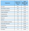
The best available data on the types and incidence of medical emergencies in oral healthcare settings is based on two independent prospective surveys.2 Over a 10-year period, 4,309 dentists documented 30,602 medical emergencies (Table 1) that fell into six major categories: (1) cardiovascular, (2) respiratory, (3) endocrine, (4) allergic, (5) neurogenic, and (6) toxic events. The data also indicate that the rate of medical emergencies per dentist per year is low (0.5/year).
Being ill prepared to respond to emerging perioperative medical events is inexcusable; being subjected to public censure or accused of negligence is an agony best prevented. Consequently, OHCP must P-R-A-Y, i.e., (1) “P”repare for the role of “first responders,” (2) “R”ecognize predisposing factors and signs and symptoms of medical emergencies, (3) “A”ct to stabilize the patient, and (4) “Y”ell for help, i.e., activate emergency medical services (EMS).2-10
OHCP must provide emergency care appropriate for the emergent setting (i.e., oral healthcare facility). At the very minimum, in the event of a life-threatening medical emergency, OHCP must feel comfortable to perform basic life support (BLS) techniques to stabilize the patient until EMS arrives. It must be emphatically stated that advanced life support (ALS) activities should not be attempted without sufficient training and maintenance of skills.
Procedure-specific Risk Factors
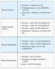
Every procedure elicits a stress-response, i.e., “surgical stress,” characterized by physiological (i.e., autocrine, endocrine, and paracrine) changes accompanied by psychological reactions (e.g., fear, anxiety, anger, tension, malaise or fatigue).11 The magnitude of these procedure-related responses is proportional to the severity of tissue trauma, duration of the procedure, volume of blood loss, fluid shifts, and changes in core body temperature.11
Based on the above criteria, procedure-related stress can be classified as high, intermediate, and low with estimated rates of associated major medical events of >5%, 1-5%, and <1%, respectively (Table 2).12 With low-stress procedures (e.g., dental procedures), the risk is negligible unless strong patient-specific risk factors are present. OHCP must identify patent-specific risk factors that may lead to medical emergencies during the perioperative period.
Patient-specific Risk Factors
“Never treat a stranger.” Identification of patient-specific risk factors is predicated on data obtained from the physical evaluation.1 Past and present illnesses; major hospitalizations; review of organ systems; family history; social history; history of drug allergies and other adverse drug effects; medications, vitamins and other dietary supplements (including special diets) currently taken by the patient must be considered in determining perioperative risk.
Since the stress-response is mediated primarily by the sympathoadrenal system, the history should also seek to determine the patient’s functional capacity (FC).13 FC relates to a person’s functional reserve, which correlates well with maximum oxygen uptake during treadmill testing and is expressed in metabolic equivalents (METs). One MET equals the resting or basal oxygen requirement (i.e., 3.5 ml of O2 per kg per minute) of a 40–year-old, 70-kg man.
A validated method to determine FC, predicated on a person’s ability to perform a spectrum of common daily activities, is presented in Table 3.14-16 FC is classified as excellent (>10 METs), good (7 METs to 10 METs), moderate (6 METs to 4 METs), or poor (<4 METs). The inability of a person to climb two flights of stairs or to run a short distance indicates poor functional capacity (<4 METs). When functional capacity is low, the risk of a medical emergency is high.13
For example, a person with no evidence of coronary artery disease (CAD), but who reports a history of sedentary lifestyle and has poor FC may benefit from a preoperative evaluation. Conversely, a patient considered high risk because of a history of CAD who is asymptomatic and runs 30 minutes daily may need no further cardiovascular testing before proceeding with planned dental procedures, i.e., when functional capacity is high, the risk of a medical emergency is low.
Physical examination is also part of risk assessment.1,13 A patient’s mental state and general appearance, e.g., cyanosis, pallor, diaphoresis, shortness of breath, tightness and/or pain in the chest with minimal activity, tremor, anxiety, and peripheral edema are signs and symptoms that provide invaluable clues regarding the patient’s overall health status. Critically, the physical examination must also include a determination of the patient’s baseline vital signs (Table 4).1,13
Predicated on patient-specific risk factors identified during the physical evaluation, the American Society of Anesthesiology (ASA) Physical Status (PS) Classification system provides a practical method to quantify perioperative risk for patients undergoing surgical (and by extension dental) procedures (Table 5).17,18 The rate of perioperative complications in medicine correlates closely to the ASA PS classification and ranges from 0.4/1000 for ASA PS I to 9.6/1000 for ASA PS IV.19
“First, Do No Harm” (Hippocratic Oath)
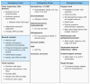
Develop a medical emergency team (Table 6). Know what to look for – be familiar with predisposing factors and signs and symptoms of medical emergencies. Be alert – monitor the patient’s physical and emotional status during treatment – look for evidence of distress or adverse reactions, particularly when drugs are being administered to the patient. Train under simulated conditions. Check regularly the status of emergency drugs and other equipment (Table 6).
Primary Survey
When faced with an emerging perioperative medical event, the following hierarchical steps must be implemented in every situation: (1) assess responsiveness, (2) check airway, (3) and, simultaneously, check breathing and pulse (Table 7). These fundamental activities comprise the primary survey (Table 6), which identifies those problems that are immediately life-threatening and must be promptly acted upon, i.e., obstructed airway, respiratory arrest, or cardiac arrest.
The unresponsive patient in an oral healthcare setting depends on the office emergency team for (1) early recognition of airway obstruction, respiratory and/or cardiac arrest and activation of EMS, (2) early high-quality CPR to delay brain damage from lack of oxygen, (3) early defibrillation to restore an effective heart rhythm, and (4) early advanced life support and post-arrest care. For each minute CPR and/or defibrillation is delayed, the patient’s chance of survival is reduced by 7 to 10 percent.
Airway Obstruction (Foreign Object)
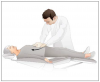
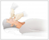
Airway obstruction can lead to respiratory and cardiac arrest if not addressed quickly and effectively. A conscious patient clutching his/her throat is showing the universal sign of choking. Encourage patient to assume a comfortable position and cough forcefully until he/she can breathe normally. If the coughs become weak and ineffective, activate EMS; place the patient in a supine position; and deliver quick, upward abdominal thrusts until the object is forced out (Figure 7).
If the patient becomes unresponsive, immediately begin CPR with chest compressions and make sure an AED is readily available. Each time the airway is opened to give ventilations, look for any visible objects in the oropharynx. If an object can be seen, remove it (if possible) using a finger sweep motion (Figure 8). If a foreign object is not visible, a blind finger sweep should not be performed; continue CPR, cycles of 30 compressions and 2 ventilations, until EMS arrives.
Secondary Survey
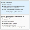
If the patient is conscious proceed with the secondary survey (Table 8). Since the pulse rate and character and the rate and character of respiration have already been determined, assess the blood pressure at this time. Next, correlate the patient’s chief complaint and signs and symptoms to a specific organ system. The goal is to identify those problems that are not immediately life-threatening, but require timely stabilization (e.g., hypoglycemia, angina pectoris, ventilation failure).
Physical signs and symptoms are produced by physical causes and must be recognized before a physical problem can be diagnosed and treated. Without at least a presumptive or working diagnosis there is nothing to treat. Based on symptoms analysis, medical emergencies can be characterized as altered consciousness; chest pain; and ventilatory, allergic (pruritus, urticaria, angioedema), neurogenic (i.e., sensory, affective, motor), and toxic events (Table 9).
Syncope
Syncope (Table 10) is defined as sudden brief loss of consciousness due to cerebral ischemia. In a young adult it is usually precipitated by a generalized, progressive autonomic discharge secondary to anxiety, pain, heat, or humidity. The initial adrenergic response to a stressor is followed by an overwhelming cholinergic surge just prior to unconsciousness. Syncope in patients over 50 years of age may likely be secondary to cardiovascular disorders (e.g., dysrhythmia, postural hypotension), hypoglycemia or cerebrovascular insufficiency.
Postural (Orthostatic) Hypotension
Postural hypotension (Table 11) is defined as a decline of ≥20 mm Hg in the systolic BP, and/or a decline of ≥10 mm Hg in the diastolic BP, or an increase of ≥20 beats/minute in pulse rate, and abrupt symptoms of cerebral ischemia (syncope) following postural change from a supine to an upright position. It may be secondary to impaired homeostatic mechanisms of blood pressure regulation; age and/or cardiovascular-disease-related physiological changes; anti-hypertensive medications; and/or recent intake of food.
Hypoglycemia
Plasma glucose concentration is closely regulated by the autonomic nervous system. Glucagon promotes hepatic glycogenolysis and gluconeogenesis and is a hyperglycemic agent. Insulin promotes cellular glucose uptake and is a hypoglycemic agent. Hypoglycemia (Table 12) is defined as sustained plasma glucose level <70 mg/dL. Heavy exercise, anxiety, and infection may cause hypoglycemia, but the most common cause is treatment with insulin and/or oral hypoglycemic agents and inadequate carbohydrate intake (delayed, decreased, or missed meals).
Angina Pectoris
Angina pectoris (Table 13) is an acute coronary syndrome associated with transient ischemia to the myocardium. Hypoxia (and at times anoxia) results from diseases and conditions which lead to atherosclerosis and obstruction of coronary arteries by fatty deposits and limits and/or impairs coronary blood flow. Precipitating factors that increase cardiac oxygen demand in the presence of decreased perfusion of the myocardium include physical exertion, emotional stress, cold, recent meal. Unstable angina pectoris may occur spontaneously at rest.
Myocardial Infarction
Myocardial infarction (Table 14) is caused by abrupt anoxia to a portion of the heart resulting in myocardial tissue necrosis. Anoxia results from conditions that lead to the formation of atherosclerotic plaques. In later stages, atherosclerotic plaques may become disrupted and contribute to thrombus formation. Atherosclerotic plaques and thrombi impair blood flow to large and medium-sized arteries of the heart. History of cardiovascular diseases, diabetes mellitus, and cerebrovascular disease increases the overall risk of perioperative MI.
Hypertensive Emergency
Hypertension is defined as a blood pressure (BP) ≥140/90 mm Hg. Hypertensive emergency (Table 15) is defined by a BP ≥180/110 mm Hg and signs and symptoms of severely elevated BP. The mechanisms that lead to severely elevated BP appear to be related to a failure of normal autoregulatory function resulting in increased vascular resistance caused by endogenous vasopressors in patients with unrecognized or under-treated hypertension; and/or following the administration of sympathomimetic drugs, such as high doses of epinephrine.
Hyperventilation
Hyperventilation (Table 16) is characterized by anxiety-related dyspnea and tachypnea. Cerebral hypoxia leads to prolonged inspiration (i.e., deep sighs), which result in low CO2 concentration and elevated arterial pH (respiratory alkalosis). Hyperventilation syndrome is common in young women. Predisposing factors include pain, and personal and environmental stress. Other causes include cardiopulmonary disease (e.g., cardiogenic shock, COPD, pulmonary edema), and central nervous system stimulants (e.g., drugs, cola, coffee, tea).
Ventilation Failure
Ventilation failure (Table 17) is defined as a rise in CO2 concentration when alveolar ventilation either falls or fails to respond adequately to increased CO2 production. The most common causes are acute exacerbation of asthma and COPD. Asthma is diffuse airway inflammation caused by household (dust mites, pets) and environmental (pollens) allergens in genetically susceptible patients resulting in reversible bronchoconstriction. COPD (chronic bronchitis, emphysema) is a reversible airway obstruction caused an inflammatory response to toxins, e.g., cigarette smoke.
Pruritus, Urticaria, and Angioedema
Pruritus or itching (Table 18) is a dermal reaction to diverse stimuli, including light touch, vibration, wool fibers, and a number of chemical mediators. Histamine, released by mast cells is one of the most significant chemical mediators. Pruritus is a common symptom of primary skin diseases including allergic contact dermatitis. Less commonly it reflects a systemic reaction to drugs (e.g., NSAIDs, penicillin, and opioids) and other allergens.
Urticaria (Table 18) is a reaction to vasoactive substances (e.g., histamine) released by mast cells in the superficial dermis resulting in intradermal edema caused by capillary and venous vasodilation. The process could be an IgE-mediated type I hypersensitivity reaction; direct non-immune-mediated-activation of mast cells by drugs; drug-induced cyclooxygenase inhibition that activates mast cells by poorly understood mechanisms; or caused by stress and anxiety.
Angioedema (Table 18) is anaphylaxis of the subcutaneous tissues. It results from mast cell and basophil activation in the deeper dermis and subcutaneous tissues and is pathogenically related to urticaria which occurs at the epidermal-dermal junction. The causes of acute angioedema, which may be accompanied by pruritus and urticaria, include drugs and other allergens. Chronic angioedema is mostly idiopathic, rarely IgE mediated, and some cases are hereditary.
Anaphylaxis
Anaphylaxis (Table 19) is a Type I hypersensitivity reaction. Initial exposure to an allergen results in antigen-specific antibody production dominated by the immunoglobulin E (IgE) isotype. Following re-exposure, IgE antibodies bind to mast cells and basophils associated with mucosal and epithelial tissues. The simultaneous binding of an antigen to adjacent IgE molecules fixed to Fc receptors triggers degranulation of mast cells and basophils resulting in the release of histamine, leukotrienes, prostaglandins, chemokines, enzymes and cytokines in target tissues.
Delayed Hypersensitivity Reaction
Delayed hypersensitivity or Type IV reactions (Table 20) are T-cell-mediated, i.e., specifically sensitized CD4+ T-lymphocytes initiate the reactions. Sensitization develops slowly requiring repeated exposures to a specific allergen. Once sensitized, upon reexposure immunologically committed lymphocytes react with the allergen (antigen) and damage tissue by direct toxic effects or through the release of cytokines, which activate eosinophils, monocytes, neutrophils, and macrophages and killer cells.
Seizures
Seizures are a group of neurological disorders caused by excessive discharge of cerebral neurons. They may lead to focal (motor, sensory somatic, visual, auditory, olfactory); psychomotor (automatisms, psychical); or generalized (myoclonic, absence, and tonic-clonic or grand mal) (Table 21) seizures. The cause may be genetic; or head trauma, hypoxia, infection (fever), pregnancy, drug or alcohol overdose or withdrawal, sensory input (e.g., sound, light, touch, and smell), hypoglycemia, circulatory disturbances, degenerative disorders, and tumors.
Cerebrovascular Accident
Cerebrovascular accident or stroke (Table 22) is a syndrome associated with the interruption of blood supply to a portion of the brain causing neurologic deficit. Most commonly, a stroke is secondary to an evolving blood clot associated with atherosclerosis that progressively blocks a cerebral artery. Alternatively, it may be due to an embolus that lodged in a cerebral artery, obstructing blood flow, or result from subarachnoid or intracerebral hemorrhage into brain tissue. Stroke-like symptoms lasting less than 1 hour are termed transient ischemia attacks (TIA).
Local Anesthetic Toxicity
Local anesthetics (LAs) are nonselective voltage-gated sodium channel blockers. This nonselective blockade is not only the source of LAs’ efficacy in blocking action potentials in Aδ and C fibers, but it is also responsible for LAs toxic properties related to the blockade of other sensory, motor, and autonomic fibers. Toxic reactions (Table 23) may result from (1) excessive dosage, (2) repeated doses, (3) rapid absorption, (4) unintentional intravascular injection, (5) low plasma protein binding, and (6) slow metabolism or elimination of the LA or its metabolites.
Summary
Preparedness in emergency medicine mandates didactic and clinical training in emergency medicine, periodic office emergency drills, and maintaining basic emergency drugs and equipment. Education and hands-on training should include issues related to prevention, recognition, and emergent-setting-appropriate management of medical emergencies with emphasis on the importance of performing a primary and a secondary survey.
References
1. Terezhalmy GT, Huber MA, Jones AC. Physical evaluation in dental practice. 1st ed., Wiley- Blackwell, 2009.
2. Malamed SF. Managing medical emergencies. J Am Dent Assoc. 1993 Aug;124(8):40-53.
3. Terézhalmy GT, Batizy LG. Urgent care in the dental office: an essential handbook. 1st ed., Quintessence Publishing Co., Inc., Chicago. 1998.
4. ADA Council on Scientific Affairs. Office emergencies and emergency kits. J Am Dent Assoc. 2002 Mar;133(3):364-5.
5. Malamed SF. Knowing your patients. J Am Dent Assoc. 2010 May;141 Suppl 1:3S-7S.
6. Haas DA. Preparing dental office staff members for emergencies: developing a basic action plan. J Am Dent Assoc. 2010 May;141 Suppl 1:8S-13S.
7. Rosenberg M. Preparing for medical emergencies: the essential drugs and equipment for the dental office. J Am Dent Assoc. 2010 May;141 Suppl 1:14S-9S.
8. Reed KL. Basic management of medical emergencies: recognizing a patient’s distress. J Am Dent Assoc. 2010 May;141 Suppl 1:20S-4S.
9. Ogle OE, Dym H, Weinstock RJ. Medical Emergencies in Dental Practice. Quintessence Publishing Co. Inc. Hanover Park, IL, 2016.
10. Porter RS, Kaplan JL, et al. The Merck Manual of Diagnosis and Therapy. 19th ed. Merck Research Laboratories, Merck & Co. Inc., Whitehouse Station, NJ. 2011.
11. Mangano DT. Perioperative medicine: NHLBI working group deliberations and recommendations. J Cardiothorac Vasc Anesth. 2004 Feb;18(1):1-6.
12. Boersma E, Kertai MD, Schouten O, et al. Perioperative cardiovascular mortality in noncardiac surgery: validation of the Lee cardiac risk index. Am J Med. 2005 Oct;118(10):1134-41.
13. Fleisher LA, Fleischmann KE, Auerbach AD, et al. 2014 ACC/AHA guideline on perioperative cardiovascular evaluation and management of patients undergoing noncardiac surgery: executive summary: a report of the American College of Cardiology/American Heart Association Task Force on Practice Guidelines. Circulation. 2014 Dec 9;130(24):2215-45. doi: 10.1161/ CIR.0000000000000105. Epub 2014 Aug 1.
14. Hlatky MA, Boineau RE, Higginbotham MB, et al. A brief self-administered questionnaire to determine functional capacity (the Duke Activity Status Index). Am J Cardiol. 1989 Sep 15; 64(10):651-4.
15. Fletcher GF, Balady GJ, Amsterdam EA, et al. Exercise standards for testing and training: a statement for healthcare professionals from the American Heart Association. Circulation. 2001 Oct 2;104(14):1694-740.
16. Reilly DF, McNeely MJ, Doerner D, et al. Self-reported exercise tolerance and the risk of serious perioperative complications. Arch Intern Med. 1999 Oct 11;159(18):2185-92.
17. Daabiss M. American Society of Anaesthesiologists physical status classification. Indian J Anaesth. 2011 Mar-Apr; 55(2): 111–115. doi: 10.4103/0019-5049.79879.
18. American Society of Anesthesiologists. ASA Physical Status Classification System – 2014. Accessed on September 8, 2016.
19. Tiret L, Hatton F, Desmonts JM, et al. Prediction of outcome of anaesthesia in patients over 40 years: a multifactorial risk index. Stat Med. 1988 Sep;7(9):947-54.
20. American Red Cross. Basic Life Support for Healthcare Providers – 2015. Accessed on September 8, 2016.
21. American Heart Association. 2015 AHA Guidelines update for CPR and ECC. Accessed September 8, 2016.
22. Kothari RU, Pancioli A, Liu T, et al. Cincinnati Prehospital Stroke Scale: Reproducibility and Validity. Ann Emerg Med 1999 Apr;33(4):373-378. doi: 10.1016/S0196-0644(99)70299-4. Accessed September 8, 2016.
ABOUT THE AUTHORS
Palma A. Freydinger, DDS
Dr. Palma Freydinger is an Assistant Professor, Department of Comprehensive Dentistry, Case Western Reserve University, School of Dental Medicine, Cleveland, OH. Dr. Freydinger received her DDS from Case Western Reserve University, School of Dental Medicine in 1992 and completed a General Practice Residency (GPR) in the Department of Dentistry, The Cleveland Clinic Foundation in 1993. Dr. Freydinger serves as Clinical Preceptor and is responsible for competency examinations and licensing board preparations.
Email: pxf17@case.edu
Fady F. Faddoul, DDS, MSD
Dr. Faddoul is Professor and Vice-Chairman of the Department of Comprehensive Care at CWRU School of Dental Medicine, as well as, the Director of both the Advanced Education in General Dentistry Program and the Faculty Practice. Additionally, he serves as the Infection Control and Safety Officer for the School of Dental Medicine. Dr. Faddoul is a 1988 graduate of the Case Western Reserve University School of Dental Medicine. He completed his Advanced Education in General Dentistry and Oral Medicine training at the Case School of Dental Medicine, as well as, his Master of Science Degree in Oral Medicine in 1992. Dr. Faddoul is an active member of organized dentistry, is a past President of the Greater Cleveland Dental Society, is a member of the commission on dental accreditation, and the North East Board of Dental Examiners. He has lectured extensively on the local, national and international arenas on the topics implants, infection control, medical emergencies, and the management of the medically compromised patients.
Email: fff2@case.edu
Disclaimer: Participants must always be aware of the hazards of using limited knowledge in integrating new techniques or procedures into their practice. Only sound evidence-based dentistry should be used in patient therapy.