You must be signed in to read the rest of this article.
Registration on CDEWorld is free. Sign up today!
Forgot your password? Click Here!
INTRODUCTION
Some of the first microorganisms studied in the dawn of microbiology originated from dental plaque. Dutch scientist Antonie van Leeuwenhoek performed some of his initial experiments on scrapings of plaque from his teeth, and these studies would establish the foundations for modern microbiology. In one of his studies, he described scraping the white material lodged between his gums and teeth, in which he observed “moving animalcules.”1 At the time, Leeuwenhoek only had the aid of a microscope to analyze the microorganisms he observed from the teeth scraping samples. Some of the organisms described by van Leeuwenhoek, though unknown at the time, were the most abundant microorganisms present in the oral cavity.
W.D. Miller, a practicing dentist in the 1890s, spent much of his time analyzing the microbes found in the oral cavity. He later wrote a book called Microorganisms of the Human Mouth, which discussed the theory that microorganisms present in the mouth were a group of bacteria working together.2 These initial studies on dental biofilms have inspired further studies of the organisms that live in the oral cavity. Today, dental biofilms are defined as a diverse community of microorganisms living as a structural unit, with complex communication pathways between species.3 These microbial colonies have also been found to cause dental caries and periodontal disease.4
Dental plaque is a well organized biofilm that attaches to the tooth surface. Its location in the mouth allows for a constant source of moisture, nutrients, warmth and surface, all of which contribute to its growth. The inhabitants of the mouth are incredibly diverse, and mutualistic relationships often take place. While some microbes occupy the niche provided by the host, other species may only thrive in the presence of the primary colonizers. Further, the developing colony may prevent competing species of bacteria from colonizing by monopolizing space and resources. This mutualistic relationship is an important aspect in the development of biofilms in general, and modern research techniques have expanded our understanding of the ecology of oral bacterial communities.
Dental plaque formation is unique from typical biofilm formation due to the nature of the oral environment. Tartar, or calculus, is a calcified deposit on the teeth that is formed by the continuous presence of plaque. The rough surface of the tartar provides an ideal place for plaque to accumulate. Almost immediately, a salivary glycoprotein film called a pellicle coats a clean tooth. The pellicle allows for the adherence of gram positive primary colonizers, which include Streptococcus mutans, Streptococcus anguis and Actinomycete species.5,6 The biofilm mass continues to increase due to the multiplication of the primary colonizers, which provides a place for other organisms to subsequently attach. In a span of 1 to 3 days, the secondary colonizers adhere to the gram positive primary colonizers. These secondary colonizers are generally gram negative species and typically include Fusibacterium nucleatum, Prevatella species and Capnocytophaga species.6 In the final stage of dental biofilm formation, the tertiary colonizers attach, and there is an overall shift from gram positive facultative microbes to gram negative anaerobes.4–6
The thriving biofilm may result in cariogenic conditions that can lead to caries, or affect the adjacent soft tissue and result in periodontal disease. Further, chronic oral infections have also been associated with systemic diseases, such as diabetes and heart disease, due to the spread of oral microbes into the blood stream, and to certain lung diseases due to the aspiration of the plaque into the respiratory system.7,8 Thus, oral biofilms have health consequences beyond infections of the mouth, and novel methods for eradication or control of these colonies are needed.
In traditional microbiology, the individual cell unit is typically the focus. However, in the case of biofilms and dental biofilms in particular, the whole organism is working together and each bacterium is dependent on the other species present.3 Therefore, typical microbiological approaches may not be sufficient for the identification or study of biofilm–forming bacteria. Treatment strategies must incorporate a more holistic, ecological approach to the control of the dental biofilm. An understanding of the etiology of diseases caused by oral biofilms first requires identification of the bacterial species involved, which is best accomplished using molecular genetic techniques. This review summarizes many of the molecular techniques that may be utilized in the detection of bacterial species in dental biofilms and discusses the future of molecular diagnostics in dental hygiene practice.
METHODS USED FOR STUDY OF BIOFILMS
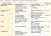
Due to the complex multispecies lifestyle of dental biofilms, unique research methods have been developed for the study of these organisms. Traditional culturing methods of bacteria are often insufficient for the analysis of biofilms, because many bacteria that are present in the oral cavity are considered viable but not culturable.9 It has been suggested that less than 1% of microorganisms can be cultured in the laboratory, meaning that the vast majority of oral bacteria evade standard microbiological detection methods.9 This has lead to the development of alternative methods to assess dental biofilms based on DNA analysis or other molecular techniques. By learning more about the genetics and biochemistry of the organism, we can derive better strategies for treating infection. Biofilm colony homeostasis is a delicate balance, and when disrupted, pathological species can predominate.5 DNA analysis can allow identification of all of the species present in an oral biofilm, of which only 1 or 2 species may be the pathological culprits. By knowing which species of bacteria are present in the oral cavity, new treatment options can be developed that would, in turn, provide better dental care. Table I summarizes each molecular technique discussed below.
Checkerboard DNA–DNA Hybridization
DNA–DNA hybridization is considered the gold standard of oral biofilm analysis. It was developed by Socransky et al for the synchronized processing of large numbers of samples and the profiling of multiple species within the same sample in a semi–quantitative manner.10 The technique relies on the binding of DNA isolated from bacterial samples to a membrane, followed by hybridization with DNA probes specific to at least 40 different bacterial species.10 This method is very useful for analyzing dental plaque because of the simultaneous processing of large numbers of samples.11 The technology has been able to detect microbes present on oral surfaces, biofilm composition in periodontal disease and bacterial prevalence in specific oral communities.12–15 Furthermore, this technique has been used to assess the outcome of therapeutic treatment.16
Because of the use of whole genome probes, DNA–DNA hybridization was originally limited only to the identification of species that can be cultured. However, a reverse capture checkerboard hybridization method was developed.17 In this modification of the traditional method, PCR–amplified 16S ribosomal RNA genes of up to 30 known bacterial species are spotted onto blots. The membrane is then hybridized with PCR–amplified 16S rRNA genes from unknown plaque samples. The primers for these targets are labeled with universal probes which are detected by chemifluorescence. This reverse capture hybridization method allows for 1,350 hybridizations simultaneously on 1 membrane.17 A disadvantage of these slot–blot methods is that they are rather laborious, and non–hybridization PCR methods are now more commonly used.
16S rRNA Gene Sequencing
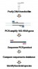
The 16S ribosomal RNA gene is highly conserved and can be used in the formation of phylogenetic trees or genetic relationships.18,19 This discovery, along with the advent of PCR techniques, has allowed the analysis of oral biofilms on a genetic level. 16S RNA is present in almost all bacterial species, with unique sequence differences allowing discrimination between species.20 Amplification methods, such as 16S rRNA sequencing, have eliminated the requirement for culture based techniques, allowing the identification of unculturable species. Identification of the species present is determined by comparing the 16S rRNA sequence derived from the unknown sample to databases of known species. Figure 1 summarizes the process of 16S rRNA sequencing.
There is some disagreement on the similarity threshold necessary to verify a species.20 A reasonable criterion for genus identification is a 97% similarity score to a known database sequence, while 99% similarity was determined sufficient to identify at the species level.21 A limitation of this method is low resolution in distinguishing between bacteria at the species level. Species may share identical 16S rRNA sequences or the differences between related species may be very small (less than 0.5%).20 Despite these limitations, 16S rRNA sequencing has yielded a wealth of new information about dental biofilms. 16S rRNA analysis has shown that there are over 300 bacterial species present in the oral cavity that were not initially identified by typical culturing methods.22,23 Furthermore, it was found that there are differences in bacterial flora present in the oral cavity of individuals with immunosuppressive diseases such as HIV.24 A total of over 700 bacterial species have been identified in the oral cavity, many of which are specific to a particular oral surface.25
Denaturing Gradient Gel Electrophoresis
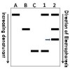
Denaturing gradient gel electrophoresis (DGGE) is a PCR and electrophoresis–based approach for analysis of microbial communities. Various marker genes, including 16S rRNA, are amplified using PCR and then analyzed on a denaturing gel. A banding pattern develops based on the denaturation characteristics determined by the sequence composition of each amplified DNA. Each band observed on a DGGE gel theoretically represents a different bacterial population within a community.26 Thus, DGGE band patterns can illustrate the complexity and diversity of a biofilm sample, and individual bands can be subsequently excised and sequenced to determine species identity. Figure 2 shows a schematic example of a DGGE gel. DGGE has been applied in the analysis of oral microbial communities in conditions such as periodontitis and severe childhood caries.27–29 A limitation of DGGE is that sequence differences greater than 1 base pair may fail to separate on a denaturing gel because of similarities in nucleotide proportions that result in identical denaturing characteristics of 2 different sequences. Therefore, excision and sequencing is necessary to confirm the identification of species present within an individual band.30
Terminal Restriction Fragment Length Polymorphism
Terminal restriction fragment length polymorphism (T–RFLP) is another PCR–based technique that can be applied to the study of oral biofilms. This technique originated from the study of bacterial diversity in environmental samples, and was later used for the analysis of oral microbial communities.31–34 T–RFLP is similar to DGGE in that certain gene markers, including 16S rRNA, are amplified by PCR using gene–specific primers labeled with a fluorescent probe. The amplified products are then digested with restriction endonucleases, and the fragments are separated by capillary electrophoresis. The fragments with the attached fluorescent probes are detected by the instrument and analyzed using fragment analysis software. When the samples are analyzed by gel electrophoresis, specific banding patterns can be assessed which represent complex microbial communities.35 This technology has been used to assess different microbial profiles in human saliva, changes in microbial communities in the oral cavity after treatment and bacteria present in infected root canals.32–34 The applications of T–RFLP are promising, but the technique is still in its infancy stages. T–RFLP requires expensive instrumentation, high computational power and very large databases to compare the genetic sequences.11
Emerging Technologies
A number of recently developed techniques have been implemented for microbial identification, and these methods show potential for future applications in the study of oral biofilms. Denaturing high–performance liquid chromatography (DHPLC) is a PCR–based method which is followed by separation based on partial denaturation of the amplified DNA. This technique can be used to detect DNA sequence changes, such as point mutations.36 DHPLC has been previously utilized in other areas of research, such as intestinal microbiota, and has more recently been applied for analysis of dental biofilms and bacteria.37,38 Techniques used in chronic wound biofilm analysis may also become useful for oral biofilm research and diagnosis. Pyrosequencing, a rapid sequencing method that can simultaneously identify microbes and detect antibiotic resistance, has been applied for the determination of bacterial diversity in chronic wound biofilms such as in diabetic foot ulcers, venous leg ulcers and pressure ulcers.39–40 Recently, the pyrosequencing method was applied to the analysis of saliva and supragingival plaque samples, and it was estimated that 19,000 different microbial species are present in the mouth.41 Studies which utilize these next–generation methods are revealing that original approximations of oral microbial diversity were highly underestimated.
DISCUSSION
The mainstream application of molecular methods in both research and clinical settings has allowed for a rapid expansion of our understanding of the oral microbial environment. As in other fields, such as chronic wound care, the future management of oral disease will benefit from adoption of molecular biofilm analysis methods. While the identification of species present within a plaque biofilm is essential for focused treatment, the understanding of the unified communication and adaptive changes that occur within the microbial community as a whole is equally important. Some future directions should include the assessment of gene expression levels in the oral biofilm. The analysis of gene expression within a biofilm can help aid in the identification of virulence factors that might make the biofilm more resistant to antibiotics or other treatment, similar to studies performed on methicillin–resistant S. aureus.42 Methods such as real–time PCR or microarray can analyze the gene expression patterns that may make a particular biofilm population inclined to cause disease. Expression data derived by such methods can be applied to analyze oral biofilms under conditions such as inflammation or immune suppression, or can be used to evaluate dental bacteria behavior before and after antibiotic treatment. This can provide insight into how the oral biofilm communicates and behaves as a whole unit.
As molecular techniques become mainstream and more widely available in clinical laboratories, the capability to obtain individual patient biofilm profiles is becoming attainable. By identifying the pathogenic bacteria in a patient, treatment can be personalized to the infection. A recently launched clinical diagnostic laboratory (OralDNA Labs) now offers molecular testing to dental practitioners for the diagnosis of periodontal disease, using PCR–based tests to identify pathogenic oral bacteria.43 Such services may help avoid the generalized use of antibiotics that are ineffective or encourage antibiotic resistance. The traditional empirical method of prescribing antibiotics in dentistry has been questioned because of unnecessary or inappropriate use of antibiotics that can lead to antibiotic resistant organisms.44,45
There are a number of obstacles preventing the immediate marriage of dentistry and molecular diagnostics. Rapid treatment and relief for the patient is a primary concern for the dental practitioner. A patient with a critical oral infection should not be denied treatment for the 48 hours or more that is required for traditional microbiological tests, thus empirical treatment has been traditionally utilized in the absence of a better option. However, the rapid nature of most molecular assays provides a vast improvement over lengthy culture methods, with many molecular techniques providing identification of organisms in a matter of a few hours. Even a turnaround time of 24 hours for reliable identification of pathogenic bacteria can allow for customized modification of the initial empirical antibiotic treatment of very ill patients, particularly for refractory forms of oral disease. There is underuse of diagnostic microbiology laboratories by dental practitioners, which may be mitigated by a greater awareness of the services provided by such laboratories.44
Other considerations for implementation of molecular diagnostics in dental practice are that of practicality and cost.46 Some of the techniques discussed above are currently cost prohibitive for routine use in the diagnosis of oral infection. The reimbursement of molecular assays by third–party payers is also complicated by lacking or ambiguous Current Procedural Terminology codes for some molecular tests. However, molecular assays are rapidly becoming higher–throughput and more standardized, and some molecular tests are kit–based and relatively inexpensive. Nonetheless, while molecular diagnostics are quickly becoming a feasible approach, laboratory diagnosis of oral disease will likely remain reserved for patients with severe periodontal disease or those who have been unresponsive to traditional treatment. Although molecular diagnostics will not take the place of the primary clinical methods of prevention and debridement, it does offer a beneficial complement to the practice of dental hygiene.
CONCLUSION
Understanding the complex interactions between bacteria that occur within an oral biofilm will provide insight necessary for improving diagnosis, treatment and prevention of periodontal disease. Dental practitioners should be aware of emerging diagnostic techniques and should strive to work in concert with researchers to harness new technologies for improving biofilm management. Molecular diagnostics of dental biofilms will allow for rapid, focused and personalized treatment, enhancing the traditional methods used by dental hygienists to control and prevent periodontal disease.
Sarah Hiyari, MS, is currently a PhD student in Oral Biology at the University of California Los Angeles, School of Dentistry. Katie Bennett, PhD, is an assistant professor in the Molecular Pathology and Clinical Laboratory Science programs at the Texas Tech University Health Sciences Center, School of Allied Health, in Lubbock, Texas.
References
1. Gest H. The discovery of microorganisms by Robert Hooke and Antoni Van Leeuwenhoek, fellows of the Royal Society. Notes Rec R Soc Lond. 2004;58(2):187–201.
2. Miller WD. The micro–organisms of the human mouth. Am J Med Sci. 1891;101(2):159.
3. Aas JA, Paster BJ, Stokes LN, Olsen I, Dewhirst FE. Defining the normal bacterial flora of the oral cavity. J Clin Microbiol. 2005;43(11):5721–5732.
4. Marsh PD. Dental plaque as a microbial biofilm. Caries Res. 2004;38(3):204–211.
5. Marsh PD. Dental plaque as a biofilm and a microbial community – implications for health and disease. BMC Oral Health. 2006;6(Suppl 1):S14.
6. ten Cate JM. Biofilms, a new approach to the microbiology of dental plaque. Odontology. 2006;94(1):1–9.
7. Li X, Kolltveit KM, Tronstad L, Olsen I. Systemic diseases caused by oral infection. Clin Microbiol Rev. 2000;13(4):547–558.
8. Akutsu Y, Matsubara H, Shuto K, et al. Pre–operative dental brushing can reduce the risk of postoperative pneumonia in esophageal cancer patients. Surgery. 2010;147(4):497–502.
9. Staley JT, Konopka A. Measurement of in situ activities of nonphotosynthetic microorganisms in aquatic and terrestrial habitats. Annu Rev Microbiol. 1985;39:321–346.
10. Socransky SS, Smith C, Martin L, Paster BJ, Dewhirst FE, Levin AE. “Checkerboard” DNA–DNA hybridization. Biotechniques. 1994;17(4):788–792.
11. Kuramitsu HK, He X, Lux R, Anderson MH, Shi W. Interspecies interactions within oral microbial communities. Microbiol Mol Biol Rev. 2007;71(4):653–70.
12. Mager DL, Ximenez–Fyvie LA, Haffajee AD, Socransky SS. Distribution of selected bacterial species on intraoral surfaces. J Clin Periodontol. 2003;30(7):644–654.
13. do Nascimento C, Barbosa RE, Issa JP, Watanabe E, Ito IY, de Albuquerque Junior RF. Use of checkerboard DNA–DNA hybridization to evaluate the internal contamination of dental implants and comparison of bacterial leakage with cast or pre–machined abutments. Clin Oral Implants Res. 2009;20(6):571–577.
14. Papapanou PN, Baelum V, Luan WM, et al. Subgingival microbiota in adult Chinese: prevalence and relation to periodontal disease progression. J Periodontol. 1997;68(7):651–666.
15. Socransky SS, Haffajee AD, Smith C, et al. Use of checkerboard DNA–DNA hybridization to study complex microbial ecosystems. Oral Microbiol Immunol. 2004;19(6):352–362.
16. Haffajee AD, Cugini MA, Tanner A, et al. Subgingival microbiota in healthy, well–maintained elder and periodontitis subjects. J Clin Periodontol. 1998;25(5):346–353.
17. Paster BJ, Bartoszyk IM, Dewhirst FE. Identification of oral streptococci using PCR–based, reverse–capture, checkerboard hybridization. Methods Cell Sci. 1998;20(1):223–231.
18. Fox GE, Stackebrandt E, Hespell RB, et al. Science. 1980;209(4455):457–463.
19. Woese CR. Bacterial evolution. Microbiol Rev. 1987;51(2):221–271.
20. Janda JM, Abbott SL. 16S rRNA gene sequencing for bacterial identification in the diagnostic laboratory: pluses, perils, and pitfalls. J Clin Microbiol. 2007;45(9):2761–2764.
21. Drancourt M, Bollet C, Carlioz A, Martelin R, Gayral JP, Raoult D. 16S ribosomal DNA sequence analysis of a large collection of environmental and clinical unidentifiable bacterial isolates. J Clin Microbiol. 2000;38(10):3623–3630.
22. Kroes I, Lepp PW, Relman DA. Bacterial diversity within the human subgingival crevice. Proc Natl Acad Sci U S A. 1999;96(25):14547–14552.
23. Paster BJ, Boches SK, Galvin JL, et al. Bacterial diversity in human subgingival plaque. J Bacteriol. 2001;183(12):3770–3783.
24. Paster BJ, Russell MK, Alpagot T, et al. Bacterial diversity in necrotizing ulcerative periodontitis in HIV–positive subjects. Ann Periodontol. 2002;7(1):8–16.
25. Paster BJ, Olsen I, Aas JA, Dewhirst FE. The breadth of bacterial diversity in the human periodontal pocket and other oral sites. Periodontol 2000. 2006;42:80–87.
26. Fischer SG, Lerman LS. DNA fragments differing by single base–pair substitutions are separated in denaturing gradient gels: correspondence with melting theory. Proc Natl Acad Sci U S A. 1983;80(6):1579–1583.
27. Fujimoto C, Maeda H, Kokeguchi S, et al. Application of denaturing gradient gel electrophoresis (DGGE) to the analysis of microbial communities of subgingival plaque. J Periodontal Res. 2003;38(4):440–445.
28. Zijnge V, Harmsen HJ, Kleinfelder JW, van der Rest ME, Degener JE, Welling GW. Denaturing gradient gel electrophoresis analysis to study bacterial community structure in pockets of periodontitis patients. Oral Microbiol Immunol. 2003;18(1):59–65.
29. Li Y, Ge Y, Saxena D, Caufield PW. Genetic profiling of the oral microbiota associated with severe early–childhood caries. J Clin Microbiol. 2007;45(1):81–87.
30. Jackson CR, Roden EE, Churchill PF. Denaturing gradient gel electrophoresis can fail to separate 16S rDNA fragments with multiple base differences. Molecular Biology Today. 2000;1(2):49–51.
31. Liu WT, Marsh TL, Cheng H, Forney LJ. Characterization of microbial diversity by determining terminal restriction fragment length polymorphisms of genes encoding 16S rRNA. Appl Environ Microbiol. 1997;63(11):4516–4522.
32. Sakamoto M, Takeuchi Y, Umeda M, Ishikawa I, Benno Y. Application of terminal RFLP analysis to characterize oral bacterial flora in saliva of healthy subjects and patients with periodontitis. J Med Microbiol. 2003;52(Pt 1):79–89.
33. Hommez GM, Verhelst R, Claeys G, Vaneechoutte M, De Moor RJ. Investigation of the effect of the coronal restoration quality on the composition of the root canal microflora in teeth with apical periodontitis by means of T–RFLP analysis. Int Endod J. 2004;37(12):819–827.
34. Sakamoto M, Huang Y, Ohnishi M, Umeda M, Ishikawa I, Benno Y. Changes in oral microbial profiles after periodontal treatment as determined by molecular analysis of 16S rRNA genes. J Med Microbiol. 2004;53(Pt 6):563–571.
35. Schütte UM, Abdo Z, Bent SJ, et al. Advances in the use of terminal restriction fragment length polymorphism (T–RFLP) analysis of 16S rRNA genes to characterize microbial communities. Appl Microbiol Biotechnol. 2008;80(3):365–380.
36. Hoogendoorn B, Norton N, Kirov G, et al. Cheap, accurate and rapid allele frequency estimation of single nucleotide polymorphisms by primer extension and DHPLC in DNA pools. Hum Genet. 2000;107(5):488–493.
37. Goldenberg O, Herrmann S, Adam T, et al. Use of denaturing high–performance liquid chromatography for rapid detection and identification of seven Candida species. J Clin Microbiol. 2005;43(12):5912–5915.
38. Jacinto RC, Gomes BP, Desai M, Rajendram D, Shah HN. Bacterial examination of endodontic infections by clonal analysis in concert with denaturing high–performance liquid chromatography. Oral Microbiol Immunol. 2007;22(6):403–410.
39. Petrosino JF, Highlander S, Luna RA, Gibbs RA, Versalovic J. Metagenomic pyrosequencing and microbial identification. Clin Chem. 2009;55(5):856–866.
40. Dowd SE, Sun Y, Secor PR, et al. Survey of bacterial diversity in chronic wounds using pyrosequencing, DGGE, and full ribosome shotgun sequencing. BMC Microbiol. 2008;8:43.
41. Keijser BJ, Zaura E, Huse SM, et al. Pyrosequencing analysis of the oral microflora of healthy adults. J Dent Res. 2008;87(11):1016–1020.
42. Plata K, Rosato AE, Wegrzyn G. Staphylococcus aureus as an infectious agent: overview of biochemistry and molecular genetics of its pathogenicity. Acta Biochim Pol. 2009;56(4):597–612.
43. Innovations in Salivary Diagnostics. OralDNA Labs [Internet]. 2010 [cited 2010 April 4]. Available from: http://www.oraldna.com/periodontal–testing.html
44. Sweeney LC, Dave J, Chambers PA, Heritage J. Antibiotic resistance in general dental practice––a cause for concern? J Antimicrob Chemother. 2004;53(4):567–576.
45. Goodchild JH, Donaldson M. Appropriate antibiotic prescribing for the general dentist. Gen Dent. 2009;57(6):626–634.
46. Pfaller MA. Molecular approaches to diagnosing and managing infectious diseases: practicality and costs. Emerg Infect Dis. 2001;7(2):312–318.