You must be signed in to read the rest of this article.
Registration on CDEWorld is free. Sign up today!
Forgot your password? Click Here!
Introduction
Typically, the term "periodontal disease" refers to gingivitis and periodontitis, both common inflammatory diseases that involve a variety of pathogenic bacterial species and an innate host response to those bacteria.1 Gingivitis, the most familiar form of inflammatory periodontal disease, has a high prevalence rate, affecting 50%-90% of adults worldwide.2,3 By definition, gingivitis is limited to an inflammation that involves only the gingival soft tissues, ie, gingival epithelium and subjacent fibrous connective tissues. In spite of its high prevalence rate and worldwide distribution, biofilm (plaque)-induced gingivitis is preventable and rather easily reversed by routine oral hygiene measures.
Inflammation that extends into the deeper tissues to involve bone, resulting in resorption of tooth supporting bone, is termed periodontitis. Concomitant with the loss of bone is the formation of a deepened space between the root of the tooth and the gingiva, a periodontal pocket. Periodontitis can present as a chronic and slowly progressing disease (most common form) or as an aggressive disease causing loss of bone over a relatively short period of time. Periodontitis of advanced severity can result in tooth mobility, occasional pain and discomfort (generally associated with abscess formation), impaired ability to masticate food, and eventual tooth loss.
Although more common to adults, epidemiologic data indicate that periodontitis can also be found in children and adolescents.4,5 In the United States, chronic periodontitis is more prevalent in men than women, and in African Americans, Native Americans, and Mexican Americans than Caucasians.2,6,7 Various epidemiology studies, when considered in aggregate, suggest a progressive decrease in the prevalence of periodontitis between the years 1988-2004.7-11 The more recent of these studies indicate a prevalence rate for moderate to advanced periodontitis ranging from approximately 5% to 15% for individuals > 18 years of age.9-11 Given the current US Department of Census projections, a 5% to 15% prevalence rate translates to 11 to 33 million US adults that may exhibit periodontitis of moderate to advanced severity.12 If one includes slight severity, the prevalence rate for periodontitis increases to approximately 30% of the US adult population, or roughly 65 million individuals.9-12 However, all epidemiology studies that have reported on the prevalence of chronic periodontitis have utilized partial-mouth examinations, which tend to underestimate prevalence, extent, and severity of disease.13-15
Microbes and Biofilm
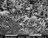
A biofilm is a complex community of microorganisms characterized by the excretion of an adhesive and protective extracellular matrix, microbe-to-microbe attachment, structural heterogeneity, genetic diversity, and complex community interactions. Dental plaque is a microbial biofilm (Figure 1). As with any biofilm, the constituent microbes are tightly adherent to each other and to an oral substrate by means of an extracellular matrix, ie, slime layer or glycocalix, into which they are embedded.16,17 The microbial populations in biofilm have 2 strategies that enable them to successfully survive within their community. The first is a high rate of reproduction for continued survival, and the second is physiologic adaptation to the available environmental resources or life-supporting capacity of the environment.18
Biofilms inherently dictate profound changes in the behavior of individual microbes, their relationship to the host, and their response to environmental conditions.19 Indeed, oral biofilms, as distinct entities, are the causative agents of biological processes such as dental caries, periodontal disease, and peri-implantitis, rather than any single microbe evading the host defense and causing disease.20 Biofilms exhibit characteristics that impact the clinical management of inflammatory periodontal disease. For example, both altered patterns of microbial gene expression and the composition and density of the extracellular matrix reduce the susceptibility of microbes to antimicrobial agents.21-23 Bacteria growing in dental biofilms display an increased tolerance to antimicrobial agents, including those used in dentifrices and mouthrinses.24-27 In addition, confocal microscopy of in situ established natural biofilms showed that chlorhexidine only affected the outer layers of cells in 24 and 48 hour plaque biofilms, suggesting either quenching of the agent at the biofilm surface or a lack of penetration.28 Further, biofilms of oral bacteria are also more tolerant of antibiotics (eg, amoxycillin, doxycycline, minocycline, and metronidazole) than planktonic cells.29-31 In this regard, biofilms of Porphyromonas gingivalis have been shown to tolerate 160 times the minimum inhibitory concentration (MIC) of metronidazole that was determined for planktonic cells.32
Over 700 species of aerobic and anaerobic bacteria have been identified in the human oral cavity.33,34 The microbes grow as complex, mixed, interdependent colonies in biofilms, and may achieve considerable thickness, achieving a thickness of 1 mm within 96 hours, if left undisturbed.16,17 Oral biofilms, like all microbial biofilms, exhibit a successional colonization with gram-positive aerobic Streptococci species (spp.) being the initial colonizers, followed in sequence by Actinomyces spp., Corynebacterium spp., Veillonella spp., and then in more mature biofilm, a variety of gram-negative anaerobic microbes such as Treponema spp., Fusobacterium spp., Porphyromonas spp., Prevotella spp., and Tannerella spp.17,35,36
As the biofilm is allowed to mature with concomitant increases in thickness, the percentage of Gram-negative anaerobic microbes increases. Specific complexes of such microbes commonly cohabit subgingival sites and are consistently associated with inflammatory periodontal diseases.35 These putative microbial pathogens include Porphyromonas gingivalis, Tannerella forsythia, and Treponema denticola.35
In the human host, the transition from gingivitis to periodontitis does not occur automatically, either in every patient or every site, but depends on 3 factors: 1) degree of host susceptibility, 2) presence and numbers of pathogenic bacteria, and 3) presence and numbers of protective bacteria.36 Pathogenic bacteria exhibit virulence features that decrease the effectiveness of the host response by inducing tissue degradation and retarding attempts at healing.
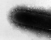
Host defense mechanisms are impaired through a variety of mechanisms. As one example, consider that Aggregatibacter (formally Actinobacillus) actinomycetemcomitans produces a leukotoxin that alters the cell membranes of neutrophils and monocytes and thereby alters chemotactic and phagocytic responses.36 Infection with Gram-negative anaerobes is accompanied by the release of epitheliotoxins, endotoxins, leukotoxins, collagenase, gellatinase, elastase, fibrinolysins, and other proteolytic enzymes.37 These bacterial toxins and enzymes are tissue irritants and/or cytotoxic and viewed by the host immune system as foreign proteins (Figure 2). The aggregate cellular/tissue insult activates the host immune system locally and is generally visualized at a clinical level as inflammation with all the inherent gingival changes, eg, vasculitis, edema and swelling, change in tissue color from white-pink to red or red-purple, and spontaneous gingival bleeding or bleeding on provocation.38
Role of the Host Immune Response
Bacteria are necessary but not sufficient by themselves to produce a destructive periodontal disease. Disease initiation and progression requires a susceptible host.38 The microbial challenge induces an immediate inflammatory and immune response in the host. The nature and magnitude of the response have an impact on the severity and rate of progression of the periodontal disease.39 Locally, bacteria and their metabolic byproducts stimulate a cellular immune response within the affected gingiva represented by a dense infiltration of neutrophils, macrophages, and lymphoid cells. These cells and host connective tissue cells within the developing inflammatory lesion are stimulated to synthesize and release proinflammatory cytokines, prostanoids, and proteolytic enzymes, eg, interleukin-1 (IL-1), interleukin-6 (IL-6), interleukin-8 (IL-8), tumor necrosis factor-alpha (TNF-α), prostaglandin E2 (PGE2), matrix metalloproteinases.38 It is this host inflammatory-immune response that ultimately leads to the clinical signs of gingivitis and chronic periodontitis and their characteristic features of fibrous connective tissue degradation, resorption of tooth supporting alveolar bone, and periodontal pocket formation.
In contrast to the epidermis of skin, the epithelial lining of the soft tissue wall of a periodontal pocket lacks a stratum corneum and stratum granulosum. Consequently, the pocket epithelium is easily ulcerated and breached by invasive subgingival pathogenic bacteria.40 In addition, endotoxins and other microbial antigens may gain access to the underlying connective tissues and gingival vasculature, leading to bacteremia and endotoxemia. There is considerable evidence that the locally produced proinflammatory cytokines and prostanoids gain access to the circulatory system and may, in turn, induce the production of liver-derived markers of a systemic inflammatory reaction, such as C-reactive protein, fibrinogen, serum amyloid-A, and haptoglobin.41-45 Elevations in both the locally generated inflammatory mediators and systemic markers of inflammation have been associated with various systemic diseases such as atherosclerosis,46 cardiovascular disease,47 ischemic stroke,48 pre-eclampsia,49 and poor glycemic control50 in diabetic patients.
Risk Factors Associated With Development of Chronic Periodontitis
In addition to the accepted associations of pathogenic microbes to the pathogenesis of inflammatory periodontal diseases, several genetic and environmental risk factors have been identified that affect the host response. It is well established that the prevalence and severity of chronic periodontitis increases with advancing age, poor oral hygiene, marginally or poorly controlled type I and II diabetes, and use of tobacco.51,52 In addition, data from twin studies indicate that about 50% of the population variance in periodontitis can be attributed to genetic factors.53,54 Several studies indicate that genetic polymorphisms (variations) in a cluster of at least 3 genes on chromosome 2q13, which control the production of proinflammatory cytokines, may affect the systemic inflammatory response in a significant percentage of people with chronic periodontitis.55,56
Scaling and Root Planing in the Control of Chronic Periodontitis
Periodontitis is a chronic and progressive inflammatory disease for which there is no known cure. It is now well-established that periodontitis is not associated with a single microorganism but rather the initiation and progression of periodontitis is the result of the host's immune response to a consortium of bacteria. For periodontopathic bacteria to initiate periodontitis, it is essential that they are able to colonize subgingival pockets and produce virulence factors that directly damage host tissue. Thus, a major goal of nonsurgical periodontal therapy is to suppress, to the extent possible, the subgingival pathogenic microbial flora and thereby significantly reduce or eliminate the associated inflammatory lesion.
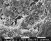
Dental calculus was the original etiologic agent associated with development of chronic periodontitis. In the 1960s and 1970s it was established that the rough, irregular surface of dental calculus was always covered with a nonmineralized microbial biofilm (Figure 3).57-59 In addition to the surface biofilm, at least one recent study has identified the presence of several viable periodontal pathogens within the mass of dental calculus, ie, Aggregatibacter actinomycetemcomitans, Treponema denticola and Porphyromonas gingivalis.60 Interestingly, the persistence of Porphyromonas gingivalis in the subgingival environment following periodontal therapy has been associated with progressive alveolar bone loss.61 In support of this observation, Offenbacher et al62 recently reported a significant association between serum immuneoglobulin G (IgG) titers against Porphyromonas gingivalis in patients that exhibit deep PDs (≥ 4 mm) and moderate (≥ 10% to < 50%) and severe (≥ 50%) bleeding on probing.
In spite of the fact that calculus can serve as a reservoir for pathogenic microbes, the role of subgingival calculus, as an etiologic agent in chronic periodontitis, was relegated to secondary status once microbial biofilm was declared the primary, extrinsic etiologic factor. Thus, the need for complete removal of subgingival calculus became a subject for debate.63 However, the traditional treatment modality of scaling and root planing (SRP) remains the "gold standard" for the nonsurgical management of periodontitis.64
The periodontal literature is replete with studies showing that treatment of periodontitis by SRP results in reductions in probing depth (eg, a mean reduction of 1.29 mm for 4-6 mm pockets and a mean of 2.16 mm for pockets of ≥ 7 mm) and subgingival bacterial loads and gains in clinical attachment.65-67 Probing depth (PD) reduction is generally greater at sites with deeper initial probing depths. The decrease in PD is the result of 2 phenomena: shrinkage of the pocket soft tissue wall manifested as recession of the gingival margin which results from a decrease in soft tissue inflammation and the inherent edema; and gain in clinical attachment. The latter usually accounts for roughly one-half of the probing depth reduction.65-67 In general, clinicians should evaluate post-SRP healing at 4 to 6 weeks following treatment. After 6 weeks, most of the healing has taken place but repair and collagen maturation may continue for an additional 9 months.67,68
Three relevant observations must be considered when deciding to use nonsurgical therapy as the primary modality for treatment of early to moderate chronic periodontitis. First, regarding SRP, clinicians must be careful when interpreting data from published clinical trials as they may not accurately reflect the private practice setting in terms of time, skill level, severity of disease, and diversity of patient population.65 For example, university-conducted clinical trials often use highly skilled clinicians, select patients for level of disease, and report spending 10 minutes per tooth when performing SRP.66,67 Ten minutes per tooth equates to about 70 minutes per quadrant. It is the experience of this author that in private practice a quadrant of SRP may be completed in approximately 60 minutes, regardless of the level of disease, and this allows approximately 10 minutes for setting of the patient and administration of anesthetic. Greenstein67 has rightfully noted that decreased time devoted to SRP in more recent studies probably accounts for the diminished results reported when to the more classic clinical trials. Second, one must remember that microbes embedded in a mature, undisturbed subgingival biofilm may exhibit an increased tolerance to antimicrobial agents.28-32 Third, even when chronic periodontitis is treated successfully, the reduction in subgingival pathogenic microbes is transitory. SRP of diseased root surfaces can open dentinal tubules, allowing invasion by periodontal pathogens into the exposed tubules, and possibly then serve as a reservoir for re-infection of the pocket.69,70 Thus, the need for follow-up treatment, usually consisting of supra- and subgingival debridement at 3 to 4 month intervals, is necessary to maintain the initially gained beneficial effects.71,72 Collectively considered, the distinct probability of less than ideal results from SRP and pocket re-infection by residual microbes is a forceful argument for the use of adjunctive treatment modalities in addition to SRP.
Clinical Implications
- The prevalence rate for chronic periodontitis (slight, moderate, and advanced severity) is approximately 30% of the US adult population or roughly 65 million individuals.
- Bacteria growing in undisturbed dental biofilms exhibit a significant increased tolerance to antimicrobial agents and antibiotics.
- The transition from gingivitis to periodontitis does not occur automatically, either in every patient or every site, but depends on 3 factors: 1) degree of host susceptibility, 2) presence and numbers of pathogenic bacteria, and 3) presence and numbers of protective bacteria.
- Even when chronic periodontitis is treated successfully, the reduction in subgingival pathogenic microbes is transitory. Thus, the need for follow-up treatment, usually consisting of supra- and subgingival debridement at 3 to 4 month intervals, is necessary to maintain the initially gained beneficial effects.
- Due to limitations of SRP and reinfection of the periodontal pocket, adjunctive treatment modalities may increase the likelihood of improvement in the periodontal condition.
Disclosure
Dr. Cobb has served as a scientific advisor and consultant for OraPharma, Inc.
References
1. Armitage GC. Development of a classification system for periodontal diseases and conditions. Ann Periodontol. 1999;4:1-6.
2. Albandar JM, Kingman A. Gingival recession, gingival bleeding, and dental calculus in adults 30 years of age and older in the United States, 1988-1994. J Periodontol. 1999; 70:30-43.
3. Albandar JM, Rams TE. Global epidemiology of periodontal diseases: an overview. Periodontol 2000. 2002;29:7-10.
4. Löe H, Brown LJ. Early-onset periodontitis in the United States of America. J Periodontol. 1991;62:608-616.
5. Jenkins WM, Papapanou PN. Epidemiology of periodontal disease in children and adolescents. Periodontol 2000. 2001;26:16-32.
6. Douglass CW, Fox CH. Cross-sectional studies in periodontal disease: current status and implications for dental practice. Adv Dent Res. 1993;7:25-31.
7. Albandar JM, Brunelle JA, Kingman A. Destructive periodontal disease in adults 30 years of age and older in the United States, 1988-1994. J Periodontol. 1999;70:13-29.
8. Brown LJ, Oliver RC, Löe H. Evaluating periodontal status of US employed adults. J Am Dent Assoc. 1990;121: 226-232.
9. Borrell LN, Burt BA, Taylor GW. Prevalence and trends in periodontitis in the USA: The NHANES, 1988 to 2000. J Dent Res. 2005;84:924-930.
10. Page RC, Eke PI. Case definition for use in population-based surveillance of periodontitis. J Periodontol. 2007; 78:1387-1399.
11. Borrell LN, Crawford ND. Social disparities in periodontitis among United States adults 1999-2004. Community Dent Oral Epidemiol 2008;36:In Press.
12. Cobb CM, Williams KB, Gerkovitch M. Is The Prevalence of Periodontitis in the United States in Decline? Periodontol 2000. 2008; In Press.
13. Kingman A, Morrison E, Löe H. Systematic errors in estimating prevalence and severity of periodontal disease. J Periodontol. 1988;59:707-713.
14. Hunt R, Fann S. Effect of examining half teeth in a partial periodontal recording of older adults. J Dent Res. 1991;70:1380-1385.
15. Eaton KA, Duffy S, Griffiths GS, Gilthorpe MS, Johnson NW. The influence of partial and full-mouth recordings on estimates of prevalence and extent of lifetime cumulative attachment loss: A study in a population of young male military recruits. J Periodontol. 2001;72:140-145.
16. Listgarten MA. Structure of the microbial flora associated with periodontal health and diseases in man. J Periodontol. 1976;47:1-18.
17. Cobb CM, Killoy WJ. Microbial colonization in human periodontal disease: an illustrated tutorial on selected ultrastructural and ecologic considerations. Scan Microsc. 1990;4:675-691.
18. Nishihara T, Koseki T. Microbial etiology of periodontitis. Periodontol 2000. 2004;36:14-26.
19. Marsh PD. Dental plaque as a microbial biofilm. Caries Res. 2004;38:204-221.
20. Caldwell DE, Atuku E, Wilkie DC, et al. Germ theory vs. community theory in understanding and controlling the proliferation of biofilms. Adv Dent Res. 1997;11:4-13.
21. Marsh PD. Dental plaque: Biological significance of a biofilm and community life-style. J Clin Periodontol. 2005;32(Suppl 6):7-15.
22. Gilbert P, Maira-Litran T, McBain AJ, Rickard AH, Whyte FW. The physiology and collective recalcitrance of microbial biofilm communities. Adv Microbial Physiol. 2002;46:203–255.
23. Stewart PS, Costerton JW. Antibiotic resistance of bacteria in biofilms. Lancet. 2001;358:135-138.
24. Marsh PD, Bradshaw DJ. Microbiological effects of new agents in dentifrices for plaque control. Inter Dent J. 1993;43:399-406.
25. Kinniment SL, Wimpenny JWT, Adams D, Marsh PD. The effect of chlorhexidine on defined, mixed culture oral biofilms grown in a novel model system. J Appl Bacteriol. 1996;81:120-125.
26. Wilson M. Susceptibility of oral bacterial biofilms to antimicrobial agents. J Med Microbiol. 1996;44:79-87.
27. Pratten J, Wilson M. Antimicrobial susceptibility and composition of microcosm dental plaques supplemented with sucrose. Antimicrob Agents Chemother. 1999;43:1595-1599.
28. Zaura-Arite E, van Marle J, ten Cate JM. Confocal microscopy study of undisturbed and chlorhexidine-treated dental biofilm. J Dent Res. 2001;80:1436-1440.
29. Larsen T. Susceptibility of Porphyromonas gingivalis in biofilms toamoxicillin, doxycycline and metronidazole. Oral Microbiol Immunol. 2002;17:267-271.
30. Socransky SS, Haffajee AD. Dental biofilms: difficult therapeutic targets. Periodontol 2000. 2002;28:12–55.
31. Noiri Y, Okami Y, Narimatsu M, Takahashi Y, Kawahara T, Ebisu S. Effects of chlorhexidine, minocycline and metronidazole on Porphyromonas gingivalis strain 381 in biofilms. J Periodontol. 2003;74:1647-1651.
32. Wright TL, Ellen RP, Lacroix JM, Sinnadurai S, Mittelman MW. Effects of metronidazole on Porphyromonas gingivalis biofilms. J Periodont Res. 1997;32:473-477.
33. Aas JA, Paster BJ, Stokes LN, Olsen I, Dewhirst FE. Defining the normal bacterial flora of the oral cavity. J Clin Microbiol. 2005;43:5721-5732.
34. Paster BJ, Olsen I, Aas JA, Dewhirst FE. The breadth of bacterial diversity in the human periodontal pocket and other oral sites. Periodontol 2000. 2006;42:80-87.
35. Socransky SS, Haffajee AD, Cugini MA, Smith C, Kent RL, Jr. Microbial complexes in subgingival plaque. J Clin Periodontol. 1998;25:134-144.
36. Sbordone L, Bortolaia C. Oral microbial biofilms and plaque-related diseases: Microbial communities and their role in the shift from oral health to disease. Clin Oral Invest. 2003;7:181-188.
37. Zambon JJ. Periodontal diseases: Microbial factors. Ann Periodontol. 1996;1:879-925.
38. Offenbacher S. Periodontal diseases: pathogenesis. Ann Periodontol. 1996;1:821-878.
39. Page RC, Kornman KS. The pathogenesis of human periodontitis: an introduction. Periodontol 2000. 1997:14:9-11.
40. Hujoel PP, White BA, Garcia RI, Listgarten MA. The dentogingival epithelial surface area revisited. J Periodont Res. 2001;36:48-55.
41. Ebersole JL, Machen RL, Steffen MJ, Willmann DE. Systemic acute-phase reactants, C-reactive protein and haptoglobin in adult periodontitis. Clin Exper Immunol. 1997;107:347-352.
42. Noack B, Genco RJ, Trevisan M, Grossi S, Zambon JJ, De Nardin E. Periodontal infections contribute to elevated systemic C-reactive protein level. J Periodontol. 2001;72:1221-1227.
43. Amar S, Gokce N, Morgan S, Loukideli M, Van Dyke TE, Vita JA. Periodontal disease is associated with brachial artery endothelial dysfunction and systemic inflammation. Arterioscler Thromb Vasc Biol. 2003;23:1245-1249.
44. Slade GD, Ghezzi EM, Heiss G, Beck JD, Riche E, Offenbacher S. Relationship between periodontal disease and C-reactive protein among adults in the Atherosclerosis Risk in Communities study. Arch Intern Med. 2003;163:1172-1179.
45. Leivadaros E, van der Velden U, Bizzaro S, et al. A pilot study into measurements of markers of atherosclerosis in periodontitis. J Periodontol. 2005;76:121-128.
46. Tonetti MS, D'Aiuto F, Nibali L, et al. Treatment of periodontitis and endothelial function. N Eng J Med. 2007;356:911-920.
47. Kinane DF, Lowe GD. How periodontal disease may contribute to cardiovascular disease. Periodontol 2000. 2000;23:121-126.
48. Grau AJ, Becher H, Ziegler CM, et al. Periodontal disease as a risk factor for ischemic stroke. Stroke. 2004;35:496-501.
49. Siqueira FM, Cota LOM, Costa JE, Haddad JPA, Lana AMQ, Costa FO. Maternal periodontitis as a potential risk variable for preeclampsia: A case-control study. J Periodontol. 2008;79:207-215.
50. Hein C, Cobb CM, Iacopino A. Report of an independent panel of experts of the Scottsdale Project. The independent study initiative for collaboration in diabetes, cardiovascular disease and periodontal disease intervention. Grand Rounds in Oral-Sys Med. 2007;2(Suppl):2-27.
51. Abdellatif HM, Burt BA. An epidemiological investigation into the relative importance of age and oral hygiene status as determinants of periodontitis. J Dent Res. 1987;66: 13-18.
52. American Academy of Periodontology. Position paper: Epidemiology of periodontal disease. J Periodontol. 1996;67:935-945.
53. Michalowicz BS, Aeppli D, Virag JG, et al. Periodontal findings in adult twins. J Periodontol. 1991;62:293-299.
54. Michalowicz BS, Diehl SR, Gunsolley JC, et al. Evidence of a substantial genetic basis for risk of adult periodontitis. J Periodontol. 2000;71:1699-1707.
55. Kornman KS, Crane A, Wang HY, et al. The interleukin-1 genotype as a severity factor in adult periodontal disease. J Clin Periodontol. 1997;24:72-77.
56. D'Aiuto F, Parkar M, Brett PM, Ready D, Tonetti MS. Gene polymorphisms in proinflammatory cytokines are associated with systemic inflammation in patients with severe periodontal infections. Cytokine. 2004;28:29-34.
57. Baumhammers A, Conway JC, Saltzberg D, Matta RK. Scanning electron microscopy of supragingival calculus. J Periodontol. 1973;44:92-94.
58. Muhleman HR, Schroeder HE. Dynamics of supragingival calculus formation. Adv Oral Biol. 1964;1:175-203.
59. Mandel ID. Dental plaque: Nature, formation and effects. J Periodontol. 1966;37:357-367.
60. Calabrese N, Galgut P, Mordan N. Identification of Actinobacillus actinomycetemcomitans, Treponema denticola and Porphyromonas gingivalis within human dental calculus: A pilot investigation. J Inter Acad Periodontol. 2007;9:118-128.
61. Chaves ES, Jeffcoat MK, Ryerson CC, Snyder B. Persistent bacterial colonization of Porphyromonas gingivalis, Prevotella intermedia, and Actinobacillus actinomycetemcomitans in periodontitis and its association with alveolar bone loss after 6 months of therapy. J Clin Periodontol. 2000;27:897-903.
62. Offenbacher S. Barros SP. Singer RE. Moss K. Williams RC. Beck JD. Periodontal disease at the biofilm-gingival interface. J Periodontol. 2007;78(10):1911-1925.
63. Robertson PB. The residual calculus paradox. J Periodontol. 1990;61:65-66.
64. Ryan ME. Nonsurgical approaches for treatment of periodontal diseases. Dent Clin N Am. 2005;49:611-636.
65. Cobb CM. Non-surgical pocket therapy: Mechanical. Ann Periodontol. 1996;1:443-490.
66. Greenstein G. Periodontal response to mechanical nonsurgical therapy: a review. J Periodontol. 1992;63:118-130.
67. Greenstein G. Nonsurgical periodontal therapy in 2000: a literature review. J Am Dent Assoc. 2000;131:1580-1592.
68. Badersten A, Nilveus R, Egelberg J. Effect of nonsurgical periodontal therapy. I: moderately advanced periodontitis. J Clin Periodontol. 1981;8:57-72.
69. Adriaens PA, DeBoever JA, Loesche WJ. Bacterial invasion in root cementum and radicular dentin of periodontally diseased teeth in humans. J Periodontol. 1988;59:222-230.
70. Giuliana G, Ammatuna P, Pizzo G, Capone F, D'Angelo M. Occurrence of invading bacteria in radicular dentin of periodontally diseased teeth: microbiological findings. J Clin Periodontol. 1997;24:478-485.
71. Listgarten MA. A rationale for monitoring the periodontal microbiota after periodontal treatment. J Periodontol. 1988;59:439-444.
72. Listgarten MA, Levin S, Schifter CC, Sullivan P, Evian CI, Rosenberg ES, Laster L. Comparative longitudinal study of 2 methods of scheduling maintenance visits: 2 year data. J Clin Periodontol. 1986;13:692-700.
About the Author
Charles Cobb, DDS, PhD, graduated from the University of Missouri-Kansas City with a dental degree, a Certificate of Specialty in Periodontics, and a Master of Science degree in Microbiology. He later earned a PhD in Anatomy from Georgetown University. He has held academic positions at Louisiana State University, the University of Alabama, and UMKC. In addition to teaching and research, Dr. Cobb has practiced periodontics full-time for 15 years in a private dental practice. Dr. Cobb recently retired from academics as Professor Emeritus at UMKC. He is a Diplomate of the American Board of Periodontology, has published over 165 peer-review articles, and presented over 120 programs at regional, national, and international meetings.