You must be signed in to read the rest of this article.
Registration on CDEWorld is free. Sign up today!
Forgot your password? Click Here!
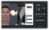
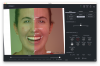
In recent years, the digital smile design concept has emerged as a pivotal workflow in dentistry, facilitated by technological advancements. This approach enables the creation of a "digital patient" (Figure 1), allowing dental professionals to digitally strategize treatments prior to performing irreversible procedures. The capacity to iterate, refine, and digitally evaluate treatments, coupled with the ability to collaborate with interdisciplinary teams visually, has enhanced both treatment planning and outcomes.1,2 Essentially, through the integration of various digital records, such as photographs, intraoral scans, digital imaging and communications in medicine (DICOM), facial scans, and implant data, a comprehensive digital replica of the patient is produced, capturing crucial facets of the patient's oral health and smile. Not only can the digital records be incorporated into the digital patient, but with such recorded technologies as jaw movement, important information for diagnostics can be added.
Platforms are available that offer the salient capability of consolidating diverse data sources into a singular digital patient profile, which can then be utilized to enhance the smile design process. Additionally, advanced technologies, notably facial analysis (eg, Smile Creator, exocad; TRIOS Smile Design, 3Shape; NemoSmile Design 3D, Nemotec), are underpinned by seminal research on facially driven smile design and facial harmony.3 Such technologies play a pivotal role in facilitating superior planning and design, with an emphasis on achieving facial harmony as opposed to mere symmetry. This convergence of long-standing expertise within a unified platform underscores how technological integration can elevate the precision of analysis, treatment planning, and design in the field of dentistry.
The primary objective of digital smile design (in the initial vision of Gurel, Calamita, Coachman, and others) is to formulate a comprehensive treatment plan that encompasses the entirety of the patient's health context, integrates all procedural facets, and renders the most precise simulation of the anticipated outcome.4,5 In addition to achieving predictable outcomes, a distinctive benefit of this approach is the facilitation of patient engagement throughout the process, enabling the patient to provide insights concerning design, dentition morphology, dimensions, and other related parameters.
Patient Study and Diagnostics
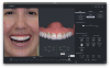
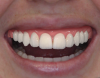
As an example, a case is presented in photographs throughout this article to demonstrate key elements of the digital smile design process. In this case, a young female patient was dissatisfied with the small size of her teeth and wanted a bigger and more appealing smile. She also disliked the anterior incisal edges of her smile, which were uneven (Figure 2).
The formulation of an extensive patient study (Figure 3) is instrumental in obtaining a holistic diagnostic overview. Sophisticated diagnostic tools for both cone-beam computed tomography (CBCT) diagnostics and x-ray examinations (eg, Diagnocat AI™, Diagnocat; Second Opinion®, Pearl, Inc.; dentalXrai Pro, dentalXr.ai; Denti.AI, Denti.AI Technology Inc.) can be utilized to enable a swift yet precise analysis of the patient's oral health status. Typically, these analyses can be completed within minutes and offer insights into various dental concerns, including caries, fractures, alveolar bone deterioration, periodontal disorders, and more. Even when initiating a case with a specific design intent, such comprehensive information is paramount in guiding the decision-making process relative to the final treatment plan.
The Digital Patient
Establishing a comprehensive digital patient profile (Figure 1) is pivotal for both the design phase and subsequent treatment plan determinations. Among the visualization techniques, the segmentation of CBCT stands out as a crucial workflow. By meticulously examining structures, such as tooth roots and bone integrity, practitioners can ascertain whether orthodontic intervention is required prior to embarking on smile restoration endeavors.
The patient can be presented with two versions of the smile design so that they have options for achieving the goals they indicated they wished to have addressed.
Version One
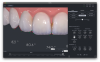
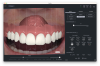
In the first version presented to the patient, no major treatment was needed. The design could be achieved with flowable composite veneers (Figure 4). With this version of the smile design, improvements would include a corrected smile incisal curve, tooth form, and incisor edges.
Version Two
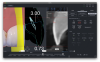
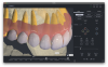
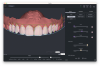
In the second version of the smile design presented to the patient, the design was based on a treatment plan that included crown lengthening to attain the desired height for the central incisors (Figure 5). Crown lengthening was used to establish the biological width required for the veneer, which was positioned 3 mm apically from the gingival margin. Concurrently, a gingivectomy would be conducted, ensuring that the incisor edge level would be minimally increased. With the use of calibrated tri-view imaging (Figure 6), reference lines facilitate accurate measurements of the bone margin, soft-tissue margin, and veneer height. After meticulous dimension adjustments, the elevation of the central incisors is augmented. The outcome is then carefully evaluated in relation to the face, adhering to principles of facially driven smile design.
The comprehensive smile design is subsequently modified to incorporate the smile analysis feature in smile simulation software that is augmented by artificial intelligence (AI) capabilities.
Zenith Analysis
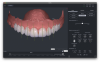
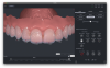
Zenith analysis elucidates the triangular configurations formed by the zenith points of the central incisors, lateral incisors, and canines (Figure 7). Ideally, these delineated lines should possess comparable lengths, and the resulting angles should manifest equivalent values. In this context, it is imperative to incorporate considerations such as lip symmetry and facial analysis. The objective is to achieve facial harmony based on the particular patient rather than mere symmetry. For instance, the curvature of the smile ought to mirror that of the lower lip. A smile is deemed harmonious when it maintains consistent curvature throughout.6
Axial Angulations
Axial angulation serves as a pivotal quality control measure within smile design, facilitating the fine-tuning of minute esthetic details (Figure 8). A thorough analysis of both the design's axial angulations and the tooth roots (via CBCT segmentation [Figure 9]) is instrumental for informed decision-making concerning potential orthodontic interventions. The precise axial angulations are paramount for achieving durability of dental prosthetics such as crowns and veneers. This precision ensures that occlusal forces are appropriately distributed, safeguarding the correct angulation upon the roots during biting actions.
Incisor Angulations in Dental Harmony
For a harmonious dental presentation, the apertures of the incisor embrasures should progressively increase in value from the central incisors outward to the canines (Figure 10). Such a gradation is deemed as exemplifying dental harmony.6
Measurements
Consistent measurements serve as a rigorous quality control mechanism, ensuring precision and accuracy at every juncture of both the design and treatment processes. Examples of elements that get measured are tooth height, amount of increase needed on incisal edges (Figure 11), and tooth width in order to analyze the proportions (ie, RED/golden) in relationship to adjusting the height or width of the teeth in the design process.
Smile Dominance
A smile is characterized as dominant when the lateral incisors are smaller in size and their incisal edges are elevated relative to the incisal edges of the central incisors (Figure 12). Conversely, when the incisal edges of both the laterals and centrals align with the smile curve, the smile is denoted as monotonic (Figure 13). This particular esthetic dimension profoundly influences facial dynamism and harmony. It is imperative to note that there is no absolute standard in this context; given the subjective nature of smiles, adaptations should be predicated upon patient feedback and preferences.7
Facial Flow and Its Role in Smile Harmony
Facial flow (Figure 14) is a term derived from the observation that most individuals (more than 95%) do not possess an impeccably linear facial midline.7 Instead, this midline can be conceptualized as a curve, delineated by four primary landmarks: the glabella, tip of the nose, philtrum, and chin. This curve elucidates a specific directional trajectory, termed as the "facial flow."
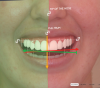
Empirical studies on patients reveal that a smile designed in alignment with the facial flow (indicated by the green region in Figure 14) is perceived as harmonious.7 In contrast, deviations from this flow (Figure 15), venturing into the red region, yield intersecting lines that manifest as visual tension. Such tension is typically deemed less harmonious in the overall facial esthetic.
Central Incisors Ratio and RED Proportions
The optimal proportional ratio for dental esthetics posits that the width of the central incisor should be 80% of its height. In other words, if the height of the central incisor is considered 100%, its width ideally should measure 80% (Figure 16). Adhering to this ratio prevents the visual appearance of the central incisors from being overly square (eg, 100% height and 90% width) or excessively narrow (eg, 100% height and 60% width). This proportionality is crucial for achieving a balanced and harmonious dental esthetic.8,9
When embarking on the reconstruction of dental proportions, one might commence with the ideal ratio, wherein the width of the central incisor is set at 80% of its height (a 100% to 80% ratio). This foundational step should be grounded in the context of the biological width and any necessary crown lengthening procedures.
Subsequently, to allocate space accurately for the lateral incisors and canines, the "recurring esthetic dental" (RED) proportions should be employed (Figure 17).8,9RED proportion is a visual reference for teeth width in which, from the frontal view, the following mathematical model is applied:
central incisor width = x
lateral incisor width = x*0.62
canine width = x*0.38
These values serve as an esthetic compass during the design phase. However, slight variations from these proportions may be made as needed to ensure flexibility and adaptability in the design process.
Smile Frame and the Papilla Curve
Within the context of dental esthetics, the papilla curve is delineated by the region between the 50% and 80% height markers of the central incisors(Figure 18).10 As part of the smile design procedure, any interventions, such as crown lengthening or gingivectomy adjustments, must ensure that the apex of the papillae falls within this designated zone. Adhering to this guideline fosters harmony between the gingival margins and the tip of each papilla. Moreover, these congruencies contribute significantly to the overall vitality and energy exuded by the smile.
VDO, Shimbashi Index - CEJ to CEJ in Centric Occlusion
Whether or not to open the patient's bite often emerges as a pivotal decision within the smile design continuum. The Shimbashi index provides an instrumental framework for initiating and scrutinizing the design, as well as any modifications necessary within the treatment plan. It suggests that the distance between the zenith points of the upper and lower central incisors should be around 19 mm. This index is used to set a new vertical dimension of occlusion (VDO) (Figure 19).11
When a clinical resolution to open the bite is finalized, bite registration is employed to discern the most optimal value, followed by a subsequent rescan of the patient's mandible in its newly ad-
justed position. Contrary to a digitally approximated bite opening, this scanning method offers a precise representation of the patient's mandibular alignment in its updated stance, ensuring greater fidelity to the actual anatomical alteration.
While the bite registration can aspire to align with the parameters set by the Shimbashi index, the definitive boundary will invariably be the posture wherein the patient experiences maximum comfort. In this intricate interplay between digital and analog techniques, the digital platform furnishes an initial blueprint, which is then fine-tuned with hands-on, analog precision.
Final Design Proposals
To craft an ideal smile using digital design requires a blend of clinical and technical experience, research, and artistic touch. However, AI tools like facial analysis and automated smile dominance are making this process easier, even for those who are newer to the field. These tools infuse the process with knowledge from decades of dental practice, allowing for better care and outcomes for patients.
While the quality of the final treatment still rests on the skills of the dentist and technician, technology is playing an increasingly vital role. Devices like implant guides simplify and refine the process.
Dentistry is evolving fast, with technology like AI shaping its future. Nonetheless, the roots of digital dentistry still lie in traditional, analog methods. The two go hand-in-hand: the solid foundation of traditional dentistry paved the way for the digital advancements seen today, improving the overall quality of dentistry.
Conclusion
Digital smile design enables dental providers to digitally plan treatments before actually implementing procedures. By integrating an assortment of digital records, a comprehensive digital replica of the patient is generated whereby vital aspects of the patient's oral condition and smile are captured. Digital smile design, a versatile conceptual tool for treatment planning in esthetic dentistry, can be used to improve diagnostic vision, enhance communication among providers and the patient, and make treatment more predictable throughout the restorative process.
About the Author
Ralph Georg, MA, MS
Creator and Founder, SmileFy Academy, Miami, Florida
Queries to the author regarding this course may be submitted to authorqueries@broadcastmed.com.
References
1. Coachman C, Sesma N, Blatz MB. The complete digital workflow in interdisciplinary dentistry. Int J Esthet Dent. 2021;16(1):34-49.
2. Coachman C, Blatz MB, Bohner L, Sesma N. Dental software classification and dento-facial interdisciplinary planning platform. J Esthet Restor Dent. 2021;33(1):99-106.
3. Blatz MB, Chiche G, Bahat O, et al. Evolution of aesthetic dentistry. J Dent Res. 2019;98(12):1294-1304.
4. Coachman C, Calamita M. Digital smile design: a tool for treatment planning and communication in esthetic dentistry. QDT. 2012. chrome-extension://efaidnbmnnnibpcajpcglclefindmkaj/https://digitalsmiledesign.com/files/Old-Website-Assets/Static/Coachman_Calamita_DSD_Eng_12.pdf. Accessed October 3, 2023.
5. Coachman C, Calamita MA, Coachman FG, et al. Facially generated and cephalometric guided 3D digital design for complete mouth implant rehabilitation: a clinical report. J Prosthet Dent. 2017;117(5):577-586.
6. Gurel G. The Science and Art of Porcelain Laminate Veneers. Berlin, Germany: Quintessence Publishing; 2003.
7. Silva BP, Mahn E, Stanley K, Coachman C. The facial flow concept: an organic orofacial analysis-the vertical component. J Prosthet Dent. 2019;121(2):189-194.
8. Murthy BV, Ramani N. Evaluation of natural smile: golden proportion, RED or golden percentage. J Conserv Dent. 2008;11(1):16-21.
9. Baghiana G, Peter D, Manju V, et al. Relevance of recurring esthetic dental (RED) proportion and golden proportion among patients attending a tertiary care center at Kochi, Kerala. J Oral Biol Craniofac Res. 2022;12(6):890-893.
10. Chu SJ, Tarnow DP, Tan JH, Stappert CF. Papilla proportions in the maxillary anterior dentition. Int J Periodontics Restorative Dent. 2009;29(4):385-393.
11. Joy TE, Kiran MS, Rahul R, et al. Evaluation of vertical facial height reduction and severity of temporomandibular joint disorders using Shimbashi number and cephalometric analysis. Cranio. 2021;39(4):287-293.