You must be signed in to read the rest of this article.
Registration on CDEWorld is free. Sign up today!
Forgot your password? Click Here!
An estimated 54,540 adults (39,290 men and 15,250 women) in the United States will have been diagnosed with an oral or oropharyngeal cancer this year.1 Worldwide, an estimated 476,125 people were diagnosed with these conditions in 2020.1
At the 2020 Greater New York Dental Meeting, the author was asked to present a synopsis in a poster presentation on oral cancer from a dental laboratory perspective.2 In 45 years as a dental laboratory technician, the author had worked with many dentists and had seen a large number of oral cancer patients who required obturator appliances from the laboratory. Some were effective, and others were not.
A certain percentage of problems related to patient compliance. However, in researching for the poster presentation, the author discovered that the protocols for the various stages of the fabrication of these appliances often were not followed correctly by the clinician and technician. This included procedure, materials, and patient compliance. This article will explain how to achieve consistent and predictable results with these cases.
Solutions
With so many presentations on digital dentistry, esthetic crown-and-bridge alternatives, and materials science at the Greater New York Dental Meeting, the author was uncertain of how a presentation on obturator appliances would be received. However, as dentists and students started to enter the exhibit hall, a concerned-looking dentist said, "I am so thankful that someone is speaking on this topic." The dentist said her brother, who had been in perfect health a year earlier before being diagnosed with oral cancer, was having a very difficult time with the obturator appliance that was fabricated for him. The author explained the various stages with obturator construction and which design options and materials were available, and ultimately received her brother's case to fabricate. Although there were some adjustments needed in the upcoming months, a functional obturator appliance was fabricated for her brother.
Adjustments are expected and changes associated with healing will continue to occur in the border areas of the defect for at least a year. Some of the challenges the patient may face include facial asymmetry and deformity, tooth loss, alveolar bone resorption, changes to the hard palate, decreased quality of life, psychological disorders, and hyper-nasal speech.2
Dental technicians can help alleviate many of these issues by properly designing the obturator appliance. Goals for the final outcome should be improving speech, aiding proper airflow, improving eating capabilities, preserving remaining dentition and tissue, and providing comfort, function, and esthetics.2
The role of the maxillofacial prosthodontist in these cases is primarily to help with the rehabilitation of surgically treated patients with maxillary cancers. Prescribing an obturator prosthesis will help with pain control, oral function (speech, swallowing, mastication, and facial esthetics), psychological issues, and quality of life.2
Oral cancer-related intraoral defects in the maxilla can occur in the form of an opening into the antrum and nasopharynx resulting from malignancies, congenital malformations, and acquired defects resulting from surgery; surgery can create anatomical defects between the oral and nasal cavity.2 The opening produced may be quite small or it may include any portion of the hard and soft palate, the alveolar ridges, and the floor of the nasal cavity.2 Many of the postsurgical maxillary defects result in hyper-nasal speech and impaired masticatory function.2 Patients have difficulties while performing normal functions such as swallowing and speaking due to these defects.2 Obturators rehabilitate the intraoral and extraoral structures and aid in the normal function of mastication, speech, and esthetics.2
Fabrication Protocol
Stage 1: Temporary or Transitional Obturator
With a temporary obturator, the patient is usually seen every 2 weeks because of the rapid soft-tissue changes that occur within the defect during healing.3 The temporary obturator is constructed from the postsurgical impression, and the closed bulb extending into the defect area is hollow. A new lining or soft-liner material is then processed to the bulb intraorally. Some clinicians put a liner on top of the old liner, but it is best to remove the entire interim lining material because of bacterial contamination, odors, and mucosal irritations.3 These temporary obturators typically need to function comfortably for as long as 6 months. The timing will vary depending on the size of the defect, the progress of healing, the function of the obturator, and the presence or absence of natural dentition.
Stage 2: Final or Definitive Obturator
The final obturator should not be fabricated until the surgical site is healed and dimensionally stable, taking into consideration the patient's physical and emotional state.3 Changes associated with healing will continue to occur in the surrounding areas of the defect for at least 1 year.3 Dimensional changes are primarily related to the peripheral soft tissues.3
Because of these defects and ongoing dimensional change, it is so important to utilize the correct materials. Utilizing a polymer with dimensional accuracy, including impact resistance and flexural strength, is a requirement. A soft-liner material that is resistant to bacteria and adapts accordingly to the temperature of the oral environment is very beneficial in overcoming dimensional change. It should also be considered that many obturator patients are very sensitive to monomer content and even the chemicals in some soft-liner materials. The final obturator may include a metal framework with acrylic and teeth or just acrylic, which replaces the palate and supports the teeth and the bulb.
It should be noted that an immediate obturator also may be utilized at the time of surgery. Any phase of treatment may be altered dependent on the nature of the disease and its staging, radiation, chemotherapy, surgical complications, and the morbidity of the disease.
Impression Protocol
The use of digital impressions continues to increase for obturator cases. Utilizing mesh-manipulating software has been very effective for designing, but scanning defective soft tissue on these case types can present challenges. Still, the impression and model can be scanned with good accuracy.
When analog impressions are utilized for these cases, the clinician takes a preliminary impression and the laboratory constructs two custom trays: one small, thin tray for the defective area and one larger, thicker tray for the full arch. During the impression taking, border molding is performed on defective margins, and the smaller tray is used for this procedure, utilizing a medium and light-body vinyl polysiloxane (VPS) material.2 An alginate impression can be taken over the smaller tray, especially with extremely difficult patients. A model is then poured in Type IV stone for stability.2
Clinical Case
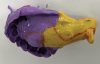
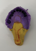
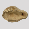
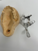
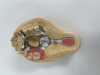
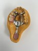
The author recently completed a case that required a cast partial framework, denture teeth, and a posterior palatal bulb. The clinician made his own custom trays, utilizing the impression method described above (Figure 1 and Figure 2). The technician expressed that the impression was less than ideal, but the clinician said several were taken and this was the best that could be done. The technician poured the impression with Type IV stone (Figure 3) for higher compressive strength.
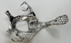
The case required a metal framework with an extension for a hollow bulb to cover the defect. The clinician had drawn an outline of the design with an indelible marker on the impression. The technician designed the frame with mesh retention so denture teeth could be processed to the frame. A sintered framework was fabricated (Figure 4 and Figure 5) and the denture teeth were set in wax for a try-in. Anatomical 33° denture teeth were utilized for better chewing capability (Figure 6). The clinician tried in the frame with the denture teeth and found everything to be acceptable, so the technician could proceed to the final prosthetic (Figure 7).
After processing the case, the technician utilized a duplicate model poured from VPS duplicating material and then processed soft-liner around the bulb to compensate for soft-tissue defects. The final case worked out perfectly and was a success with great fit and function.
Relining Existing Cases
Last year, a dentist was seeking help for a rabbi who had previously led a busy lifestyle but was now living with oral cancer and wearing an obturator appliance. His son had to speak for him during services at the synagogue due to hyper-nasal speech. The author visited the patient chairside and asked the clinician to take a light-body wash impression in the bulb area. The appliance was then able to be relined with a soft-liner material that adapted to the patient's oral defects based on the temperature of his mouth. With this relined appliance, the patient could speak with normal phonetics and regained his former lifestyle. Being chairside to witness this life-changing moment for the patient was an emotional and rewarding experience.
Relining the final obturator prosthesis is at times necessary at regular intervals.2 These intervals rely solely on the condition of the soft tissue and whether any denture teeth needed to be added to the existing obturator after the final insertion.2 The following laboratory reline procedure has proven very successful. First, border mold the margin areas of the obturator by applying adhesive and a medium-body VPS material, and insert in the patient's mouth. After border molding, apply a light-body VPS material over the medium-body material and have the dentist take a wash impression where the soft-tissue defect is. If additional denture teeth need to be added to the appliance, then a pickup alginate impression is needed. Bead the defective areas in the VPS impression, pour a stone model, and make a putty matrix of the position of the appliance. Remove the impression material and prepare the obturator for the soft liner. A traditional reline jig also may be used for this. Process a high-quality soft-liner material where the impression material was present. Process, finish, and polish.
Conclusion
Following the correct protocol on obturator fabrication can help patients overcome the problems associated with oral cancer and congenital defects. The basic principles of the design of removable partial dentures and full dentures should be reviewed when designing an obturator appliance. Post care for a definitive obturator is very important for the rehabilitation of a maxillary defect. This includes the successful relining of the prosthesis, patient counseling, and hygiene maintenance.
About the Author
Dennis Urban, CDT
Director of Operations and
Clinical Education
MicroDental New York
Hicksville, NY
References
1. Oral and Oraopharyngeal Cancer: Statistics. Cancer.Net. https://www.cancer.net/cancer-types/oral-and-oropharyngeal-cancer/statistics. Published February 2023. Accessed July 31, 2023.
2. Urban D. Successful Obturator Solutions. Poster presented at: Greater New York Dental Meeting; November 2021; New York, NY.
3. Singh M, Bhushan A, Kumar N, Chand S. Obturator prosthesis for hemimaxillectomy patients. Natl J Maxillofac Surg. 2013;4(1):117-20.