You must be signed in to read the rest of this article.
Registration on CDEWorld is free. Sign up today!
Forgot your password? Click Here!
New polymers and resin-based composite materials are continually being introduced for use in the production of provisional and permanent dental restorations and prostheses. Among other factors, this is largely in response to the rapid expansion of the field of digital dentistry. This article reviews the many types of resin-based materials that are currently available for CAD/CAM milling and 3D printing workflows in dentistry.
Resin-Based Materials for Subtractive Manufacturing
Subtractive manufacturing (ie, milling) has undergone tremendous refinement and adaptation to become a primary means of producing indirect dental restorations. Although blocks made entirely of ceramics are the most popular, there are many resin-based materials available. These resin matrix ceramics (RMCs) are subdivided into two categories: dispersed filler resin-based ceramics, which are basically composites, and polymer-infiltrated ceramic network (PICN) materials.1 It has been suggested that when compared with all-ceramic CAD/CAM blocks, RMC CAD/CAM blocks have higher load capacity and a modulus of elasticity that is closer to that of natural tooth structure, which is possibly better for implant restorations; are easier to mill; and are better at facilitating intraoral repairs.2,3 In addition, staining and finishing is relatively easy because these resin-based systems do not require any post-milling heat treatment processes. However, RMCs tend to be weaker and less wear resistant than all-ceramics.4
The first commercially available RMC designed for CAD/CAM dentistry (Paradigm™ MZ100, 3M) was introduced in 2001. It was simply a photopolymerized block of the company's composite that was popular at the time. Subsequently, RMC CAD/CAM blocks were produced under high temperature and pressure (typically greater than 100°C and 150 MPa)1 to enhance their mechanical properties through densification and high polymer conversion and to reduce their potential to release unreacted monomers.3 Resin-based ceramics are made by mixing glass filler particles of various compositions and sizes, which range from less than 100 nm up to several micrometers, with the same dental dimethacrylate monomers that are used in direct-fill composites, such as bisphenol A-glycidyl methacrylate (Bis-GMA), ethoxylated bisphenol-A dimethacrylate (Bis-EMA), urethane dimethacrylate (UDMA), and triethylene glycol dimethacrylate (TEGDMA). In 2013, an alternative material concept was introduced (VITA ENAMIC®, VITA North America), which involved the sintering of compacted glass or ceramic particles to produce a ceramic scaffold that could be infiltrated with a curable resin and polymerized at high temperature. Presumably, the microstructure allows the material to distribute stress more uniformly when compared with a dispersed particle composite.1 This type of material, which is referred to as a PICN material or hybrid ceramic, has a high percentage of inorganic content that may be accompanied by slightly higher mechanical properties. Manufacturers often describe their materials as hybrid ceramics; however, many of these products are actually dispersed particle resin-based ceramics that are cured with heat. Polymethyl methacryate (PMMA) blocks also exist, but they are weaker than the RMCs and are predominantly used for provisional restorations.
A significant difference between the two types of RMCs relates to the protocols used to bond them with resin cement. Because the aforementioned hybrid ceramic contains feldspathic porcelain as its main component, it is recommended to be treated with hydrofluoric acid followed by a silane coupling agent. In contrast, dispersed particle resin-based ceramics should be treated with air abrasion using silica-coated alumina particles followed by a silane agent.1,2
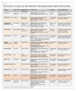
The properties of these indirect composite materials are largely influenced by the composition, quantity, and particle size of their fillers. Generally, materials with filler particles that are smaller on the nanoscale produce the glossiest polished surfaces, and materials with larger percentages of filler content demonstrate greater mechanical properties. The type of monomer used in dispersed particle resin-based ceramics may also affect their properties because the matrix that supports the reinforcing fillers is a continuous polymer network. Table 1 provides manufacturer data regarding the clinical applications of some of the resin matrix ceramics available for milling indirect restorations. It should be noted that the manufacturer-reported values for properties such as flexural strength and elastic modulus are often higher than what is reported by independent research in the literature.4 Research has also indicated that the wear resistance of milled RMCs is typically lower when compared with that of milled ceramics.4
In general, milled indirect composite materials, due to their enhanced cure, have been shown to be more color stable than direct composites but still less so than all-ceramics, which suggests that discoloration over time in the oral environment is both inevitable and material dependent.5
Clinical Applications and Considerations for Subtractive Manufactured Materials
Resin-based milling blocks are pre-polymerized under high heat; therefore, they exhibit superior mechanical properties when compared with direct composite resins. In addition, the indirect technique allows for a well-fitting restoration to be fabricated extraorally and then bonded intraorally, which offers many benefits.
Posterior Restorations
Taking these factors into consideration, resin-based milling materials are best suited for large restorations that will be subjected to high occlusal forces, restorations for which cuspal coverage is indicated, or cases in which access to the cavity is compromised, such as those involving patients whose ability to open their mouths is limited.6 The survival rate of posterior restorations fabricated from resin-based CAD/CAM blocks has been reported to be over 80% after 10 years, which is a reasonable clinical outcome.7 However, due to the relatively similar clinical outcomes achieved by these restorations when compared with direct restorations, selection of these materials remains mostly case dependent. Generally, a milled composite-based restoration is considered to be an intermediate solution between a direct composite restoration and a ceramic restoration, combining low cost, low abrasiveness to the opposing dentition, satisfactory marginal fit and physical properties, and long-term clinical performance that is acceptable despite being less than that of all-ceramics.6
Veneers
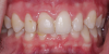
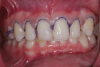
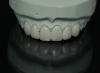
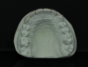
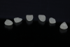
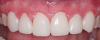
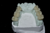
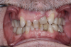
There is insufficient evidence regarding the use of indirect composites as a laminate veneer material. Indirect composite laminate veneers, both heat- and photopolymerized, have been reported to have a lower survival rate (75%) when compared with ceramic veneers (100%).8 Composite veneer failures have been attributed to either debonding or restoration fracture. In addition, the shade match, surface quality, and restoration wear of indirect composite veneers have all been shown to be inferior when compared with ceramic veneers. For these reasons, indirect composite veneers are only indicated for a limited number of clinical scenarios, such as long-term provisional/interim treatment with a plan for eventual replacement with ceramic material (Figure 1 through Figure 6) or when finances are a concern. If this approach is chosen, the process of obtaining informed consent from the patient should include an explanation about the increased risk for future esthetic and/or mechanical complications.
Full- and Partial-Coverage Restorations
Resin-based CAD/CAM materials are often being used for full- or partial-coverage restorations as an alternative to ceramic materials, especially in cases in which patient finances are limited. There are only a few clinical studies on the long-term survival rate of composite crowns that can be used to compare their performance to ceramic crowns. A clinical study that assessed the performance of milled composite partial coverage crowns at 1 year and 2 years reported success rates of 95% and 85.7%, respectively, with a cumulative 5% failure rate.9 Over the study period, the clinical outcomes that exhibited the highest deterioration were crown anatomy and marginal adaptation. The researchers emphasized the importance of following a strict bonding protocol to maximize success rates for these types of restorations.
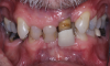
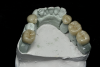
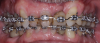
Composite-based crowns appear to have the capacity to successfully withstand average axial occlusal forces; however, they tend to exhibit higher stress on their intaglio surface under lateral forces, which makes their long-term use prohibitive for patients with parafunction, such as bruxism (Figure 7 through Figure 11).6,10
Resin-Based Materials for Additive Manufacturing
During the past decade, the use of additive manufacturing in dentistry has grown exponentially. The advantages of this method over subtractive ones include the ability to reuse materials, reduce waste, support sophisticated hierarchical multilevel architectures, and shorten overall chair time. Among the different technologies available, vat photopolymerization, material extrusion, and material jetting seem to be most suitable for 3D printing indirect restorations from dental polymers.11
Advances in the development of polymer-based 3D printing dental materials have played a key role in accelerating the bench-to-chair process of additive manufacturing. The commercially available array of resin-based composites for 3D printed indirect restorations has been rapidly broadening, and most of these materials are compatible with digital light processing (DLP) or stereolithography (SLA) technologies. Although the exact formulations of these materials are proprietary, generally speaking, they are similar to those of conventional composites that are based on (meth)acrylate monomers, photoinitiators, and inorganic fillers. Table 2 provides manufacturer data regarding the clinical applications of some of the resin-based composites available for 3D printing indirect restorations.
In the organic matrices of these materials, the ratios of low and high viscosity monomers are balanced to achieve proper viscosity for satisfactory recoating and printing accuracy and to endow the final polymer with toughness. Most of the dental composites available for 3D printing are composed of multifunctional acrylates and methacrylates to ensure high reactivity as well as blends of monomers and oligomers to mitigate polymerization shrinkage.12,13 High concentrations of photoinitiators that range from 1 to 10 wt% are added to the formulations of 3D printing composites to allow them to reach gelation from smaller doses of energy, which leads to faster printing speeds and facilitates rapid prototyping. The most common photoinitiators used are the type I radical initiators diphenyl(2,4,6-trimethylbenzoyl)phosphine oxide (TPO) (λ = 380 to 425 nm) and phenylbis(2,4,6-trimethylbenzoyl)phosphine oxide (BAPO) (λ = 350 to 440 nm), which is largely due to their compatibility with 3D printers based on higher wavelength lamps, color stability, and excellent photobleaching behavior.12,13 However, despite the high reactivity of the (meth)acrylates and photoinitiators in composites for 3D printing, washing and thermal and/or light-curing post-processing steps are required to remove potential harmful unreacted compounds and maximize the degree of conversion and mechanical properties of the 3D printed restorations.12 This is required in rapid prototyping because the irradiation of the material is heterogeneous due to the printing directions or the focus of the light scanning patterns being mostly on the outline of each layer. The high concentration of photoinitiators added to the formulations in combination with the short irradiation exposure times also contributes to the presence of unreacted monomers in the product. Moreover, because additive manufacturing is based on a layer-by-layer buildup approach, oxygen is commonly incorporated both within and between the layers, which results in the formation of oxygen-inhibition islands and pellicles, respectively.
The inorganic content of 3D printing dental resins is mainly composed of silica, titanium dioxide, aluminum oxide, and zirconium dioxide, and ranges from 30 to 50 wt% (higher for materials designed for permanent restorations).11 Although the incorporation of filler particles is critical to endow the 3D printed restorations with mechanical resistance, the size, refractive index, and ratios of these fillers need to be fine-tuned to result in an appropriate overall viscosity and minimize light scattering during the printing process, which is critical to ensuring high resolution and accuracy.12,13 In addition, it is important to ensure that the fillers are homogenously dispersed into the organic matrix because otherwise they can easily concentrate at the bottom of the reservoir. The most recent developments and directions in dental resin-based composites for 3D printing include formulations that:
• are compatible with printers based on different technologies;
• are endowed with antibacterial properties, enhanced toughness, maximized biocompatibility, shape-memory, and lower polymerization shrinkage;
• contain photoinitiators that are sensitive to long wavelengths and capable of undergoing "living" free radical polymerization;
• are based on alternative non-acrylate compounds; and
• have minimal post-processing requirements.
Clinical Applications and Considerations for Additively Manufactured Materials
To support these advances in the chemistry of 3D printing resins, improvements have been made to the firmware and hardware of desktop 3D printers. This has resulted in final products with properties that are similar to or better than their conventional and milled counterparts. In general, material selection for indirect restorations depends on ease of use, biocompatibility, esthetics, color stability, and acceptable mechanical properties. 3D printing resins are either "FDA cleared" or "FDA 510(k) exempt," of which the latter means that the manufacturer is responsible for ensuring biocompatibility. In either case, reviewing the safety data sheets of materials before considering their clinical use is highly advisable.14
Provisional Restorations
The discoloration of provisional restorations over time is a common occurrence.15 Some of the contributing factors include surface roughness, incomplete polymerization, and water sorption. According to research, the color stability of 3D printed provisional restorations follows a similar trend to that of milled and conventional PMMA provisional restorations, with maximum discoloration occurring at approximately 8 weeks.15 However, 3D printing resins have demonstrated high microhardness values when compared with their milled and conventional PMMA alternatives, which is advantageous when testing vertical dimension changes with provisional restorations.16 In addition, the flexural strength of 3D printed provisional fixed dental prostheses has been shown to be similar to that of milled restorations and higher than that of conventional PMMA-based provisional restorations.17 Contrasting results between different studies may be attributed to differences in the aging protocols and fabrication processes used as well as the chemistry of the resins. Regardless, the strength of 3D printed provisional restorations is higher than what is minimally required.17 Attempts have been made to further improve the mechanical properties of these restorations by adding filler particles such as zirconium and titanium dioxides, silica, and carbon fibers to the resins. The results that have been achieved are promising and may encourage the use of 3D printing resins for long-term provisionalization.
Definitive Restorations
Research regarding the use of additive manufacturing for dental ceramics is limited. Although there are several different technologies available for the additive manufacture of definitive ceramic restorations, the most popular is SLA printing of a resin-coated ceramic slurry. Ultraviolet light is used to photoactivate the resin material one layer at a time based on the shape of the restoration. Once printing is complete, the restoration undergoes post-processing and is sintered in a porcelain furnace.15 In general, there are no significant differences in fracture toughness between milled and SLA printed restorations; however, in some research, zirconia showed higher values of biaxial flexural strength when milled versus SLA printed. Interestingly, the Weibull modulus, which quantifies structural reliability and homogeneity, has been shown to follow an opposite trend with higher values for SLA printed restorations than those for milled ones.15 Pressed alumina consistently shows significantly lower mechanical performance than milled and SLA printed restorations. Although no direct comparison study has been performed, evidence suggests that there is little difference in the quality of the final products produced by different additive manufacturing technologies.
Printing and Post-Processing Considerations
Proper calibration of the printer, including resolution and orientation, and appropriate post-processing protocols play important roles in the success of additively manufactured dental restorations. The resolution of each print is determined by the clinical application. A higher resolution of approximately 25 to 50 µm is more critical when 3D printing indirect restorations as opposed to diagnostic models.14,18 Some printers have a fixed resolution and preset calibration, which limits the customization of printing protocols. This makes it crucial to follow the manufacturer's recommendations regarding material-specific post-processing protocols, timely replacement of resin tanks and cartridges, and regular firmware and software updates to ensure the precision and accuracy of final prints.18 With respect to post-processing, curing time has been proven to be essential in achieving desirable mechanical properties and minimizing color alterations.19 Similarly, the wash time affects the surface roughness, hardness, and biocompatibility of printed components.19 The specifics of both curing and wash time protocols are material-dependent, and not following them may be detrimental to the mechanical properties and longevity of 3D printed restorations. Finally, research indicates that the print orientation seems to affect the overall accuracy, with studies showing that a 90-degree orientation results in better accuracy when compared with 0-, 15-, and 45-degree orientations. Although the mechanical properties may also be affected by the printing orientation, they have a stronger association with the printing resolution.20
Conclusion
Based on the many options available and the limited published research comparing resin-based materials for the subtractive and additive manufacturing of dental restorations, clinicians are faced with difficult choices. However, positive outcomes are being achieved, and as the technologies and materials continue to improve, the expectation is that in the future, these systems will be universally used in dental offices to fabricate all types of permanent restorations.
Queries regarding this course may be submitted to authorqueries@broadcastmed.com
About the Authors
Esha Mukherjee, BDS, MSD
Assistant Professor
Department of Oral Rehabilitation and Biosciences
School of Dentistry
Oregon Health & Science University
Ana Paula Piovezan Fugolin, DDS, MS, PhD
Assistant Professor
Department of Oral Rehabilitation and Biosciences
School of Dentistry
Oregon Health & Science University
Despoina Bompolaki, DDS, MS
Associate Professor
Director, Clinical
Restorative Dentistry
Department of Oral Rehabilitation and Biosciences
School of Dentistry
Oregon Health & Science University
Jack L. Ferracane, PhD
Professor, Chair
Department of Oral Rehabilitation and Biosciences
School of Dentistry
Oregon Health & Science University
References
1. Mainjot AK, Dupont NM, Oudkerk JC, et al. From artisanal to CAD-CAM blocks: state of the art of indirect composites. J Dent Res.2016;95(5):487-495.
2. Blatz MB, Conejo J. The current state of chairside digital dentistry and materials. Dent Clin North Am. 2019;63(2):175-197.
3. Ruse ND, Sadoun MJ. Resin-composite blocks for dental CAD/CAM applications. J Dent Res. 2014;93(12):1232-1234.
4. Stawarczyk B, Liebermann A, Eichberger M, Güth JF. Evaluation of mechanical and optical behavior of current esthetic dental restorative CAD/CAM composites. J Mech Behav Biomed Mater. 2015;55:1-11.
5. Paolone G, Mandurino M, De Palma F, et al. Color stability of polymer-based composite CAD/CAM blocks: a systematic review. Polymers (Basel). 2023;15(2):464.
6. Bompolaki D, Lubisich EB, Fugolin AP. Resin-based composites for direct and indirect restorations: clinical applications, recent advances, and future trends. Dent Clin North Am. 2022;66(4):517-536.
7. Bustamante-Hernández N, Montiel-Company JM, Bellot-Arcís C, et al. Clinical behavior of ceramic, hybrid and composite onlays. A systematic review and meta-analysis. Int J Environ Res Public Health. 2020;17(20):7582.
8. Gresnigt MMM, Cune MS, Jansen K, et al. Randomized clinical trial on indirect resin composite and ceramic laminate veneers: up to 10-year findings. J Dent. 2019;86:102-109.
9. Zimmermann M, Koller C, Reymus M, et al. Clinical evaluation of indirect particle-filled composite resin CAD/CAM partial crowns after 24 months. J Prosthodont. 2018;27(8):694-699.
10. Duan Y, Griggs JA. Effect of elasticity on stress distribution in CAD/CAM dental crowns: glass ceramic vs. polymer-matrix composite. J Dent.2015;43(6):742-749.
11. Schweiger J, Edelhoff D, Güth JF. 3D printing in digital prosthetic dentistry: an overview of recent developments in additive manufacturing. J Clin Med. 2021;10(9):2010.
12. Bagheri A, Jin J. Photopolymerization in 3D printing. ACS Appl Polym Mater. 2019;1(4):593-611.
13. Ligon SC, Liska R, Stampfl J, et al. Polymers for 3D printing and customized additive manufacturing. Chem Rev. 2017;117(15):10212-10290.
14. Methani MM, Revilla-León M, Zandinejad A. The potential of additive manufacturing technologies and their processing parameters for the fabrication of all-ceramic crowns: a review. J Esthet Restor Dent. 2020;32(2):182-192.
15. Song SY, Shin YH, Lee JY, Shin SW. Color stability of provisional restorative materials with different fabrication methods. J Adv Prosthodont. 2020;12(5):259-264.
16. Revilla-León M, Morillo JA, Att W, Özcan M. Chemical composition, Knoop hardness, surface roughness, and adhesion aspects of additively manufactured dental interim materials. J Prosthodont. 2021;30(8):698-705.
17. Park SM, Park JM, Kim SK, et al. Flexural strength of 3D-printing resin materials for provisional fixed dental prostheses. Materials (Basel).2020;13(18):3970.
18. Mukherjee E, Malone L, Tackett E, et al. Monitoring the calibration of in-office 3D printers. Dent J (Basel). 2023;11(1):20.
19. Soto-Montero J, de Castro EF, Romano BC, et al. Color alterations, flexural strength, and microhardness of 3D printed resins for fixed provisional restoration using different post-curing times. Dent Mater. 2022;38(8):1271-1282.
20. Tahayeri A, Morgan M, Fugolin AP, et al. 3D printed versus conventionally cured provisional crown and bridge dental materials. Dent Mater.2018;34(2):192-200.