You must be signed in to read the rest of this article.
Registration on CDEWorld is free. Sign up today!
Forgot your password? Click Here!
The American Cancer Society estimates that 1,638,910 new cancer cases and 577,190 cancer deaths will occur in the United States in 2012.1 The good news is that both of these statistics are decreasing. The bad news is that many patients will still get cancer and have significant oral complications due to the treatment of their cancer. Many patients may not receive appropriate guidance to help manage these complications. This article discusses the potential oral problems related to cancer treatment and suggests some strategies to help avoid or treat these problems.
Types of Treatment for Cancer
Many patients will have chemotherapy (CT) to manage their cancer. Oral complications will differ by the type of chemotherapy agent, and the type and severity of these complications can vary from patient to patient.2,3 Chemotherapy has become much more sophisticated throughout the years; however, the main goal of the treatment is to kill cancer cells. As research has improved, the drugs used in CT have changed and target the specific cancer cells, such as with antibodies that are designed to attack the individual’s type of cancer cells (this is sometimes referred to a “targeted” or “directed” chemotherapy).4 Unfortunately, many patients will still receive CT agents that target both the rapidly dividing cells of the tumor and normal cells in the mucosa, bone marrow, and immune system (sometimes called cytotoxic chemotherapy). Adverse events of common cytotoxic CT agents include myelosuppression (thrombocytopenia [an abnormally low platelet count]/bleeding and neutropenia [an abnormally low neutrophil count]/oral infections), nausea and vomiting, oral mucositis, and neurotoxicity (Table 1).3–––– Dysgeusia (distorted taste) is also reported as a complication that may be related to the neurotoxicity, the inability to clean one’s mouth because of the pain of mucositis, infections such as candidosis, or the CT agent itself as it perfuses into oral fluids.5
Patients with some types of malignancies, such as leukemia or lymphoma, may undergo hematopoietic stem cell or bone marrow transplant. Oral complications, including mucositis and myelosuppression, can occur at several stages of the transplant (Table 2).6 Another rare oral finding is graft-versus-host disease (GVHD).
Patients with head and neck cancer will likely have some radiation treatments (RTs), with resulting oral complications. Mucositis, neurotoxicity and dysgeusia, xerostomia, trismus, and other functional changes may occur in the short term, with the potential for osteoradionecrosis or developmental delays in the long term.7 These complications have been greatly reduced by use of intensity modified radiation therapy.8 However, if the tumor is in close proximity to salivary glands, oral complications are still possible.9
Some patients with cancer will undergo surgical treatment, such as the removal of tumors of the breast, colon, or other solid organs. These patients will likely be intubated for general anesthesia, and there may be risks associated with less-than-perfect oral health due to the aspiration of loose teeth, plaque, or calculus during the surgery.10
How the Oral Healthcare Team Can Help
In a perfect world, a patient who receives a cancer diagnosis will be in excellent oral health; in reality, many patients have potential dental problems just waiting to worsen, or “blow up,” when they are least able to manage it. A comprehensive oral examination is needed to determine if any oral health problems must be addressed before the patient undergoes cancer treatment.
After the patient has received a cancer diagnosis, the oral healthcare team should work quickly so that the cancer treatment is not delayed. The team should have an understanding of the type of cancer and treatment plan (eg, specific medications, radiation, surgery), as well as any other medical conditions that the patient has and medications that the patient is taking.
For patients with leukemia and other cancers, recent laboratory test results are necessary to assist in the treatment planning of any dental emergencies. Results from the complete blood count with differential will provide information about the patient’s ability to fight any infections as well as whether the patient has enough platelets to have good hemostasis after surgery. The prothrombin time/international normalized ratio also helps determine the blood’s clotting ability.10
The pretreatment oral examination must include a complete extraoral and intraoral examination, as well as panoramic and full-mouth series of radiographs. Careful attention must be given to the presence of any existing carious lesions, broken or unserviceable restorations, periodontal or endodontic infections, other soft tissue problems, and any potential problems that could be exacerbated during the cancer treatment (Table 3).10
The timely management of these problems will keep the patient comfortable during cancer treatment, as well as reduce the risk of having to stop the cancer treatment in order to manage a dental emergency. An untreated dental abscess may become a serious systemic infection when the patient’s immune system is compromised due to CT and/or conditioning prior to stem cell or bone marrow transplant. Another consideration is to maintain proper nutrition by assuring that the patient is able to eat, including the patient’s having well-fitting appliances (eg, dentures).
The pretreatment dental visit also allows the oral health care team to communicate with the patient about what to expect, provide some solutions, and reinforce the importance of good oral hygiene. The patient’s ability to tolerate the oral complications of the cancer treatment will allow the patient to continue with the complete regimen. For some patients, just knowing what to expect makes it easier to handle.
For patients who will have radiation to the head and neck, the extraction of infected or problematic teeth approximately 2 weeks before the start of the radiation will allow for the healing to start. This will also help reduce the chance for development of osteoradionecrosis. These patients may benefit from the fabrication of custom fluoride trays to help prevent caries that accompany the loss of saliva.2,5,7
Loose anterior teeth must be removed before general anesthesia, so that the patient can tolerate the intubation procedure. Aspiration pneumonia is a known risk for patients who aspirate loose teeth, plaque, or calculus during the surgery.10
Common Oral ComplicationsDuring Cancer Treatment
Dysgeusia
In patients, dysgeusia, or the distortion of taste, may be multifactorial. The medications used in CT may spread throughout the body and into the saliva and gingival fluids. These CT agents may have a metallic or chemical taste and a degree of neurotoxicty.5,11
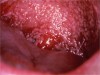
In many patients, the presence of candida infection leads to dysgeusia. Myelosuppression may have led to the use of systemic antibiotics, which allows candidosis (Figure 1) to develop. Also, xerostomia secondary to CT or RT can allow candida to grow.5,12
Graft-Versus-Host Disease
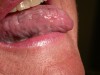
Graft-versus-host disease (GVHD) can occur in a patient (the host) who receives a bone marrow or hematopoietic stem cell transplant (the graft). The new immune system actually attacks the patient; essentially, the “graft” tries to eliminate the foreign tissue of the “host.” This can appear on the oral mucosa as a mild lichenoid reaction (white plaques) or be very serious with erosive, or ulcerative, lesions. If GVHD occurs in the first 100 days after the transplant, the condition is considered acute and may need to be treated with antirejection drugs such as cyclosporine or corticosteroids. Occurrence of GVHD after the 100-day mark, is called chronic13 (Figure 2).
Patients who take antirejection drugs have other oral issues to manage, such as gingival overgrowth, or immunosuppression.14
Impaired Nutrition
The primary cause of impaired nutrition for patients with cancer is mucositis (see below). Xerostomia may make existing appliances uncomfortable for the patient to wear. If teeth were extracted just before cancer treatment began, there may not have been adequate healing time to fabricate an appliance to replace the teeth.2,5
Mucositis
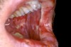
Mucositis can range from simple redness to widespread bleeding and ulceration (Figure 3). It can be limited to the oral cavity or occur in the whole gastrointestinal tract. In a very severe case, the patient may need to briefly stop taking CT. Antimicrobial medications, antiemetics, and/or the placement of a feeding tube may be needed to help ensure proper nutrition.5,15
Treatment usually consists of topical anesthetics (diphenhydra-mine solution or viscous lidocaine) or coating agents (liquid antacids or sucralfate). The patient should be advised to continue good oral hygiene; however, the use of a cotton swab or very soft toothbrush is recommended in order to reduce soft tissue trauma. For some patients, brushing without toothpaste may be reasonable. Rinsing with a sodium bicarbonate/salt mixture may help soothe the tissues (8 ounces of warm water, one half teaspoon of baking soda, and one half teaspoon of salt).5,6,16
Another way to soothe the tissues is to advise the patient to melt ice chips in the mouth, providing some anesthesia as well as water to wash away debris.16
Myelosuppression
Many CT agents will cause immune system suppression, which may mean dampening the ability to fight infections and reducing the presence of adequate thrombocytes (platelets) to prevent bleeding. For most patients, CT will be given for a number of days, with a break, or holiday, to avoid these serious complications. For many patients, injections of bone-marrow stimulating agents will help increase the numbers of white and red blood cells and platelets.10
Neurotoxicity
Several CT agents are known to cause neurotoxicity (eg, cisplatin, vincristine), although experiences will vary between patients. Radiation exposure may lead to changes to the nerves. Symptoms include dysgeusia, paresthesia, tooth pain, and temporomandibular joint pain. The dentist should rule out any odontogenic sources of the pain and refer the patient to the physician to manage neurogenic issues.7,17
Nausea and Vomiting/Diarrhea
Similar to mucositis, the patient may have gastrointestinal upset because of the CT action on the rapidly dividing cells in the stomach and intestines. Vomiting can cause excessive wear to the teeth, so the patient should be advised not to brush the teeth immediately after vomiting as the enamel may be softened by the stomach acid.5
Osteoradionecrosis
Osteoradionecrosis (ORN) occurs after RT that reaches a certain threshold of radiation exposure to the jaws. The mandible is most often affected, and ORN typically develops when extractions are necessary in the area of the jaw that has been radiated. Improvements in the aim of the radiation beam have reduced this problem, but prevention is the best way to avoid it; any hopeless teeth should be removed before the start of RT. The use of hyperbaric oxygen treatments remains controversial for prevention of ORN if the patient needs extractions after RT.7
Trismus
Trismus, or limited opening of the mouth, may occur if the muscles of mastication are in the field of RT. Fibrosis of the muscles due to exposure to radiation is a permanent change; however, jaw exercises can help the patient retain flexibility. The newer intensity modified radiation therapy techniques have essentially eliminated this problem.7,18
Xerostomia
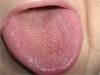
As with ORN and trismus, the most xerostomia symptoms have been seen with RT, especially when the parotid glands were in the field of radiation (Figure 4). Most CT regimens are not likely to cause permanent changes to the salivary glands, although there may be a temporary loss of saliva during the induction phase of treatment with bone marrow transplants.2,8
Conclusion
The oral healthcare team is a valuable part of helping patients with cancer. The patient’s quality of life can be improved, as well as the compliance with the cancer treatment plan. By supporting the patient with better oral health, the overall long-term prognosis will be improved.
References
1. Siegel R, Naishadham D, Jemal A. Cancer statistics, 2012. CA Cancer J Clin. 2012;62(1):10-29.
2. Meurman J, Grönroos L. Oral and dental health care of oral cancer patients: hyposalivation, caries and infections. Oral Oncol. 2010;46(6):464-467.
3. Sharma R, Tobin P, Clarke SJ. Management of chemotherapy-induced nausea, vomiting, oral mucositis, and diarrhoea. Lancet Oncol. 2005;6(2):93-102.
4. Watters AL, Epstein JB, Agulnik M. Oral complications of targeted cancer therapies: a narrative literature review. Oral Oncol. 2011;47(6):441-448.
5. Mosel DD, Bauer RL, Lynch DP, Hwang ST. Oral complications in the treatment of cancer patients. Oral Dis. 2011;17(6):550-559.
6. Hupp WS, Migliorati CA, Brennan M. Clinician’s guide to the dentist’s role in the management of the cancer patient. American Academy of Oral Medicine. 2011.
7. Sciubba JJ, Goldenberg D. Oral complications of radiotherapy. Lancet Oncol. 2006;7(2):175-183.
8. Jensen SB, Pedersen AM, Vissink A, Andersen E, et al; Salivary Gland Hypofunction/Xerostomia Section, Oral Care Study Group, Multinational Association of Supportive Care in Cancer (MASCC)/International Society of Oral Oncology (ISOO). A systematic review of salivary gland hypofunction and xerostomia induced by cancer therapies: prevalence, severity and impact on quality of life. Support Care Cancer. 2010;18(8):1039-1060.
9. Peterson DE, Doerr W, Hovan A, et al. Osteoradionecrosis in cancer patients: the evidence for treatment-dependent frequency, current management strategies, and future studies. Support Care Cancer. 2010;18(8):1089-1098.
10. Brennan MT, Woo SB, Lockhart PB. Dental treatment planning and management in the patient who has cancer. Dent Clin North Am. 2008;52(1):19-37.
11. Hovan AJ, Williams PM, Stevenson-Moore P, et al; Dysgeusia Section, Oral Care Study Group, Multinational Association of Supportive Care in Cancer (MASCC)/International Society of Oral Oncology (ISOO). A systematic review of dysgeusia induced by cancer therapies. Support Care Cancer. 2010;18(8):1081-1087.
12. Lalla RV, Latortue MC, Hong CH, et al; Fungal Infections Section, Oral Care Study Group, Multinational Association of Supportive Care in Cancer (MASCC)/International Society of Oral Oncology (ISOO). A systematic review of oral fungal infections in patients receiving cancer therapy. Support Care Cancer. 2010;18(8):985-992.
13. Schubert MM, Correa ME. Oral graft-versus-host disease. Dent Clin North Am. 2008;52(1):79-109, viii-vix.
14. Epstein JB, Raber-Durlacher JE, Wilkins A, Chavarria MG, Myint H, et al. Advances in hematologic stem cell transplant: an update for oral health care providers. Oral Surg Oral Med Oral Pathol Oral Radiol Endod. 2009;107(3):301-312.
15. Elting LS, Cooksley C, Chambers M, Cantor SB, Manzullo E, Rubenstein EB. The burdens of cancer therapy. Clinical and economic outcomes of chemotherapy-induced mucositis. Cancer. 2003;98(7):1531-1539.
16. Lalla RV, Sonis ST, Peterson DE. Management of oral mucositis in patients who have cancer. Dent Clin North Am. 2008;52(1):61-77, viii.
17. Epstein JB, Hong C, Logan RM, et al. A systematic review of orofacial pain in patients receiving cancer therapy. Support Care Cancer. 2010;18(8):1023-1031.
18. Bensadoun RJ, Riesenbeck D, Lockhart PB, Elting LS, et al. A systematic review of trismus induced by cancer therapies in head and neck cancer patients. Supp Care Cancer. 2010;18(8):1033-1038.
Suggested Readings
The National Institute of Dental and Craniofacial Research (www.nidcr.nih.gov) has a series of free fact sheets called “Oral Health, Cancer Care, and You,” which include information for oral health care providers and patients.
For Health Professionals:
- Dental Provider’s Oncology Pocket Guide
- Oral Complications of Cancer Treatment: What the Dental Team Can Do
- Oral Complications of Cancer Treatment:What the Oncology Team Can Do
For Patients:
- Chemotherapy and Your Mouth
- Head and Neck Radiation and Your Mouth
- Three Good Reasons to See a Dentist BEFORE Cancer Treatment
About the Authors
Wendy S. Hupp, DMD
Assistant Professor of Oral Medicine
Department of General Dentistry and Oral Medicine
University of Louisville School of Dentistry
Louisville, Kentucky
Katharine Ciarrocca, DMD, MScD
Assistant Professor, Department of Oral Rehabilitation
Director, Division of Geriatric Dentistry, Oral Health and Diagnostic Sciences
Georgia health Sciences University
College of Dental medicine
Augusta, Georgia
Scott S. DeRossi, DMD, DABOM
Chairman, Department of Oral Health and Diagnostic Sciences
Director, Clinical Center for Oral Medicine
Associate Professor of Oral Medicine
Associate Professor of Otolaryngology/Head & Neck Surgery
Associate Professor of Dermatology
Georgia Health Sciences University
Augusta, Georgia
F. John Firriolo, DDS, PhD
Professor of Oral Diagnosis and Oral Medicine
University of Louisville School of Dentistry
Louisville, Kentucky