You must be signed in to read the rest of this article.
Registration on CDEWorld is free. Sign up today!
Forgot your password? Click Here!
Consistent and predictable management of the soft tissue is critical. In contemporary adhesive dentistry, this control will make the difference between an excellent restoration and a clinical failure. Partly technique and partly material selection, effective soft-tissue management is a required skill for any clinician.
Successful control of the soft tissue has always challenged dentists in creating indirect restorations. For the purposes of restorative procedures, soft tissue control refers to the clinical management of the marginal gingival tissues, crevicular fluids, and localized bleeding in such a manner as to create a clean and visible operating field. This enables the capture of clinically acceptable dental impressions as well as the successful use of adhesive dental materials. The literature has consistently demonstrated that inadequate impressions appear far too often in the dental laboratory.1-4 Dentists may mistakenly believe that the impression material is the problem. Yet these products are among the most developed and reliable of all dental materials.1 Often, the dentist may not be adequately controlling the soft tissue. Taking the time to manage this soft-tissue interface using established techniques and the proper materials can make the difference between an excellent impression or one that requires a laboratory technician to make approximations or otherwise be “creative” when trimming the die. However, today’s dentist can provide tremendous accuracy to help avoid this.
Many ceramic and indirect composite materials require the use of adhesive materials for cementation. No crevicular fluids or blood should be present on the tooth preparation because contamination will negatively affect the final bond and cause the restoration to fail. Therefore, proper soft-tissue management is critical during cementation.
Good soft-tissue management techniques are also important when placing many direct resin restorations, including Class II, Class III, Class IV, and Class V type dental fillings. Because composite resins are held in place by the use of adhesive bonding, no blood or crevicular fluids should contaminate the operative field during the placement of the resin material. This article reviews the techniques and materials available to assist in proper soft-tissue management, which will lead to a better final restoration.
Soft-Tissue Management and the Final Impression
The Two-Cord Technique
The use of a two-cord technique is a time-proven way to properly deflect and control the soft tissue to capture the margin of the tooth preparation.1-11 The clinician will leave in place the initial retraction cord used during tooth preparation, and then position a second retraction cord on the first. Typically, the first cord is smaller than the second. Baharav et al demonstrated that this second cord in the two-cord technique should remain for 4 minutes.12 Afterward, the top cord is pulled and the final impression taken. This technique has been proven to consistently produce excellent impressions.
The Single Retraction Cord Technique
A single-cord technique can work well with tooth preparations that terminate supragingivally or at the tissue height. Today’s all-ceramic dentistry has often found that the placement of subgingival margins is not always necessary. The translucency and vitality present in current all-ceramic materials, especially porcelain veneers, can allow for supragingival placement of the restoration margins. The cord used to displace the tissue during tooth preparation remains in place for the final impression.11 Often, this technique will work well, provided the soft tissue and the related fluids in the area are controlled during the preparation.
Soft-Tissue Control via Soft-Tissue Alteration
Clinical situations can arise in which the soft tissue is too bulky and/or too inflamed to enable visualization of the preparation margin. Mechanical removal of the soft tissue may be indicated. The use of electrosurgery or laser surgery provides the opportunity to remove excess soft tissue, which will allow the clinician to see where to place the margin of the final restoration. The tissue removal will also enable exposure of the preparation margin for taking the final impression. Gingivectomy with either an electrosurge or laser can be practical for dealing with excessive and/or irritated soft tissue (Figure 1). However, the dentist should keep in mind the concept of biological width.6 Excessive or inappropriate removal of gingival tissue impinging on the biological width can lead to long-term failure of the final restoration.
Chemical Management of Soft Tissue and Crevicular Fluids
Often, bleeding is managed chemically. Ferric sulfate, aluminum chloride, and epinephrine are the most commonly used. These will cause peripheral blood vessels to constrict, resulting in a transient shrinkage of the surrounding tissues.5,13,14
For years, ferric sulfate has been the most common hemostatic agent used and has been proven to be highly effective in stopping sulcular bleeding.15,16 However, ferric sulfate may leave a black organic residue on the tooth preparation.2,5 This may be problematic if placing an all-ceramic restoration or composite resin restorations.
Aluminum chloride, while not quite as effective as ferric sulfate, is another popular option for controlling localized bleeding. Aluminum chloride leaves no dark residue on the restoration. This makes it the chemical of choice when the final restoration is made of an all-ceramic or indirect composite.
Epinephrine stops localized bleeding through vasoconstriction. While effective, the potential for systemic interactions5,17 makes it the least desirable hemostatic agent. However, when used with a local anesthetic in a 1:50,000 concentration, it can be highly effective at controlling localized bleeding for a short period by directly infiltrating the area.
Soft-Tissue Management for Direct Composite Restorations and Bonded Indirect Restorations
All the materials and techniques described above can be used when placing a direct composite restoration and during cementation of a bonded indirect restoration. The clinician must identify the area and the places where tissue/fluids may present an issue during a bonding procedure. Then, once this site has been noted, the dentist should address it appropriately in order to establish control.
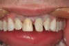
Frequently, retraction cord is used for Class III and Class V composite resin restorations as the nature of the decay associated with these restorations brings them into proximity of the gingival tissues.18 Often, during the placement of porcelain veneers and/or anterior all-ceramic crowns, the margins have been positioned slightly below the gingival margin. During cementation, gingival tissues must be retracted again to properly isolate this area to ensure that this critical part of the restoration is bonded correctly (Figure 2).
Conclusion
Proper soft-tissue management is the art and science of meticulously controlling the operative field in order to create the best possible final restoration. Dentistry has both the materials and techniques available to accomplish this. Dentists are urged to use all these tools to provide their patients with the best service possible.
Acknowledgement
The author would like to thank Dr. Robert A. Lowe for his assistance.
References
1. Christensen GJ. The state of fixed impressions: room for improvement. J Am Dent Assoc. 2005;136(3):343-346.
2. Christensen GJ. Laboratories want better impressions. J Am Dent Assoc. 2007:138(4):527-529.
3. Miller MB. Impression taking–is it a lost art. Gen Dent. 2007; 55(5):392-393.
4. Radz G. Impression materials today and in the near future. Dental Products Report. May 2009. Available at: http://www.dentalproductsreport.com/articles/show/dpr0509_cl_first-impressions. Accessed June 25, 2010.
5. Lee E. Impression-taking considerations for predictable indirect restorations. Pract Proced Aesthet Dent. 2003;15(6):454-457.
6. Vakay RT, Kois JC. Universal paradigms for predictable final impressions. Compend Contin Educ Dent. 2005;26(3):199-206.
7. Terry DA. The impression process: part III–technique. Pract Proced Aesthet Dent. 2007;19(1):27-29.
8. Lowe RA. Predictable fixed prosthodontics: technique is the key to success. Compend Contin Educ Dent. 2002;23(3 suppl 1):4-12.
9. McDonald TR. The ‘second-cord technique’ for taking great impressions. Inside Dentistry. 5(8):92-94
10. Cloyd S, Puri S. Using the double-cord packing technique of tissue retraction for making crown impressions. Dent Today. 1999;18:54-59.
11. Azzi R, Tsao T, Carranza FA Jr, et al. Comparative study of gingival retraction methods. J Prosthet Dent. 1983;50(4):561-565
12. Baharav H, Laufer BZ, Langer Y, et al. The effect of displacement time on gingival crevice width. Int J Prosthodont. 1997;10(3):248-253.
13. Woychesshin FF. An evaluation of the drugs used for gingival retraction. J Prosthet Dent. 1964;14:769-776.
14. Council on Dental Therapeutics of the American Dental Association. Hemostatics and astringents. In: Accepted Dental Therapeutics. 40th ed. Chicago, Ill: American Dental Association; 1984:334-41.
15. Bailey JH, Fischer DE. Procedural hemostatis and sulcular fluid control: fulcrum to modern dentistry. Ultradent Restorative Monograph. 1995:31-41.
16. Radz GM. Veneers and the 10 most common mistakes. Dental Economics. 2007:94-96,149.
17. Pallasch TJ. Vasoconstrictors and the heart. J Calif Dent Assoc. 1998;26(9):668-673
18. Lowe RA. Using Expasyl for other clinical uses. Inside Dentistry. 2010;6(5):108.