You must be signed in to read the rest of this article.
Registration on CDEWorld is free. Sign up today!
Forgot your password? Click Here!
LESSON
Dentistry, like many other professions, is consistently seeing revolutionary technological changes. Product improvements and the techniques that employ them are allowing dental professionals to provide more efficient dental care while satisfying the esthetic concerns of their patients. The esthetic indirect restoratives category of dental techniques has seen tremendous growth, especially in the bonding and cementation segments of the procedures.1 The standard selection of porcelain ceramic materials has been marred by reputed high clinical fracture rates and poor clinical longevity when used in conjunction with traditional acid-based cementation techniques.2 However, there is significant in vivo and in vitro evidence to support the notion that adhesive resin cements can improve clinical longevity and fortify indirect ceramic restorations.3 In fact, the adhesive nature of some restorations has been said to reinforce the remaining tooth structure by increasing fracture toughness and stiffness, and evidence suggests that adhesive resin cements may boost the clinical longevity of porcelain prosthetics by decreasing the probability of crack initiation from the internal aspect of the restorations.4
This new information, coupled with the esthetic trends in dentistry, has increased the study of various product lines involved with indirect restorations, particularly dental cements. Permanent resin cements are touted as the material of choice for metal-free restorations, with an emphasis on self-etching products, which are increasingly more popular because of their ability to offer a simplified protocol, good bond strength, and improved esthetics.5,6
RESIN CEMENTS
Adhesive resin cements are able to bond both to tooth structure and restorative material but they have some critical differences. Resin cements typically are used in conjunction with bonding agents to facilitate micromechanical attachment to both structures through bonding, thus allowing for increased retention.8 These products were developed to reduce postoperative sensitivity, microleakage, marginal staining, and caries; also, they are known to have a stronger ability than traditional cements to fill voids under restorations. 9,10 In today's dental practice, there are three main categories of resin cements:
- cements that use a total-etch adhesive system (separate etch, adhesion, and cement systems);
- cements that use an acidic self-etching adhesive system - not phosphoric acid - to demineralize the tooth (combined etch and bonding steps followed by application of the cement); and
- a select group of self-etch cements that reportedly have self-adhesive characteristics embedded in the cement, which negates the necessity for a separate adhesive application to the tooth.11
Dental cements have evolved to optimize bonds to dental substrates, simplify the application procedure, and reduce the vulnerability to the clinician's technique.12 The trend to eliminate steps from dental procedures has been questioned in the bonding agent arena and researchers have found it necessary to test bond strengths in the new category of self-etch cements. Recent research conducted at the University of Frankfurt in Germany in the Department of Prosthodontics demonstrated that simplifying cementation by eliminating the number of procedural steps does not negatively affect the bond to dentin.13 Manufacturers are aiming to simplify processes and eliminate steps while still maintaining the bond quality between substrates.
SELF-ETCHING CEMENTS
Self-etching cements contain etchant, primer, and bonding components in one delivery device. These cements appeal to modern clinicians as they allow the practitioners to fill the restoration with resin cement, seat the restoration, clean the excess, and cure; these cements also allow for a chemical cure if desired. The inherent features of self-etching cements are paramount to give clinicians who perform fewer nonmetal restorations the ability to fulfill their patients' esthetic demands easily and predictably. This is unlike traditional adhesive resin cements that require an etching gel for the enamel and dentin, primer and adhesive components in multiple bottles, or unidose carriers followed by an application of cement.14 A self-etching bonding system may be used in conjunction with the cement to provide added insurance against postoperative sensitivity and insufficient bond quality if clinicians would rather use a more traditional approach.
With simpler, shorter cementation techniques and computer-aided prosthetic design, indirect procedures have never been easier or quicker to accomplish. For certain dental cases without periodontal symptoms and with proper tissue management, prosthetic fabrication and insertion can be completed with little waiting time for the patient. This article will focus on using a more simplistic self-etch cementation protocol with a chairside in-office CAD/CAM restorative system to provide the ultimate in dental predictability and customization.
{C}CASE REPORT


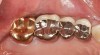
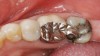
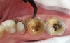
A middle-aged woman presented with the chief concern of seven existing full-coverage restorations that required replacement because of recurrent decay and compromised margins (Figure 2');" rel="imagepop" rem="#ip:figure1 and Figure 2">Figure 1 and Figure 2). She underwent an oral examination and medical history review, after which it was concluded that she had no medical or dental contraindications to treatment. The teeth to be replaced were the upper left bicuspids and first and second molars, and the lower second bicuspid and first and second molars. It was decided that these new restorations would be fabricated in-office with the author's chairside CAD/CAM restorative system (CEREC® 3D, Sirona Dental Systems Inc, Charlotte, NC).
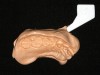
A preoperative impression was taken to assist in the fabrication of the provisionals. An alginate alternative was used because of its ease of use and dimensional stability over time as compared with traditional powder alginate (Figure 3).6 The teeth were covered with a talc-like powder (which functions as a contrast medium for taking the optical impressions) and the preoperative virtual impressions were taken. The existing restorations were removed and the presence of decay was verified with a caries detector and a spoon excavator.

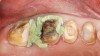
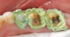
All decay was removed from the teeth and any necessary buildups were placed to eliminate undercuts or gain proper retention form in the resulting preparations. Before the optical scanning of the preparations for restoration design, the soft tissue was retracted in the subgingival areas with a putty-type retraction system (Figure 5');" rel="imagepop" rem="#ip:figure4 and Figure 5">Figure 4 and Figure 5). A putty retraction system was specifically chosen over traditional retraction cord or other hemostatic agent because of these two methods' potential for tissue (epithelial attachment) damage and recession.15-17
After 2 minutes, the putty retraction material was thoroughly rinsed away with air/water spray and dried. The preparations were powdered and several optical images of the preparations were taken. Subsequently, the teeth were temporized with a bis-Acryl material, and cemented with a resin-based temporary cement.

Restoration Fabrication
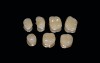
The in-office CAD/CAM restorative system was chosen because of its high success rate18 and for its esthetic properties, inherent strength, and fracture resistance of the porcelain blocks used after oven glazing.19 After the optical images of the preparations were obtained, the restorations were fabricated using the CEREC 3D Correlation mode. The Correlation mode takes the preoperative condition, reproduces it over the preparations, and allows it to be further modified by the clinician to obtain the desired occlusal and esthetic result. After the restorations were milled, they were stained and glazed in a porcelain oven to obtain the desired surface gloss (Figure 6). Preparing the internal aspect of the restorations involved micro-abrading with a micro-ether containing 50- m m aluminum oxide powder to prevent damage to the margins, and rinsing with water and drying for approximately 5 seconds. A 9.5 % hydrofluoric acid gel was applied for 1 minute, rinsed with water for approximately 20 seconds, and dried thoroughly. Before the advent of resin adhesive products, cements typically were formed by an acid/base reaction in which the acidic liquid and basic powder were combined; the powders commonly were either zinc oxide or aluminosilicate glasses while the liquids were phosphoric acid, polyacrylic acid, or eugenol.7
The choice of luting cement is often dictated by its chemical and mechanical properties and the clinical situation at hand. Cement performance preferences typically include: low viscosity and film thickness, long working time with rapid set at oral temperatures, low solubility, high compressive and tensile strengths, adhesion to tooth structure and restorative materials, anticariogenic properties, and biocompatibility.


Insertion Appointment

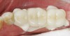
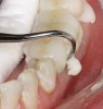
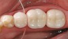
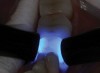
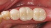
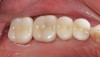
Tissue management was then obtained by the application of the same putty-type retraction system (Figure 8). After 2 minutes, the putty retraction material was thoroughly rinsed away with air/water spray and dried. The teeth were then cleaned again with 2% chlorhexidine, rinsed, and dried without reaching desiccation.20 The restorations were filled with a dual-cure resin cement system, Maxcem™ (Kerr Corporation, Orange, CA), seated, and remained undisturbed for 90 seconds before light-curing (Figure 9).6 This waiting period was mandatory for the self-adhesive chemistry in the cement to be fully effected. The cement reached a gel state and was easily removed with a handscaler (Figure 10). To ensure no movement of the restorations while cleaning cement interproximally, the restorations were tacked down mid-cervically on the buccal with a 4-mm turbo tip in a LED curing light. Interproximal cleaning was accomplished with waxed floss (Figure 11) and final curing was performed on both the buccal and lingual aspects with two LED lights simultaneously for 20 seconds (Figure 12), followed by occlusal curing for 20 seconds. Marginal finishing was performed with an 8-fluted carbide bur and composite points and cups. Interproximal finishing involved using medium- and fine-composite finishing strips. Occlusion was checked and slight adjustments were performed before the final polishing of these areas with porcelain polishing points. The final results are shown in Figure 13 and Figure 14.


CONCLUSION
Because of the high esthetic demands of patients, dentists are performing a larger number of tooth-colored procedures, especially with regard to bonded porcelain restorations. Therefore, it behooves clinicians to seek education about the latest improvements to ensure that patients receive the most advanced form of treatment. In cases using newer self-etching, dual-curing resin cements the dentist has the ability to place a highly esthetic porcelain (or resin) restoration with very predictable results. The results are predictable not only in longevity but also with regard to lack of sensitivity and other technique related issues. There are two theories to explain the decrease in postoperative sensitivity after the use of a self-etching cementation system. The first is based on the idea that etching and the priming are done consecutively, theoretically rendering it impossible to etch deeper than the primer can penetrate.6 The second theory postulates that because the collagen is supported during the whole process, sensitivity is eliminated or kept to a minimum.21 When enamel is still present or unprepared, most manufacturers recommend etching the enamel with phosphoric acid for 15 seconds before applying the self-etching cement.22
Recent advances in the self-etching arena have enabled the clinician to place an all-ceramic restoration in a very similar fashion to a conventional cemented porcelain-fused-to-metal restoration. This is because of the simplistic nature of the dual-curing resin cement system. With this combination, inlays, onlays, and full-coverage all-ceramic restorations can be placed with confidence and meet the high esthetic demands of today's dental patient.
References
1. Kugel G, Ferrari M. The science of bonding: from first to sixth generation. J Am Dent Assoc. 2000;131(Suppl):20S-25S.
2. Malament KA, Socransky SS. Survival of Dicor glass-ceramic dental restorations over 14 years: Part I. Survival of Dicor complete coverage restorations and effect of internal surface acid etching, tooth position, gender, and age. J Prosthet Dent. 1999;81(1):23-32.
3. Pagniano RP, Seghi RR, Rosenstiel SF, et al. The effect of a layer of resin luting agent on the biaxial flexure strength of two all-ceramic systems. J Prosthet Dent. 2005;93(5):459-466.
4. Caron GA, Murchison DF, Cohen RB, Broome JC. Resistance to fracture of teeth with various preparations for amalgam. J Dent. 1996;24(6):407-410.
5. Pegoraro TA, da Silva NR, Carvalho RM. Cements for use in esthetic dentistry. Dent Clin North Am. 2007;51(2):453-471.
6. Miller MB. Dental adhesives. In: Miller MB, ed. REALITY: The Information Source for Esthetic Dentistry. Vol. 20. Houston, TX: REALITY Publishing Co; 2006.
7. Anusavice KJ. Phillips' Science of Dental Materials. W.B. Saunders Co: Philadelphia, Pa; 1996:555-581.
8. El-Mowafy O. The use of resin cements in restorative dentistry to overcome retention problems. J Can Dent Assoc. 2001;67(2):97-102.
9. Burke FJ, Watts DC. Fracture resistance of teeth restored with dentin-bonded crowns. Quintessence Int. 1994;25(5):335-340.
10. Dietschi D, Maeder M, Meyer JM, Holz J. In vitro resistance to fracture of porcelain inlays bonded to tooth. Quintessence Int. 1990;21(10):823-831.
11. Burgess JO, Latta MA, White RC. Dual Cure Resin-based Cements: Expert Panel Discussion. ADA Professional Product Review. [serial online] 2006;1(2):1-12.
12. Ferrari M, Tay FR. Technique sensitivity in bonding to vital, acid-etched dentin. Oper Dent. 2003;28:3-8.
13. Piwowarczyk A, Bender R, Ottl P, Lauer HC. Long-term bond between dual-polymerizing cementing agents and human hard dental tissue. Dent Mater. 2007;23(2):211-217.
14. Freedman G. Fifth-generation bonding systems. Predictable posterior composite restorations. Dent Today. 1996;15(11):68-75.
15. Azzi R, Tsao TF, Carranza FA Jr, Kenney EB. Comparative study of gingival retraction methods. J Prosthet Dent. 1983;50(4):561-565.
16. Donovan TE, Gendara BK, Nemetz H. Review and survey of medicaments used with gingival retraction cords. J Prosthet Dent. 1985;53(4):525-531.
17. Shaw DH, Krejci RF, Kalkward KL, Wentz FM. Gingival response to retraction by ferric sulfate (Astringedent). Oper Dent. 1983;8(4):142-147.
18. Mormann W. The evolution of the CEREC system. Paper presented at: The CEREC 20th Anniversary Experience; October 13, 2006; Las Vegas, NV.
19. Chen HY, Hickel R, Setcos JC, Kunzelmann KH. Effects of surface finish and fatigue testing on the fracture strength of CAD/CAM and pressed ceramic crowns. J Prosthet Dent. 1999;82(4):468-475.
20. Gwinnett AJ. Effect of cavity disinfection on bond strength to dentin. J Esthet Dent. 1992;4(Suppl):11-13.
21. Perry RD. Clinical evaluation of total-etch and self-etch bonding systems for preventing sensitivity in Class 1 and Class 2 restorations. Compend Contin Educ Dent. 2007:28(1):12-14.
22. Lopes GC, Thys DG, Klaus P, et al. Enamel acid etching: A review. Compend Contin Educ Dent. 2007;28(1):18-25.