You must be signed in to read the rest of this article.
Registration on CDEWorld is free. Sign up today!
Forgot your password? Click Here!
It has long been shown in the scientific dental literature that the appearance of a person's face and teeth has a profound impact on how they are perceived and judged by others.1-5 Teeth and smiles that are considered esthetically pleasing are associated with kindness, popularity, intelligence, and high social status.6 In addition, the degree of satisfaction one has with their own smile is directly correlated with their self-perception.7 In a clinical study, tooth malpositioning and discolorations as well as extensive gingival exposure during a smile (ie, "gummy smile") were the most displeasing factors when patients judged their own smiles.8 Moreover, an unattractive smile was correlated with the personality characteristics of neuroticism and poor self-esteem, impacting overall well-being and health.
However, the ethical bases for extensive esthetic or elective cosmetic dental treatment typically have been discussed controversially, as inadequate, unnecessary, unsuccessful, and overly invasive, and excessive treatment can have severe detrimental consequences on the attractiveness and well-being of a patient.9A complicating factor is that every face and smile is different, and esthetic needs and the awareness of harmony and beauty vary among people.10Furthermore, there may be vast differences in the perception of esthetics among patients and dentists, and even among dental professionals themselves.1,11 Therefore, understanding and realizing a patient's esthetic vision and individual needs are arguably the greatest challenges in esthetic dentistry.
Digital Smile Design
Digital technologies have become invaluable tools used to bridge these gaps and design individual smiles that are both based on each patient's specific needs and independent of the clinician's or technician's skills and understanding of esthetic parameters.
Intra- and extraoral optical scanners and, when needed, cone-beam computed tomography (CBCT) facilitate detailed 3-dimensional (3D) evaluation of oral structures and tissues. Specific software tools enable digital planning and visualization of anticipated esthetic outcomes. They suggest the type and sequence of necessary periodontal, restorative, orthodontic, surgical, and multi-disciplinary treatment and even fabrication of specific guides for precise surgical and restorative interventions according to the plan.12 In fact, cloud-based tools and chat rooms have become essential for interdisciplinary consultations and treatment planning, allowing for asynchronous remote communication between the involved clinicians and dental technicians.
Digital smile analysis and planning software tools were introduced in the early 2000s, and the first fully facially guided digital smile design protocol, based on a series of facial, extraoral, and intraoral photographs, was developed by Coachman and coworkers in 2008.13 Digital scan files of natural teeth, tooth morphologies, and smiles from a natural algorithm library facilitate and simplify the "digital wax-up" to create customized and natural esthetics. Integrating individual facial features ("facial flow") into the smile design is key for esthetic success, and 3D face scans merged with intra- oral scans, model scans, and CBCTs have become critical components of a truly digital workflow.1 Current smile design software also incorporates digital articulators and jaw tracking devices to include functional parameters in the digital planning process. Machine learning and artificial intelligence tools are increasingly being applied to automate and simplify esthetic evaluation and digital smile design steps.
Despite all of these advances and possibilities with virtual try-in software, evaluation of the planned design in the patient's mouth through mock-ups or provisional restorations before finalization of the restorations is still an absolute necessity. The verified digital design can easily be transferred into any definitive restoration and material.
This article describes and illustrates interdisciplinary esthetic restorative treatment with current digital tools.
Clinical Case
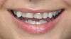
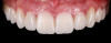
A 24-year-old female patient presented with a chief complaint related to unsatisfactory smile esthetics (Figure 1). She was aware of several malpositioned teeth and decided to first consult with an orthodontist, who had suggested interdisciplinary treatment. After clinical examination, the patient was informed about her condition and the need for interdisciplinary care to achieve optimal esthetic and functional outcomes. The interdisciplinary care would include caries treatment, tooth extractions, orthodontics, and prosthetic treatment. Medical examination did not reveal any conditions that would preclude or limit dental treatment.
Facial analysis revealed balanced proportions with a minor deviation of the tip of the nose to the right but without clinical significance. The profile did not show any noticeable aberrations. Lips were in balanced size and equilibrium, while a minor asymmetry was noticed on the upper lip, with the right side having less volume.
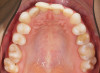
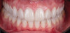
Intraoral evaluation showed compromised oral health with generalized gingivitis and plaque accumulation (Figure 2). Many teeth had already been restored, and several restorations were failing. The maxillary dental midline coincided with the facial midline with a minor inclination. The lower midline deviated approximately 3 mm to the left side in reference to the upper midline. There was moderate crowding in the maxilla, mainly on the right side, and severe crowding in the mandible. Canines on the right were in Angle class III malocclusion, while molars and canines on the left were in mild class II malocclusion. Radiographic evaluation revealed crowding, missing teeth, and failing restorative as well as endodontic treatment (Figure 3).
Treatment Plan
The following treatment goals were set and discussed with the patient: re-establish oral health, improve smile esthetics and masticatory function, eliminate tooth crowding in both arches, establish Angle class I relationship, match the facial and dental midlines, improve the level of gingival margins, open space to increase the mesiodistal size of the maxillary right lateral incisor, extract the first molars in the mandible and move the second and third molars anteriorly, and, finally, extract the maxillary left third molar.
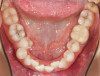
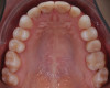
The interdisciplinary treatment plan to establish good oral health, esthetics, and function included prophylaxis and oral hygiene instructions as first steps. Respective teeth were extracted and orthodontic treatment was carried out. Figure 4 and Figure 5 depict the intraoral situation before orthodontic treatment with fixed appliances and ceramic brackets; Figure 6 through Figure 8 show the situation after orthodontic treatment, which lasted 18 months.
Restorative Treatment
Visual control during restorative treatment is vital for success. Independent of technologies or materials applied, the use of high magnification, ideally achieved with a surgical microscope, is necessary during all clinical steps to achieve maximum precision. As was used in this case, the microscope facilitates thorough inspection of preparation margins, cracks, fit of restorations, and many other details that cannot be sufficiently seen with the naked eye.
The restorative treatment steps in this case were as follows: (1) photo/video documentation, CT scan, intraoral teeth re-evaluation; (2) 3D restoration design in the laboratory and fabrication of a printed model for mock-up and preparation control; (3) mock-up test drive and photo/video documentation; (4) tooth preparations for ceramic restorations; (5) intraoral scanning and fabrication of provisional restorations; (6) final design of veneers and crowns in the laboratory; (7) CAD/CAM milling of ceramic restorations; (8) 3D ceramic staining; (9) isolation and bonding of restorations in the oral cavity; (10) crown lengthening of the four maxillary incisors; and (11) final documentation and recall.
Data Collection
Design of a customized facially driven smile requires collection of a substantial amount of data. The use of a designated clinic area for photography and video documentation is helpful. CBCT and other digital scans facilitate the creation of a "digital patient copy" for further 3D design. An important part of data collection comes from intraoral scans, which are used for documentation purposes but also to establish libraries of natural tooth shapes and smiles for future smile designs.
Based on the patient's facial features and esthetic expectations (natural shapes with dominant central incisors), tooth shapes were selected for the digital wax-up and setup (Figure 9). A model was 3D-printed from the digital smile design to fabricate an intraoral direct mock-up with a silicone index and bis-acryl material. This mock-up allows the clinician and the patient to evaluate and verify the smile design (Figure 10). It also serves as an excellent tool to motivate the patient and instill confidence in both the patient and dentist in the planned treatment. Documentation with photographs and videos is essential.
Minimally Invasive Tooth Preparation
The tooth preparation procedure should create sufficient space for the ceramic material while preserving as much tooth structure as possible. Transferring the final design prototype to the patient's mouth with another mock-up (Figure 11) before starting the preparation facilitates a minimally invasive procedure. Preparing into the mock-up with the respective depth cutting burs limits tooth reduction to the areas where necessary (Figure 12). In the authors' experience, use of the surgical microscope supports better ergonomics during preparation and maximum visual control, with magnification up to 23x.
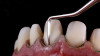
After making "depth cuts" through the mock-up, a gingivectomy was performed on the four maxillary incisors. Fine diamonds, gentle pressure, and copious water irrigation limited trauma to vital teeth during preparation. Smooth preparation surfaces and margins enabled optimal fit of the restorations, which is directly correlated with superior clinical success.14 Similarly, the widespread notion that enamel surfaces prepared with coarse diamonds may improve adhesion has long been defied; smooth enamel preparation surfaces, before phosphoric acid-etching, provide significantly better bond strength.15 Enamel chisels were used to finalize micro-marginal zones, making them sharp and visible for the CAD designer and for optimized marginal fit (Figure 13). Proper soft-tissue retraction is needed for an ideal digital impression with an intraoral scanner to create a high-definition and precision digital model. Provisional restorations were inserted after the preparation.
Laboratory CAD/CAM
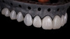
For the definitive restorations design, the CAD designer used the same library of teeth selected for the mock-up, ensuring precise realization of the smile design that the patient had verified and approved (Figure 14). With the software's ability to focus closely on the marginal areas for high precision of fit, microscopic and digital dentistry tools complement each other extremely well. Current milling machines can mill 12 monolithic ceramic units within a few hours with excellent precision. In this case, multi-layer leucite-reinforced feldspathic ceramic blocks were used. Adhering to the patient's individual preferences, the milled restorations were characterized through staining and glazing (Figure 15).
Final Insertion
Rubber dam is necessary for adequate moisture control and to ensure optimal isolation for successful resin bonding procedures. Precision of fit of each restoration was verified (Figure 16), and the restorations were bonded in place using common resin bonding protocols for silica-based ceramics: use of a composite resin luting agent after hydrofluoric acid-etching and silanization of the ceramic and a total-etch bonding protocol on the enamel. In the authors' experience, all of these procedures are best performed under the microscope.
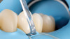
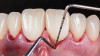
After insertion, a soft-tissue flap was partially elevated on the four maxillary incisors, and a crown lengthening procedure was done with ultrasonic diamond instruments to create the proper biologic width and symmetric gingival contours (Figure 17). This sequence gives the tissue the ability to adapt to the newly created emergence profile of the new restorations. It also eliminates the typical healing phase of 6 months or more between crown lengthening and final preparation as suggested in traditional treatment protocols. All of the above restorative treatment steps were done within 3 days.
Figure 18 and Figure 19 depict the outcome 1 year after treatment. A nightguard and professional hygiene every 3 months were recommended.
Conclusion
Current digital tools allow for predictable functional and natural esthetic restorative outcomes based on the patient's individual needs and preferences. When such tools are combined with enhanced visualization through high magnification, optimal precision, which is a primary predictor for long-term success, can be achieved on a consistent basis. Digital technologies not only facilitate treatment planning, smile design, and restoration fabrication but provide an interactive platform for asynchronous collaboration among clinicians and dental technicians for best-possible interdisciplinary patient care.
About the Authors
Nazariy Mykhaylyuk, DDS
Founder of M.Vision clinics, lab, and study center; Private Practice, Prosthodontist, Kiev, Ukraine
Bogdan Mykhaylyuk, CDT
Dental Technician and Owner of M.Vision lab, Ivano-Frankivsk, Ukraine
Nuno Sousa Dias, DDS
Private Practice, Orthodontist, Porto, Portugal
Markus B. Blatz, DMD, PhD
Professor of Restorative Dentistry, Chair, Department of Preventive and Restorative Sciences, Assistant Dean for Digital Innovation and Professional Development, University of Pennsylvania School of Dental Medicine, Philadelphia, Pennsylvania
Queries to the author regarding this course may be submitted toauthorqueries@aegiscomm.com.
References
1. Blatz MB, Chiche G, Bahat O, et al. Evolution of aesthetic dentistry. JDent Res. 2019;98(12):1294-1304.
2. Root WR. Face value. Am J Orthod. 1949;35(9): 697-703.
3. Rhodes G. The evolutionary psychology of facial beauty. Annu Rev Psychol. 2006;57:199-226.
4. Anderson JN. The value of teeth. Br Dent J.1965; 119:98-103.
5. Newton JT, Prabhu N, Robinson PG. The impact of dental appearance on the appraisal of personal characteristics. Int J Prosthodont. 2003;16(4):429-434.
6. Shaw WC, Rees G, Dawe M, Charles CR. The influence of dentofacial appearance on the social attractiveness of young adults. Am J Orthod. 1985;87(1):21-26.
7. Davis LG, Ashworth PD, Spriggs LS. Psychological effects of aesthetic dental treatment. J Dent. 1998;26(7):547-554.
8. Van der Geld P, Oosterveld P, Van Heck G, Kuijpers-Jagtman AM. Smile attractiveness: self-perception and influence on personality. Angle Orthod. 2007;77(5):759-765.
9. Liebler M, Devigus A, Randall RC, et al. Ethics of esthetic dentistry. Quintessence Int. 2004;35(6):456-465.
10. Arndt EM, Travis F, Lefebvre A, et al. Beauty and the eye of the beholder: social consequences and personal adjustments for facial patients. Br J Plast Surg. 1986;39(1):81-84.
11. Brisman AS. Esthetics: a comparison of dentists' and patients' concepts. J Am Dent Assoc. 1980;100(3):345-352.
12. Zimmermann M, Mehl A. Virtual smile design systems: a current review. Int J Comput Dent.2015; 18(4):303-317.
13. Coachman C, Calamita MA, Sesma N. Dynamic documentation of the smile and the 2D/3D digital smile design process. Int J Periodontics Restorative Dent. 2017;37(2):183-193.
14. Li YQ, Wang H, Wang YJ, Chen JH. Effect of different grit sizes of diamond rotary instruments for tooth preparation on the retention and adaptation of complete coverage restorations. J Prosthet Dent. 2012;107(2):86-93.
15. Jung M, Wehlen LO, Klimek J. Surface roughness and bond strength of enamel to composite. Dent Mater. 1999;15(4):250-256.