You must be signed in to read the rest of this article.
Registration on CDEWorld is free. Sign up today!
Forgot your password? Click Here!
Much has been written about the advancement of computer aided design/computer aided manufacturing (CAD/CAM) technology over the past 30 years. Beginning as a niche experiment in single-unit restoration workflows, CAD/CAM has evolved to become a major part of the current dental landscape. Dr François Duret, considered by many to be the father of digital dentistry,1was indeed correct when he wrote in 1991, "CAD/CAM … will be a part of routine dental practice [in] the 21st century."2 As this technology has become more broadly accepted, its applications have been increasingly fine-tuned to the needs of dental practitioners and patients alike. This article will review the development of CAD/CAM applications in dentistry and explore how recent advances are helping foster greater access to dental care for vulnerable populations in the United States.
HISTORY OF CAD/CAM IN DENTISTRY
Dental applications in CAD/CAM technology began in the early 1970s, just as the computer revolution was gaining momentum.3 Pioneers in the development of dental CAD/CAM systems took what initially seemed to be a strictly theoretical approach to the fabrication of ceramic restorations, and within one decade brought it into commercial production.4 These efforts were aimed at providing patients with restorations of the highest quality that could be produced in a single visit, within less than 1 hour of fabrication time, if possible.5
Early developers of this technology pursued a number of different approaches to create a viable workflow, which ultimately resulted in a system consisting of the following steps: 1) data acquisition, 2) design of the dental restoration (CAD), and 3) fabrication of the restoration (CAM).5 This basic approach to digital dentistry has served as the foundation on which the technology continues to grow today.
Data Acquisition
Various methods have been used for image capture of the target tooth, including image triangulation, stereophotogrammetry, confocal imaging, and active wavefront sampling. Using active or passive light capture, these methods have been employed for the recording of the data necessary for model and die reconstruction.6 Examples of early acquisition units include specially modified cameras with dual lenses to capture the needed data, and laser probes that had been repurposed from other industries.5
Design of the Dental Restoration (CAD)
Several CAD programs were initially developed for the design of indirect dental restorations, including the Sopha Bioconcept System (Sopha Bioconcept, Inc) and Chairside Economical Restoration of Esthetic Ceramic (CEREC®, Sirona Dental Systems GmbH).7 Integrating the information from the data acquisition unit, early CAD systems worked much the same way they do today. The margin would be marked on the restoration and an ideal crown proposal made based on the location of the tooth in the mouth. Automatic and custom adjustments could then be made to the crown before fabrication. However, early versions of the CEREC software allowed only for design of inlays and onlays, owing to the restrictions in milling that existed during that period.4
Fabrication of the Restoration (CAM)
Early fabrication of CAD/CAM restorations used subtractive manufacturing techniques similar to those used in many milling machines today. Preformed blocks were inserted into the milling machine, and the CAM processes were run to create the final restoration. This portion of the software would calculate the toolpaths necessary to remove the excess material in the incremental reduction of material.8
COMMERCIAL VIABILITY OF CAD/CAM SYSTEMS
Today there are more than a dozen CAD/CAM systems available for in-office use.9 Initially, the CEREC 1 system (Sirona Dental Systems GmbH) was the first commercially viable product on the market.4,7,8 While the subsequent iterations of CEREC have produced a more refined system, much of the currently available CAD/CAM systems is still the same as it was with the first CAD/CAM units that were created more than 30 years ago. As materials have continued to evolve over the past four decades, the options for restoration creation have expanded from simple indirect fixed restorations to include a much wider selection of restorations.7
Currently, additive manufacturing (eg, 3D printing) has become a growing feature of CAD/CAM dentistry, with current resin profiles being planned for long-term restorations in the mouth.10 Advances in chip manufacturing and materials development are allowing laboratory technicians and clinicians to provide patients with restorations that were previously available only through an analog workflow. Although these analog approaches have been tested and have proved to be reliable, they are often labor intensive and time consuming. With the recent improvements in CAD/CAM workflows, viable restorations can be made not only much more quickly than with analog counterparts, but at a fraction of the cost.
While there are many ways this new technology can be leveraged, perhaps the most impactful potential outcome will be providing greater access to dental care for all patients.
ACCESS TO CARE
Access to dental care has been a growing concern in the United States for the past 20 years.11 The term "access to care," popularly used in public health policy, may be used to refer to the availability of a sufficient supply of healthcare services and, more particularly, to an individual's ability to utilize those services, especially those needed for general well-being.11 Access to care has been described as comprising four interrelated aspects: affordability of services, physical accessibility of services, acceptability of services, and adequacy of supply.11 Regarding dental healthcare in the United States, all of these factors play a role in the composite well-being of the communities in which dental professionals serve.
Affordability
Among the four barriers to access to care, the inability of some patients to afford healthcare looms especially large.12 Surveys have found that up to two out of five people report that they limit or delay dental treatment because of financial concerns.13The Centers for Disease Control and Prevention (CDC) reports that only half of Americans have dental insurance, and those without dental insurance coverage are much less likely to have had a dental visit than those with coverage.14
Physical Accessibility
Based on figures from the US Health and Human services, only about 30% of individuals located in Health Professional Shortage Areas (HPSAs)-ie, geographic areas that have insufficient primary care, dental, and mental healthcare providers-have adequate access to care.15 This means that of the 60 million Americans who live in these designated areas, 42 million Americans do not have physical access to dental care.
While the number of general practitioners in HPSAs throughout the United States is limited, access to specialists in these regions is even more restricted. To see the same number of patients as general practitioners, specialists would require a referral area that is much broader, owing to the specialized nature of their care services; as a result, the services of specialists tend to be concentrated in more densely populated areas. Vulnerable populations outside of these regions are thus forced to travel long distances for healthcare services or are left without adequate care.
Adequacy of Supply
According to the HPSA report of 2020, more than 10,000 dental providers are needed to meet the demand of care.15 Based on these data, even if providers are physically located in the region where need is greatest, there may still not be enough clinicians to treat all patients who need care. The report calculates that a ratio of one practitioner per 4,000 patients can be considered as meeting the need of an area. Although populations within HPSAs are most likely not receiving two dental hygiene visits per year, theo-
retically 8,000 appointments per year would be required to satisfy the hygiene needs of a patient population. For a given clinic, it would be necessary for four chairs to run eight 1-hour appointment 5 days per week for 50 weeks out of the year to satisfy this need. In many states, this would not even be legal, given the current laws established by state dental boards.16,17
A Technological Solution to Access to Care Issues
Over the past decade, health policy reforms have been aimed at improving access to care for the greatest number of Americans possible. While access to dental care has likely improved as a result of these reforms, many believe that the nation's dental care system needs to undergo significant changes and improvements in order to achieve the desired outcomes relating to care access.18 Well-intentioned policy will probably take years to implement, and even then it may still not be able to solve access to care issues reliably across the board. Individual practitioners will likely need to make changes to their practices to help achieve any type of sustainable solution.
Flexible solutions are needed to combat the problems posed by the barriers to care.19 The author suggests that one potential solution is the adoption and adaptation of CAD/CAM technology as a means of providing the highest quality of dental care more quickly and efficiently, and at a lower cost to the patient. Although the economics of investing in in-office CAD/CAM workflows may seem daunting at first, a closer examination of the details shows that an investment in digital dental technology can provide not only financial benefits to the practice and the patient, but improve clinical outcomes as well.20,21
Case Study
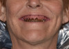
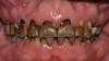
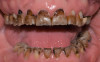
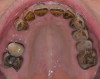
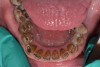
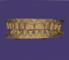
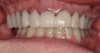
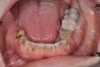
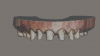
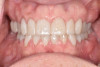
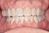
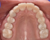
At a clinic treating patients in an underserved population, a 54-year-old female patient presented requesting extraction of all of her teeth and fabrication of dentures for her long-term restorations. The patient had a history of breast cancer, which had been treated with chemotherapy. She believed that full dentures would be an appropriate treatment option for her, owing to the poor state of her oral health and dentition and because she had limited financial resources and did not think that she could afford restoration of her teeth. The patient reported that she had experienced depression and alcohol abuse because of her cancer diagnosis, and that she had neglected her dental care during the course of her cancer treatment, resulting in generalized moderate-to-severe caries with moderate wear due to bruxism (Figure 1 through Figure 5). She also presented with significant staining of her remaining dentition due to the alcohol abuse.
After clinical evaluation and treatment planning, it was determined that the state of the patient's dentition was not hopeless and that her teeth could be restored. The patient decided that she would like to try to preserve her natural teeth with a full-mouth crown and bridge rehabilitation, and to have any salvageable teeth treated with root canal therapy instead of extraction, if possible. After a thorough examination, it was determined that one tooth would need to be extracted (tooth
No. 19) and one would need root canal therapy (tooth No. 6). Twenty-four units of crown and bridge were needed. In order to save the patient the cost of sending her crowns out to a laboratory for fabrication, it was agreed that the restorations would be made in-house. A fully digital workflow was implemented for all planning and restorative work, where possible.
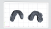
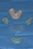
After a dental prophylaxis cleaning, the patient's mouth was scanned with an intraoral scanner for preoperative models (Figure 6). An upper and lower dentate scan was obtained with corresponding bite records. The bite was recorded at maximum intercuspation. Tooth length measurements were made, and it was found that the maxillary central incisors were only 8 mm in length, owing to prolonged bruxism and incisal wear.
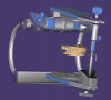
The digital articulator function was used to open the patient approximately to 1.5 mm (Figure 7), and digital wax-up was then created for ideal esthetic and functional restorations using a CAD software (Figure 8). This ideal wax-up was then printed using a 3D printer, and a putty matrix was created over the model for mock-up in the patient's mouth (Figure 9). All existing decay was then treated, and build-ups were performed on any needed teeth.
Once the existing decay was treated, the ideal wax-up was transferred to the patient's mouth via a putty matrix and tested for occlusal harmony and patient comfort using a bis-acryl temporary crown material (Figure 10). The patient wore the temporary ideal wax-up for 2 weeks, and occlusal adjustments were made where necessary to calibrate the occlusion. The patient reported that she was satisfied with the function and esthetics of the ideal wax-up, and the teeth were then prepared, starting with the lower arch.
Tooth preparation was done in segments, with the transferred wax-up still remaining on the teeth to preserve the opened vertical dimension of occlusion (VDO) (Figure 11). Starting with the lower anterior sextant, the teeth were prepared for full coverage restorations, and a progressive bite registration was used to maintain the occlusal record (Figure 12). The remaining sextants were prepared and bite records taken. A double-cord retraction technique was used, and the prepared teeth were scanned with an intraoral scanner (Figure 13). The bite record was placed on the right side of the mouth while the bite scan was completed, and the same steps were then performed on the other side of the mouth. In this way, the entire arch was able to be scanned while maintaining the VDO. The upper jaw was then prepared in a similar manner, and digital data were imported into a CAD software.
In the CAD software, restorations were designed based on the ideal wax-up, and adjustments were made based on the actual fit in the patient's mouth. Because of her history of bruxism and severe staining, a 3Y zirconia material was chosen for the final restorations. After the designs were completed, a nesting software was used to generate the toolpaths for the five-
axis Versamill 5x200 (Axsys Dental Solutions). This type of mill boasts the advantages of being able to mill many different materials, including lithium disilicate, zirconia, polymethyl methacrylate, and even titanium. In contrast to most in-office milling systems that mill from single-unit blocks of material, five-axis mills often are able to mill out of a single puck, which allows faster milling times and a reduction in the cost of materials. Other examples of five-axis milling machines include DWX-52D (Roland), Ceramill® Motion 2 (Amann Girrbach), and inLab MC X5 (Dentsply Sirona).
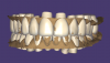
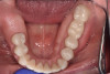
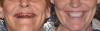
After the restorations were milled, they were sintered and polished for final effect. It was decided that these restorations should not be glazed to avoid the added abrasivity that accompanies this finish to the restoration.22 The patient was then scheduled for temporary crown removal. The final crowns were fitted on the prepared teeth, and the contacts adjusted. The patient reported that she was pleased with the feel and esthetics of the restorations, and the restorations were cemented (Figure 14 through Figure 19).
Discussion
This case study illustrates how CAD/CAM technology can be used to achieve life-changing results for individuals who would otherwise have limited or no access to appropriate dental care. This patient, a woman living in an underserved community, had believed that full-mouth dental extractions would be necessary to achieve functional and esthetic rehabilitation of her dentition.
Due to the comprehensive needs in this particular case, treatment was broken up into four different appointments. While this schedule suited the patient because she had the necessary transportation, chairside same-day restorations could also be completed depending on patient needs. Compared with traditional workflows that would require cases to be sent out to external laboratories, in-office CAD/CAM workflows can allow for greater flexibility in scheduling of patients, which can provide a distinct advantage when working with vulnerable patient populations.
Financial Considerations with CAD/CAM Systems
Comparison of Open Systems and Closed Systems
In digital dentistry, there are two broad categories of CAD/CAM systems: open systems and closed systems.23 Systems that are considered "open" are defined as such because the different components of the workflow can be paired with products from different manufacturers.24Open systems have the advantage of being flexible and can be used for multiple different applications. Numerous workflows are available with the open system architecture, as dentures, custom implant abutments, night guards, and other dental indications are easily fabricated with training.25Open systems also tend to be less expensive than closed systems.
Closed systems, on the other hand, are tailored to guide the user from start to finish, and generally tend to be slightly less complex. Usually, one company will manufacture all of the equipment needed for the fabrication process and bundle it together.24 The disadvantage of closed systems is that they tend to be more rigid, being capable of use for only a fraction of the applications used with their open-system counterparts. Closed systems also tend to be more expensive. The individual clinician must ultimately decide which system will best suit his or her needs.
Costs and Return on Investment
Acquiring and learning to use the different components that are necessary to complete a CAD/CAM system for in-office use by the general practitioner may seem daunting at first. However, while it does take time to gain the skills and knowledge necessary to utilize these new workflows, it can be well worth the investment. It is also worthwhile to consider training an interested dental assistant in these workflows, to help offload any additional work that the dentist may need to take on to fully implement these systems. A hybrid dental assistant/lab technician is a role that is increasingly being incorporated into dental practices, thus helping to increase the value of the practice's investment in CAD/CAM systems.
A breakdown of the costs of the equipment used in the case study described earlier is shown in Table 1. As shown in this breakdown, the total cost of a CAD/CAM system can be approximately $80,000. While such a capital outlay may seem a heavy cost at first glance, it is important to note that not all system components need to be acquired at once, and that even before all the components are in place, laboratory costs may be reduced for the practice. Design services from digital laboratory technicians exist through which a crown can be designed within a matter of minutes for as little as $5.00. If the clinician does not feel confident in taking on this portion of the digital workflow, such design services can serve as a great way to begin implementing CAD/CAM technology in-office in a gradual manner. Additionally, dental laboratories or dental 3D printing centers can print models for minimal costs if the clinician does not want to take on that aspect of the workflow as well.
Although much has been written elsewhere on the topic,26, 27 a full analysis on the return on investment (ROI) seen with implementation of CAD/CAM systems in dentistry is beyond the scope of this article. However, the following is a simple example of a potential ROI that can occur with CAD/CAM for in-office use:
A puck of zirconia costs $125, and therefore 20 restorations can be milled from a single puck at a cost of $6.25 per unit. After calculating the costs of the equipment and the labor involved in the creation of the restorations, the cost of processing a single-unit zirconia crown would be approximately $25. If it is assumed that the dental office is scheduling 30 crowns per month, with a laboratory cost of $100 per unit, this equates to a savings of more than $27,000 per year. In this scenario, which does not include consideration of the additional restorations that can be fabricated with these systems, it would take less than 3 years to break even on the investment.
For the case study discussed earlier, in which the patient required 24 units of crown and bridge, if each unit cost $25, the total cost would be $600-or, a savings of $1,800.
Conclusion
The introduction of CAD/CAM systems to dentistry has been one of the major developments in the profession of the past 30 years. Every year, improvements in these systems have created more flexible and predictable treatment options. Additionally, the costs associated with the implementation of CAD/CAM systems are decreasing as the technology continues to advance. CAD/CAM dentistry allows the practitioner to change the dynamics of the traditional model of dentistry, and thereby change what we as a profession can offer to our patients. The ability of these tools to improve access to care for underserved patients, by providing them with restorations quickly and affordably, is a potential significant benefit of this technological advancement that requires further exploration.
References
1. Duret F. An interview with Dr. François Duret. Inside Dental Technology. 2013;4(3). Accessed from: www.aegisdentalnetwork.com/idt/2013/03/an-interview-with-dr-francois-duret.
2. Duret F, Preston JD. CAD/CAM imaging in dentistry. Curr Opin Dent.1991;1(2):150-154.
3.Berkeley EC. The Computer Revolution.Garden City, NJ: Doubleday & Co.; 1962.
4.Moörmann WH. (2006). The evolution of the CEREC system. J Am Dent Assoc. 2006;137(Suppl):7S-13S.
5.Crawford R. Computers in dentistry. Part one. CAD/CAM: the computer moves chairside. J Can Dent Assoc. 1988;54(9):661-663.
6.Richert R, Goujat A, Venet L, et al. Intraoral scanner technologies: a review to make a successful impression. J Healthc Eng.2017;2017:8427595.
7.Liu P-R. A panorama of dental CAD/CAM restorative systems. Compend Contin Educ Dent.2005;26(7):507-513.
8.Zarina R, Jaini JL, Raj RS. Evolution of the software and hardware in CAD/CAM systems used in dentistry. Int J Prevent Clin Dent Res. 2017;4(4): 284-291.
9.Strub JR, Rekow ED, Witkowski S. Computer-aided design and fabrication of dental restorations: current systems and future possibilities. J Am Dent Assoc. 2006;137(9): 1289-1296.
10.Son K, Lee JH, Lee KB. (2021, August). Comparison of intaglio surface trueness of interim dental crowns fabricated with SLA 3D printing, DLP 3D printing, and milling technologies. Healthcare.2021;9(8):983.
11.Gulliford M, Figueroa-Munoz J, Morgan M, et al. What does ‘access to healthcare' mean? J Health Serv Res Policy.2002;7(3):186-188.
12.Vujicic M, Buchmueller T, Rachel K. Dental care presents the highest level of financial barriers, compared to other types of health care services. Health Aff (Milwood). 2016;35(12):2176-2182.
13.Americans limit or delay dental care due to financial concerns, survey finds. DentalProductsReport website. www.dentalproductsreport.com/view/americans-limit-or-delay-dental-care-due-financial-concerns-survey-finds. Published May 11, 2015. Accessed November 4, 2021.
14.Blackwell DL, Villarroel MA, Norris T. Regional variation in private dental coverage and care among dentate adults aged 18-64 in the United States, 2014-2017. NCHS Data Brief No. 336, May 2019. Centers for Disease Control and Prevention website. www.cdc.gov/nchs/products/databriefs/db336.htm#:~:text=Data%20from%20the%20National%20Health,throughout%20the%20past%2012%20months. Accessed November 4, 2021.
15.Shortage areas. Health Resources & Services Administration website. https://data.hrsa.gov/topics/health-workforce/shortage-areas. Updated November 3, 2021. Accessed November 4, 2021.
16.The State Dental Act and Rules of the Board. Oklahoma Board of Dentistry website. www.ok.gov/dentistry/Statutes_&_Rules/index.html. Published November 1, 2019. Accessed November 4, 2021.
17.Ohio Laws & Administrative Rules, Legislative Service Commission. Rule 4715-9-05. Practice when the dentist is not physically present. https://codes.ohio.gov/ohio-administrative-code/rule-4715-9-05. Published September 12, 2016. Accessed November 4, 2021.
18.Vujicic M. Our dental care system is stuck: and here is what to do about it. J Am Dent Assoc. 2018;149(3):167-169.
19.Skillman SM, Doescher MP, Mouradian WE, Brunson DK. The challenge to delivering oral health services in rural America. J Public Health Dent. 2010;70(Suppl 1):S49-S57.
20.Dickens N, Haider H, Lien W, Simecek J, Stahl J. Longitudinal analysis of CAD/CAM restoration incorporation rates into navy dentistry. Military Med. 2019;184(5-6):e365-e372.
21.Beuer F, Schweiger J, Edelhoff D. Digital dentistry: an overview of recent developments for CAD/CAM generated restorations. Br Dent J.2008;204(9):505-511.
22.Janyavula S, Lawson N, Cakir D, Beck P, Ramp LC, Burgess JO. The wear of polished and glazed zirconia against enamel. J Prosthet Dent. 2013;109(1):22-29.
23.Steinmassl P-A, Klaunzer F, Steinmassl O, Dumfahrt H, Grunert I.
Evaluation of currently available CAD/CAM denture systems. Int J Prosthodont. 2017;30(2):116-122.
24.Uzun G. An overview of dental CAD/CAM systems. Biotechnol Biotechnol Equip. 201422(1):530-535.
25.Srinivasan Mi, Schimmel M, Naharro M, O'Neill C, McKenna G, Müller F. CAD/CAM milled removable complete dentures: time and cost estimation study. J Dent.2019;80:75-79.
26.Nazzal R. Return on investment for CAD/CAM. Inside Dental Technology. 2013;4(8):18-19.
27.Frye C. Proof is in the numbers: ROI for the modern dental lab. https://info.whipmix.com/the-proof-is-in-the-numbers-roi-for-the-modern-dental-lab. Published April 22, 2016. Accessed November 21, 2021.