You must be signed in to read the rest of this article.
Registration on CDEWorld is free. Sign up today!
Forgot your password? Click Here!
A worn dentition is one of the more challenging scenarios a practitioner can face. An aging population and esthetically driven patient demands have created an increased need for dentists to be able to offer different treatment modalities for this condition. Epidemiological studies show that the proportion of adults with severe tooth wear generally rises from approximately 3% among young people in their early 20s to 17% among those aged 70 and older.1
Classifications of Tooth Wear
It is important to note that "wear" is not a diagnosis; it is merely an objective indication of a medical fact. A diagnosis is the determination of the cause of the wear, which can be of functional, chemical, or abrasive origin.2 These distinct classifications reinforce the belief that these processes occur individually; however, in clinical reality, it is much more likely that the practitioner will face a combination of etiologies.3
Attrition
Attrition is defined as tooth wear that results from tooth-to-tooth contact. Physiological (ie, normal) tooth wear occurs at a rate of about 20 to 40 µm per year. Pathological (ie, abnormal) tooth wear is the result of bite problems, such as a frictional chewing envelope with too much contact of the front teeth during functional chewing movements, which typically results in accelerated wear of the front teeth; a dysfunctional chewing envelope with enlarged chewing strokes due to uneven posterior contacts, which typically results in anterior and posterior wear4; or the result of parafunction, which is brain mediated, can be caused by certain medications, and is often related to sleep-disordered breathing.5 Abnormal attrition will typically result in posterior and lateral wear. A correct diagnosis of abnormal tooth wear requires a detailed dental and medical history, a clinical examination, and a functional analysis.
Abfraction
Abfraction is the result of biomechanical loading forces. Tooth flexure arising from cyclic, eccentric occlusal forces results in the formation and progression of defects in vulnerable cervical regions of the teeth. Abfraction expresses as wedge-shaped defects at the cementoenamel junction of the teeth. The etiology, however, is controversial, because the lesion progression is most likely accelerated by abrasive components. In addition to a detailed medical and dental history, correct diagnosis will require a clinical examination with a focus on areas of possible occlusal overload.6
Erosion
Erosion is defined as the loss of dental hard tissues from non-bacteriogenic acid, which can be either dietary or gastric. Evidence suggests that erosion may be becoming more prevalent due to the increased popularity of carbonated beverages, particularly among the younger generation, and gastric acid-related issues, namely gastroesophageal reflux disease (GERD) and bulimia. These conditions cause more severe expressions of tissue loss due the much lower pH of gastric acid when compared with dietary acids. Depending on the etiology, the hard-tissue loss from erosion presents at different sites. Dietary acid-related erosion will mostly affect the buccal and occlusal tooth surfaces, particularly when combined with a swishing habit or overzealous brushing. Gastric acid erosion tends to present more on the palatal surfaces of the upper anterior segment, particularly in cases involving bulimia, in which the tongue will rub against the upper front teeth during vomiting (ie, perimylolysis).7 Erosion from GERD will often present more severely on one side of the mouth, correlating with the patient's sleeping posture. All gastric acid-related issues can also affect the occlusal surfaces of the posterior teeth. Erosion and attrition often occur concomitantly, and a clinical presentation of dentinal cupping will typically exist when erosion is outpacing attrition. In addition to a medical and dental history and a clinical examination, diagnosis will include a dietary analysis. It has to be stressed that a gentle questioning approach is required for cases in which bulimia is suspected.
Abrasion
Abrasion describes the pathological and mechanical wearing of dental hard tissues by foreign objects that are repeatedly introduced into the mouth. Depending on the etiology, it may present as localized (eg, caused by toothpicks) or diffuse. The most common clinical presentation is brushing abrasion, which can be caused by overbrushing and abrasive dentifrices. A dental and medical history, as well as the intraoral presentation on examination, will aid in a correct diagnosis of dental abrasion.8
Treatment Considerations
The successful management and treatment of tooth wear, which depends on a correct diagnosis, the amount of hard-tissue loss, symptoms (eg, sensitivity, pain), and the age of the dentition (ie, deciduous, permanent), can range from lifestyle modifications (eg, dietary acid intake, brushing technique, choice of dentifrice), medications, and counseling (eg, in cases involving gastric acid) to simple restorative treatment (eg, noncarious cervical lesions) and very complex restorative treatment plans in cases with more advanced attrition.3
When wear is more advanced, particularly in the anterior segment, patients often want to simply restore their teeth to their appropriate appearance and can be baffled when told that to accomplish this, more complicated dentistry, possibly including orthodontics or the treatment of at least one full arch, may be required. In order to phase treatment and provide patients with a lower cost entry point, transitional composite bonding can be utilized. This article describes the successful treatment of such a case using a mixture of direct and indirect composite bonding techniques.
Case Report
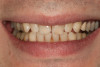
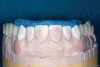
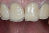
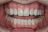
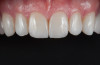
A 34-year-old male patient with no medical issues presented to the office with the chief complaint of a failing crown on tooth No. 12. The patient was also unhappy with his bite and the appearance of his teeth (Figure 1 and Figure 2).
Many years earlier, he had noticed marked wear on his upper front teeth and sought treatment. The treating practitioner diagnosed parafunction as the cause and fitted the patient with a variety of occlusal devices, none of which seemed to stop the attrition. The patient felt that this succession of different appliances-some for the mandible and some for the maxilla, some hard and some soft, and some full cover and some midline-had actually resulted in gradual bite opening, which by his late 20s, prevented him from being able to fit his teeth together at all. Because male jaw growth can continue into the mid-20s9and anterior or posterior bite opening related to the use of some midline appliances or mandibular repositioning devices has been documented,10 the patient's explanation for his current bite situation seemed plausible.
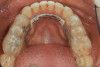
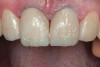
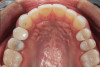
Ultimately, the patient developed joint and muscle symptoms due to his lack of posterior support and sought the help of a specialist. His bite was analyzed, and he was fitted with a dental orthotic that allowed his teeth to touch in centric relation (Figure 3). This was intended to be a temporary measure to stabilize the patient's bite and make him comfortable, but before any permanent treatment could be carried out, he moved abroad and had been living with the device for more than four years before the time of the consultation. He stated that his bite was now very stable with the orthotic, but he was looking for a more permanent solution and also wanted to improve the overall appearance of his smile.
Clinical Examination and Findings
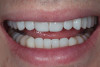
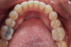
A comprehensive examination was carried out, and preoperative photographs were taken (Figure 1 through Figure 4). The patient presented with fair oral hygiene and slight, generalized tissue inflammation. Caries and defective restorations were detected on teeth Nos. 4, 5, 13, and 14. The crown on tooth No. 12 was showing signs of leakage, and although the endodontic access cavity had been temporarily restored with composite, this endodontic re-treatment was acceptable and the tooth was otherwise symptom-free. Erosion was present on most of the posterior teeth and the cuspids, and abrasion was noted on teeth Nos. 4, 5, 10, 11, 20, 21, 22, 28, and 29. An examination of the patient's muscles, joints, and bite revealed no joint sounds, a normal range of motion, and negative joint load and immobilization tests.
The patient's removable dental orthotic covered the lower bicuspids and first molars. With the orthotic in place, there was shim stock contact on all posterior teeth and on the second molars that were not covered by the appliance. However, there was no incisal overlap and a lack of anterior guidance.11 When the orthotic was removed, solid shim stock contacts were present on the second molars and the right lateral incisor. These contacts were reproduced every time the appliance was removed. The same contacts were found on the patient's mounted models, which confirmed that the joints were in centric relation. Whether the lower second molars had overerupted as a result of wearing the appliance or had already been in this position before the orthotic was fitted could not be established. Apart from these three contacts, no other teeth touched, and there was a 2-mm space between the posterior teeth, making mastication difficult. Interestingly, wear facets were present on the palatal surfaces of the upper central and lateral incisors, indicating that at some point in time, there had been significant overlap of the anterior teeth and a possible constriction of the chewing envelope. Analysis of a cephalometric radiograph showed a class II growth tendency with a reduced mandibular plane angle, which supported the theory that the patient had at one time possessed a class II occlusion.
The dentofacial examination revealed a low smile line with no incisor display when the lips were in repose. The anterior teeth were chipped and worn, and their overall color was darkened. The buccal corridors were deficient, and the failing crown on tooth No. 12 was visible in a full smile.
Treatment Plan
Periodontal therapy consisting of scaling and polishing would be provided, with a focus on increasing home care and maintaining 6-month recall intervals. Treatment of the decay, coverage of all of the eroded and abraded surfaces, and a new crown on tooth No. 12 would also be required. It is important to note that once dentin is exposed, it will wear at a rate of at least three times the average wear rate of enamel, even in the absence of causative erosive factors.12,13
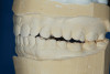
The patient's bite was obviously unsatisfactory, but with the orthotic in place, he was completely symptom-free and stable, and therefore, considered to be in acceptable function. However, he was considered to be in occlusal dysfunction without it because he lacked simultaneous, equal intensity posterior contacts. Deprogramming the patient and confirming centric relation was not considered to be required at this stage because as soon as he removed the orthotic, his occlusal situation was similar to wearing a tripod deprogrammer with only one anterior and two posterior contacts, and these had been confirmed to be reproducible every time when checked intraorally and on the mounted models (Figure 5). The use of a Kois Deprogrammer is one way to obtain centric relation, but other ways, such as bimanual manipulation or the use of a Lucia jig, a leaf gauge, a bite plane, or an occlusal splint have been discussed by various authors.14
Equilibration to close the bite was not possible because the posterior open bite was about 2 mm, which would have necessitated a drastic amount of tooth removal from the second molars. Orthodontic treatment to close the bite was also deemed an inappropriate option because closing the bite would not have eliminated the subsequent need to cover most of the posterior occlusal surfaces with restorations due to the existing erosion. In turn, this would have necessitated removal of tooth structure, which would increase the biomechanical risk of the teeth. Given these factors, the decision was made to close the posterior open bite and create anterior guidance using additive means in the patient's current maximum intercuspation, which was considered equivalent to centric relation. He would then be equilibrated to ensure even, simultaneous posterior contacts and canine guidance.
Restorative material options, namely porcelain and composite, were discussed with the patient. Because he preferred to have no tooth structure at all removed during this stage, and porcelain onlays would have necessitated some preparation of the posteriors because of the minimal thickness requirements,15 the decision was made to restore him with additive composite onlays for his posterior teeth to establish occlusion and composite veneers for his upper anterior teeth and bicuspids to address his esthetic needs and cover the existing buccal abrasions. Only tooth No. 12 would receive a porcelain restoration.
The reduced cost of these material choices was an important factor in the patient's decision-making process. To further minimize cost, the posterior units would be produced using a time-saving wax-up duplication technique by a dental technician. It was explained to the patient that the life expectancy of the posterior composite onlays was limited, and that he would eventually have to switch to a more durable option, such as porcelain. It should be noted that some authors have shown that using a composite for posterior full occlusal coverage can have quite reasonable durability.16,17
In addition, the lower anterior teeth would be bleached, and the abrasions on the lower bicuspids would be covered with composite restorations.
Treatment
Accurate vinyl polysiloxane impressions were taken of the upper and lower arches. Two sets of models were obtained and mounted in a semi-adjustable articulator. A diagnostic wax-up to close the posterior open spaces and incorporate the desired esthetic changes was carried out on one of these sets. The posterior waxed-up surfaces were then duplicated with a clear silicon matrix in quadrants. These matrices were loaded with a slightly warmed microhybrid composite and seated onto the corresponding second model, which had been isolated with a separating agent. Warming composites increases monomer conversion, thereby decreasing curing time and shrinkage.18 Once fully cured, the occlusion was adjusted, and the onlays could easily be separated from the models.
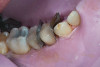
At the initial appointment, all of the patient's carious lesions were treated, and tooth No. 12 was fitted with a provisional crown made from a bis-GMA temporary material. At the next appointment, the patient's tissues were anesthetized, and the posterior teeth were isolated with a rubber dam in quadrants. The teeth were cleaned with air abrasion and etched with phosphoric acid. Studies have shown that air abrasion using 27 µm aluminum oxide at 40 psi greatly improves bond strength when compared with cleaning tooth surfaces using hand instruments, a slurry mixture of pumice and water, or air abrasion using 50 µm aluminum oxide at 40 psi.19 After silane and a bonding agent were applied, the onlays were loaded with a flowable composite and individually cemented (Figure 6). Minimal occlusal adjustments were then carried out to ensure simultaneous posterior contacts.
During subsequent appointments, teeth Nos. 4 through 13 were restored with direct composite veneers in stages. Teeth Nos. 6 through 12 were treated first, followed by teeth Nos. 4, 5, and 13, respectively. Lingual and facial silicone putty indices were fabricated from the wax-up (Figure 7), and the lingual matrix was marked with a sharp probe at the existing incisal edge. Next, low-pressure air abrasion was used to clean the area and roughen the enamel to be bonded. Teeth Nos. 4 and 12 were isolated using polytetrafluoroethylene plumber's tape, and a 37.5 % phosphoric acid etching gel was applied and rinsed off. A layer of a fifth-generation bonding agent was applied and air-thinned, then a second layer was applied, air-thinned, and cured. The application of two layers has been shown to produce an increase in microtensile bond strength.20 After placement of the bonding agent, a clear enamel shade of microhybrid composite was applied into the lingual matrix in a thin layer and drawn slightly over the previously marked line. The composite was thickened toward the facioincisal line angle, which creates a natural "halo effect." The matrix was then adapted to the teeth, and the lingual shelf was briefly cured (Figure 8).
Any excess composite on the lingual surface of the teeth was removed before the lingual shelf was fully cured. Next, a microhybrid composite in a dentin shade was layered onto the teeth and the lingual shelf, drawn out into mamelon shapes with the aid of a sable brush, and fully cured. Using a hybrid composite for the lingual shelf and dentin layer provides wear resistance and strength for the restoration.21,22 Next, a small amount of material in a translucent blue shade was used in between and around the previously created mamelons in order to "lock in" the natural translucency and create a lower value appearance between the dentinal lobes (Figure 9). Finally, a layer of microfill in shade B1 was applied to full contour and cured. A matrix pull-through technique was used in the interproximal areas. Final curing was performed after the application of a glycerin gel to ensure a fully cured restoration surface. The decision to use a microfill in the visible areas that would not be placed under occlusal load was based on its superior polishability.22 A similar incremental layering technique was used on the cuspids and bicuspids.
With the patient sitting up, the incisal edges were finished parallel to the interpupillary line. After placing the patient back into a supine position, refinement of the primary anatomy was carried out with a fine grit, flame-shaped diamond bur. Coarse discs were then used to shape the cervical, middle, and incisal facial contours and open the incisal embrasures. The facioincisal line angles were marked with a pencil to ensure that proper placement was maintained during the shaping and finishing procedure. The proximal line angles, which were also marked with a pencil, were finished using medium coarse discs to establish light reflecting and light deflecting areas.
Once the primary anatomy was finished, secondary anatomy was created by first marking off the desired position of developmental grooves with pencil and then placing them carefully with a flame-shaped diamond bur and abrasive points. The patient was also fitted with a bleaching tray for the lower arch and instructed to bleach only the lower anterior teeth.
The final polish was performed using a series of fine grit discs in various sizes, and a glass-like luster was achieved by applying aluminum oxide polishing paste with felt discs and points under light pressure.
After the composite veneers were finalized, impressions were taken for a monolithic lithium disilicate crown on tooth No. 12. This was placed during a subsequent appointment, and occlusal equilibration was carried out. The patient returned for final photographs a couple of weeks later (Figure 10 through Figure 14).
Conclusion
The use of transitional bonding techniques can allow complex cases to be treated in a cost-effective and timely manner. Furthermore, occlusal and esthetic designs can be evaluated while the maximum amount of tooth structure is conserved.
About the Author
Sandra Hulac, DDS
Private Practice
Hong Kong, China
References
1.Van't Spijker A, Rodriguez JM, Kreulen CM, et al. Prevalence of tooth wear in adults. Int J Prosthodont. 2009;22(1):35-42.
2. Hattab FN, Yassin OM. Etiology and diagnosis of tooth wear: a literature review and presentation of selected cases. Int J Prosthodont. 2000;13(2):101-7.
3. Hannif A, Rashid H, Nasim M. Tooth surface loss revisited: Classification, etiology, and management. J Res Dent. 2015;3(2):37-43.
4. Kois JC. New challenges in treatment planning: incorporating the fundamentals of patient risk assessment - Part 2. J Cosmet Dent. 2011;27(1):110-121.
5. Rouse JS. The Bruxism Triad: Sleep bruxism, sleep disturbance, and sleep-related GERD. Inside Dentistry. 2010;6(5):32-44.
6. Antonelli JR, Hottel TL, Garcia-Godoy F. Abfraction lesions--where do they come from? A review of the literature. J Tenn Dent Assoc. 2013;93(1):14-9; quiz 20-21.
7. Winter R. Bulimia: Complex Etiology, Challenging Treatment. Dent Today. 2015;34(7):119-20, 122-3.
8. Grippo JO, Simring M, Coleman TA. Abfraction, abrasion, biocorrosion, and the enigma of noncarious cervical lesions: a 20-year perspective. J Esthet Restor Dent. 2012;24(1):10-23.
9. Heij DG, Opdebeeck H, van Steenberghe D, et al. Facial development, continuous tooth eruption, and mesial drift as compromising factors for implant placement. Int J Oral Maxillofac Implants. 2006;21
(6):867-78.
10. Bereznicki T, Barry E, Wilson NHF. Unintended changes to the occlusion following the provision of night guards. Br Dent J. 2018;225(8):715-722.
11. Ash MM. Current concepts in the aetiology, diagnosis and treatment of TMJ and muscle dysfunction. J Oral Rehabil. 1986;13(1):1-20.
12. Larson TD. Tooth wear: when to treat, why, and how. Part two. Northwest Dent. 2009;88(6):19-28.
13.Yip KH, Smales RJ, Kaidonis JA. Differential wear of teeth and restorative materials: clinical implications. Int J Prosthodont. 2004;17(3):350-6.
14. Dawson PE. Functional Occlusion: From TMJ to Smile Design. St. Louis, MO: Mosby, Inc; 2007.
15. Ivoclar Vivadent Inc. 2009. All-Ceramic Chairside Preparation Guide for IPS e.max [Brochure]. Published by Ivoclar Vivadent.
16. Schmidlin PR, Filli T, Imfeld C, et al. Three-year evaluation of posterior vertical bite reconstruction using direct resin composite--a case series. Oper Dent. 2009;34(1):102-8.
17. Van Dijken JW. Direct resin composite inlays/onlays: an 11 year follow-up. J Dent. 2000;28(5):299-306.
18. Freedman G. Clinical benefits of pre-warmed composites. Private Dent. 2003;8(5):111-14.
19. Chalyabutr Y, Kois JC. The effects of tooth preparation cleansing protocols on the bond strength of self-adhesive luting cement to contaminated dentin. Oper Dent. 2008;33(5):556-63.
20. Soares CG, Carracho HG, Braun AP, et al. Evaluation of bond strength and internal adaptation between the dental cavity and adhesives applied in one and two layers. Oper Dent. 2010;35(1): 69-76.
21. Hervás-García A, Martinez-Lozano MA, Cabanes-Villa J, et al. Composite Resins. A review of the materials and clinical implications. Med Oral Patol Oral Cir Bucal. 2006;11(2):E215-20.
22. Lambrechts P, Vanherle G. Structural evidences of the microfilled composites. J Biomed Mater Res. 1983;17(2):249-60.