You must be signed in to read the rest of this article.
Registration on CDEWorld is free. Sign up today!
Forgot your password? Click Here!
A digital revolution is occurring that is rapidly changing the world of dentistry for dentists, laboratories/digital technicians, smile designers, and patients. Major advancements in digital technology have significantly improved the dentist's and the dental technician's ability to create esthetic and functional results for the patients whom they are treating. In many ways, digital dentistry has brought the future to the present.
The use of CAD/CAM technology has allowed for more predictability in fabricating full-mouth implant-supported restorations. Guided implant surgery has become mainstream with the aid of CBCT. Digital intraoral impression scanning and laboratory scanning have revolutionized the impression taking process.1 With the assistance of computer-aided framework and prosthesis design and high-precision computer-guided milling and 3D printing, the field of prosthodontics has been altered forever.2 CAD/CAM technologies have been successfully applied to treat dentate, partially edentulous, and fully edentulous patients with a myriad of prostheses, including implant-assisted and implant-supported prostheses.3,4,5
Much of the focus of these technological advances has been on the development of full-arch prosthodontics, and now with the emergence of 3D extraoral facial scanning, these procedures can be further enhanced. Dentists and technicians also have been able to capitalize on these advancements for all dental prosthetics.
3D extraoral facial scanning brings the missing link of treatment planning and design into the arena. Digitization of the patient via CBCT, intraoral scanning, and extraoral scanning allows for all of the data needed for evaluation, design, and treatment planning to be precisely aligned and merged together for a completely digital representation of the patient.6 This workflow places the digital patient on the computer screen, allowing the dentist, designer, and technician to evaluate, manipulate, and treatment plan in a way that was heretofore not possible with analog and two-dimensional workflows (Figure 1).7,8
The face has been a critical missing link in digital dentistry because it is essential to the predictability and efficiency of developing an esthetic and functional result.9 Digitizing the teeth and the bony architecture are important, but without the face and the facial data to reference, it proves to be a bit like working on a dentoform.
Furthermore, technological developments in 3D facial scanning have allowed for the realization of economic, compact, portable, in-office systems that integrate with software platforms that are geared toward prosthetic dentistry.10,11,12 Coupled with the aforementioned CAD/CAM systems, 3D facial scanning has made it more viable to virtually design and fabricate full-arch prostheses, single-tooth restorations, and all dentistry in between using a completely digital approach.5,13
3D Facial Scanning
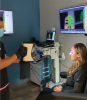
An increasing number of 3D scanners on the market have been designed for purposes such as robotic mapping, industrial design, reverse engineering, prototyping, and quality control. Several companies have recently adapted these commercially available scanners to be utilized for medical and dental applications. Newer software has been developed to adapt some of these scanners to create precise and accurate 3D representations of the face and teeth14 (Figure 2). The adaptation of these scanners and their related software can produce enough precision, accuracy, and trueness to be utilized in dentistry to design and fabricate simple and complex prostheses, and well-fitted and well-adapted restorations that seat with the appropriate passivity to be predictable and long-lasting.15,16,17
Facially Generated Treatment
The face and facial data become the missing link in treatment planning and smile design once the patient has left the operatory.18 Two-dimensional photographs can assist the dentist and technician in representing this information to the designer, but many limitations remain. Unlike a photograph, the 3D scan allows the designer to manipulate the image. A photograph, if taken off-angle, cannot be uprighted. The midline of the face, occlusal cant, profile representation, inferior and superior views of the teeth, and lip positions cannot be viewed simultaneously without clicking between photos. Consequently, combining them is left to the artistic interpretation of the designer.
Natural head position (NHP) and natural head orientation (NHO) also have proven difficult to obtain and reproduce with two-dimensional techniques.19,20 Multiple techniques and devices have been developed to attempt to communicate this information to the technician, but they are often laborious and lack predictable accuracy. Once the models have been fabricated and articulated, the bench top often becomes the "horizon." Without an accurate reference, an occlusal cant is often created, reproduced, and/or enhanced.21
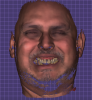
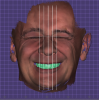
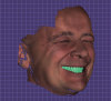
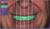
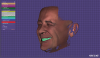
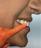
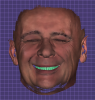
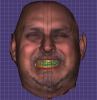
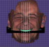
With the utilization of 3D extraoral facial scans, the face is reintroduced into the design protocols. The midline of the face is easily discernible and able to be viewed from different angles, as the patient's head can be rotated and tilted to any angle, showing the designer the complete facial anatomy preoperatively, during the design phase, and in the post-design (Figure 3 and Figure 4). The occlusal cants can be verified and altered to the NHP with grid lines on the computer to ensure the proper position of the teeth in the face, in real-time (Figure 5). The profile, as well as the inferior and superior views, can be referenced simply by turning and tilting the patient's head on the screen, giving the designer complete control for a facially generated smile design (Figure 6).
Patient Acceptance and Co-Diagnosis
A key element of restorative dentistry and prosthodontics is gaining patient acceptance of treatment plans, and the technologies utilized for diagnostics can help in this regard. Many patients favor optical impressions, which have minimized the need for conventional analog impressions with trays and materials such as alginate, polyvinylsiloxane, and poly-ether. The majority of patients have never received a facial scan at the dentist and perceive a "wow" factor with this level of treatment. The significant other and/or caregiver or companion are able to remain in the room during the scans and experience the same level of interest and excitement as the patient. The scans also can be used as a patient education tool to explain findings and create engagement.
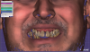
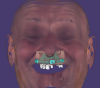
Digitizing the patient's face and teeth and displaying these scans on a large monitor in the office allows the patient to see their teeth and their smile in a new way (Figure 7). Seeing their teeth magnified, the patient can express their desires; they can point out aspects that they are pleased with and what they would like to see altered, assisting the clinician with a co-diagnosis (Figure 8). The patient can "own their condition." Treatment options and planning can then be proposed and all involved parties can have the opportunity to plan any restorations to meet the patient's needs and desires while minimizing any disappointments that may occur with the delivery of the definitive treatment. This allows the clinician to customize the treatment, design, and end results to more confidently meet patient expectations.22
The need for multiple impressions and various forms of try-ins and verifications are virtually eliminated. The patient is able to view their smile and the proposed smile design virtually in their face, allowing them to request changes that can be addressed digitally, prior to any addition or subtraction to their actual dentition. Once the design is completed, the patient can approve the design and have confidence in the result because they can see it virtually in their face, eliminating the "guess-work" by the clinician (Figure 9).22
Alignment and Merging of Scan Data
The virtual models from 3D facial scans allow technicians to create dental prostheses that rival and potentially surpass the accuracy of many analog techniques.23,24,25,26
Utilizing commercially available design software—such as exocad, 3Shape Dental System, and Blue Sky Bio Plan—STL, PLY, and OBJ files can be imported and aligned, integrating each of the individual scans together to create a digital representation of the patient in a one-to-one format and allowing the digital technician to design the smile directly in the virtual version of the patient's face27 (Figure 10). Multiple scans can be imported and merged to display multiple lip positions6 (Figure 11). The digital design can then be carried out utilizing a Duchenne (exaggerated) smile, natural smile, repose lip position, or any other lip position to accurately place the incisal edge position of the maxillary and mandibular arches, as well as to visualize the occlusal plane of the posterior teeth (Figure 12). Once the files are precisely aligned, the designer can utilize the intraoral and extraoral scans to design the smile to the most esthetic and functional position from a facially generated approach, merge the CBCT data, and design surgical guides to facilitate precise prosthetically generated implant positions, along with a multitude of other prosthetic and functional appliances13,18(Figure 13).
Laboratory Applications
In a traditional analog and two-dimensional workflow, the laboratory technician has minimal ability to manipulate images. Models, mock-ups, provisional prostheses, and verification appliances are often fabricated to give the clinician and the patient an idea of what is possible. With full-arch treatment, the laboratory technician traditionally attends the appointment to assist with the conversion and/or alteration of the prosthesis.
Once the patient is digitized and placed virtually on the screen, the technician can complete all of this work at the laboratory with the same level of accuracy, saving valuable time and expenses. During the design phase, the technician can view the scans and immediately give feedback to the clinician.
Discussion
CAD/CAM technology is no longer a novelty, and 3D extraoral scanning is allowing the dentist, the patient, and the technician to have all of the data readily available not only to display and educate, but to connect, discuss, plan, and develop the ideal customized treatment.
Utilization of digital dentistry creates a higher level of efficiency for all involved, capitalizing on the advantages of the CAD/CAM procedures. These include the elimination of multiple clinical evaluation visits and procedures, thereby reducing chair-time and treatment costs while allowing better communication with the dental laboratory regarding prosthesis design and increased ability to realize an individualized tooth arrangement.
With any technology, there is an initial outlay of capital expenditure. With an increase in efficiency, accuracy, and precision afforded by 3D facial scanning, the savings in time and remake percentages can quickly make the investment a worthwhile expense.
The nearly unlimited nonrestorative uses of digital technology are redefining how intraoral and extraoral scanners are being used without compromising the level of treatment being provided.
About the Authors
Jeff Bynum, DDS
Clinical Instructor/Course Facilitator
Kois Center
Fernando Polanco
CAD/CAM Digital Dental Technologist
References
1. Cicciù M, Fiorillo L, D'Amico C, et al. 3D Digital Impression Systems Compared with Traditional Techniques in Dentistry: A Recent Data Systematic Review. Materials (Basel). 2020;13(8):1982. Published 2020 Apr 23. doi:10.3390/ma13081982
2. Alghazzawi TF. Advancements in CAD/CAM technology: Options for practical implementation. J Prosthodont Res. 2016 Apr;60(2):72-84.
3. Wei Y, Chen G, Han B, Hu XY, Zhang HP, Deng XL. Three-dimensional imaging for quantitative evaluation of facial profile of edentulous patients before and after complete dentures restoration. Beijing Da Xue Xue Bao. 2014; 46(1): 100-103.
4. Papadiochou S, Pissiotis AL. Marginal adaptation and CAD-CAM technology: A systematic review of restorative material and fabrication techniques. J Prosthet Dent. 2018 Apr;119(4):545-551.
5. Lucio Lo Russo, Claudio Di Gioia, Angelo Salamini, Laura Guida, Integrating intraoral, perioral, and facial scans into the design of digital dentures, The Journal of Prosthetic Dentistry, Volume 123, Issue 4, 2020, Pages 584-588
6. Alexa, M. 2003. Differential coordinates for local mesh morphing and deformation. The Visual Computer. 19(2):105-114.
7. Revilla-León M, Raney L, Piedra-Cascón W, Barrington J, Zandinejad A, Özcan M. Digital workflow for an esthetic rehabilitation using a facial and intraoral scanner and an additive manufactured silicone index: A dental technique. J Prosthet Dent. 2020 Apr;123(4):564-570.
8. Stefano Granata, Lorenzo Giberti, Paolo Vigolo, Edoardo Stellini, Adolfo Di Fiore, Incorporating a facial scanner into the digital workflow: A dental technique, The Journal of Prosthetic Dentistry, Volume 123, Issue 6, 2020, Pages 781-785
9. Lane C, Harrell WJ. Completing the 3-dimensional picture. Am J Orthod Dentofacial Orthop. 2008; 133(4): 612-620
10. Nightingale, R.C., Ross, M.T., Allenby, M.C., Woodruff, M.A. and Powell, S.K. (2020), A Method for Economical Smartphone‐Based Clinical 3D Facial Scanning. Journal of Prosthodontics, 29: 818-825.
11. George Petrides, JonathAn R. Clark, Hubert Low, Nigel Lovell, Timothy J Eviston, Three-dimensional scanners for soft-tissue facial assessment in clinical practice, Journal of Plastic, Reconstructive & Aesthetic Surgery, Volume 74, Issue 3, 2021, Pages 605-614
12. Umut Özsoy, Rahime Sekerci, Arzu Hizay, Yilmaz Yildirim, Hilmi Uysal, Assessment of reproducibility and reliability of facial expressions using 3D handheld scanner, Journal of Cranio-Maxillofacial Surgery, Volume 47, Issue 6, 2019, Pages 895-901
13. Christian Coachman, Marcelo Alexandre Calamita, Francis Gray Coachman, Robert Gray Coachman, Newton Sesma, Facially generated and cephalometric guided 3D digital design for complete mouth implant rehabilitation: A clinical report, The Journal of Prosthetic Dentistry, Volume 117, Issue 5, 2017, Pages 577-586
14. Toma AM, Zhurov A, Playle R, Ong E, Richmond S. Reproducibility of facial soft tissue landmarks on 3D laser-scanned facial images. Orthod Craniofac Res. 2009; 12(1): 33-42.
15. Ma, Lili & Xu, Tianmin & Lin, Jiuxiang. (2009). Validation of a three-dimensional facial scanning system based on structured light techniques. Computer methods and programs in biomedicine. 94. 290-8.
16. Zhao YJ, Xiong YX, Wang Y. Three-Dimensional Accuracy of Facial Scan for Facial Deformities in Clinics: A New Evaluation Method for Facial Scanner Accuracy. PLoS One. 2017;12(1) Published 2017 Jan 5.
17. Chung How Kau, Stephen Richmond, Alexei I. Zhurov, Jeremy Knox, Ivor Chestnutt, Frank Hartles, Rebecca Playle, Reliability of measuring facial morphology with a 3 dimensional laser scanning system, American Journal of Orthodontics and Dentofacial Orthopedics, Volume 128, Issue 4, 2005, Pages 424-430.
18. Racich, M.J. The Basic Rules of Facially Generated Treatment Planning. Palmeri Publishing; 2014:1-79
19. Meiyappan N, Tamizharasi S, Senthilkumar KP, Janardhanan K. Natural head position: An overview. J Pharm Bioallied Sci. 2015;7(Suppl 2):S424-S427.
20. Verma SK, Maheshwari S, Gautam SN, Prabhat K, Kumar S. Natural head position: key position for radiographic and photographic analysis and research of craniofacial complex. J Oral Biol Craniofac Res. 2012 Jan-Apr;2(1):46-9.
21. Weber DW, Fallis DW, Packer MD. Three-dimensional reproducibility of natural head position. American Journal of Orthodontics and Dentofacial Orthopedics. 2013; 143(5): 738-744.
22. Liu YS, Ye HQ, Gu M, Lv LW, Sun YC, Zhao YJ, et al. Application of patient-participated digital design in esthetic rehabilitation of anterior teeth. Beijing Da Xue Xue Bao. 2014; 46(1): 90-94.
23. Ali Modabber, Florian Peters, Kristian Kniha, Evgeny Goloborodko, Alireza Ghassemi, Bernd Lethaus, Frank Hölzle, Stephan Christian Möhlhenrich, Evaluation of the accuracy of a mobile and a stationary system for three-dimensional facial scanning, Journal of Cranio-Maxillofacial Surgery, Volume 44, Issue 10, 2016, Pages 1719-1724
24. Zhao YJ, Xiong YX, Yang HF, Wang Y. Evaluation of measurement accuracy of three facial scanners based on different scanning principles. Beijing Da Xue Xue Bao. 2014; 46(1): 76-80.
25. Zhao YJ, Xiong YX, Yang HF, Lv PJ, Sun YC, Wang Y. Quantitative evaluation for the measurement accuracy of three-dimensional facial scanner. Shi Yong Kou Qiang Yi Xue Za Zhi. 2016; 32(1): 104-109Zhao YJ, Xiong YX, Yang HF, Wang Y. Evaluation of measurement accuracy of three facial scanners based on different scanning principles. Beijing Da Xue Xue Bao. 2014; 46(1): 76-80.
26. Xiong YY, Chen XB, Sun J, Zhang FQ, Xi JT. Accuracy of three dimensional facial measurement system based on structured light projection. Shanghai Jiaotong Daxue Xuebao. 2010; 30(01): 66-69.
27. Oancea L, Burlibasa M, Petre AE, Panaitescu E, Cristache CM. Predictive Model for Occlusal Vertical Dimension Determination and Digital Preservation with Three-Dimensional Facial Scanning. Applied Sciences. 2020; 10(21):7890
28. Duchenne de Boulogne GB. Mécanisme de la Physionomie Humaine: Texte. Paris: Renouard; 1862.