You must be signed in to read the rest of this article.
Registration on CDEWorld is free. Sign up today!
Forgot your password? Click Here!
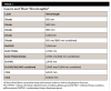
The word laser is an acronym. It stands for Light Amplification by Stimulated Emission of Radiation. A laser is simply a more powerful (amplified) beam of light energy. The textbook definition of a laser is a device that emits an intense coherent directional beam of radiant energy.1 Light energy has been used as a therapeutic modality since the time of the ancient Greeks.2 Each laser has a unique wavelength, and how well that wavelength is absorbed by its target tissue determines how well a particular laser creates a therapeutic effect, if any, on its target tissue.3 Both surgical and nonsurgical wavelengths are available in the United States. This article will limit itself to a discussion of surgical devices. Many different surgical laser wavelengths are sold in this country. Table 1 lists the different surgical wavelengths currently available.4
When Can Soft-Tissue Lasers Be Used?
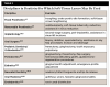
For most procedures in which a cold piece of steel (a scalpel or pair of scissors) can be used, a laser could also be used. According to the American Academy of Periodontology (AAP), a laser is a viable alternative to a scalpel.5 Table 2 lists the disciplines in dentistry in which soft-tissue lasers may be used.6-25 The list is not comprehensive; it merely presents some of the more common uses of some soft-tissue lasers. Not all soft-tissue lasers can perform all procedures, and each laser performs procedures with different degrees of success, at different speeds, and with different postoperative sequelae.
This article will be limited to a discussion of diode lasers. A complete discussion of all wavelengths can be found in textbooks26,27 or, even better in the author's opinion, a 2-day participation course where a minimum of three different laser manufacturers or three different types of lasers (eg, diode, CO2, and erbium) are available during the hands-on part of the course. In the author's opinion, any course that presents less than three wavelengths for attendees to use on pig mandibles is more than likely a sales course, rather than a bona fide educational program.
How Do Lasers Work?
To answer that question, two other questions must be answered. First, why are light colors worn in the summer and dark colors in the winter? The answer is light (and heat) absorption from the sun. In the summer, light colors are worn to reflect light/heat energy and keep people cool. In the winter, when there is less heat and light from the sun, dark colors are worn to absorb as much of the sunlight as possible. Second, why are lead shields placed on patients before radiographs are taken? The answer, once again, is absorption. The lead apron absorbs the x-rays (ultra-short ultraviolet [UV] wavelength invisible light energy), which prevents the damaging UV light energy from hitting patients' bodies. Therefore, the main word when discussing how lasers (light energy) work is absorption. Another word that must be discussed is chromophore. The definition of a chromophore is a light-absorbing compound or molecule that absorbs specific wavelengths of light energy.28 Laser-tissue interaction is based on the chromophore concept. The light energy is either absorbed by the target tissue, resulting in a therapeutic effect, or not absorbed, in which case there will be no therapeutic effect. Chromophore must be the first concept introduced to dentists at any introductory laser course. Each laser is absorbed preferentially by different chromophores in the oral cavity.
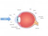
Lasers are optical devices: photons are absorbed by the chromophores of the target tissue and perform therapeutic effects. Lasers, generally, are non-contact devices. The photons leave the handpiece, are absorbed by the chromophores of the target tissue, and perform the therapeutic procedure. When ophthalmologists perform retinal surgery, they do not push the laser fiber into the back of the eyeball until it touches the target tissue; the laser energy is emitted from the laser out of contact with the eyeball and is absorbed by the chromophores of the retina, thereby producing a therapeutic effect (Figure 1).29
In medicine, when lasers are used in contact, it is usually to coagulate or melt tissue, rather than optically ablate tissue.30,31 Lasers are not initiated in medicine when performing therapeutic procedures. The purpose of initiation is to place a black material (never articulating paper or cork) on the tip of the laser fiber to prevent the photons from coming out of the laser handpiece. Black absorbs all light energy; therefore, when placing something black on the tip of a laser, the photons cannot come out of the laser handpiece. The photons are absorbed by the black material, which causes the tip of the laser to heat up.
Diode lasers, when initiated, are hot glass tips. Therefore, when used with initiated tips, diode lasers are not used as optical (photon-emitting) devices; they are used as thermal devices. Diode lasers are classified as lasers because they produce a beam of collimated, coherent, monochromatic light; however, they are not used as true optical devices, because they prevent the light energy from leaving the tip of the handpiece. How hot is the tip of a diode laser? Peer-reviewed literature states that the temperature at the tip of a diode laser is 750 to 1200° C.32,33
Erbium and CO2 lasers, which are never initiated and never touch tissue, work optically. Their chromophore, generally, is water; the chromophore for these lasers is the water in oral mucosa. Oral mucosa is 70% or more water. These lasers heat up the water in the tissue until the water evaporates, and the tissue is vaporized.34 The boiling point of water is 100° C. Erbium and CO2 lasers, therefore, work optically and need to heat up tissue only to 100° C to vaporize tissue.
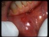
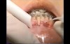
Diodes melt oral tissue at a minimum of 750° C. Diode users are familiar with the material that accumulates at the end of that fiber-melted tissue. Diode absorption is unaffected by chromophore content, except in rare instances where diodes can be used uninitiated, out of contact (treating an aphthous ulcer, herpetic stomatitis, venous lake, or hemangioma) (Figure 2).35 When diodes are placed onto tissues without initiation, they immediately become initiated with a globule of melted tissue (Figure 3). This method of initiation is less than ideal. However, it does not mean that diode lasers should not be used; it just means that a diode user must be educated on how to use a hot glass tip at 750 to 1200° C on oral mucosa.
Diode Laser Dentistry Technique
Because diode lasers must be used as hot glass tips in contact with tissue, there is a specific technique that must be used to prevent too much melted tissue from accumulating on the tip. The width of the incision made by a diode laser is a function of the tip diameter. For example, a 200-micron diameter tip will create an incision 200 microns wide; a 300-micron tip will create an incision 300 microns wide, and so forth. When a globule of melted tissue forms on the diode laser tip, the resulting incision will then be the width of the widest part of that globule. Therefore, it is imperative to constantly clean the diode laser tip. Because diodes are nothing more than hot glass tips, the technique for using a diode is different from that of a true optical device (CO2 or erbium). The more contact time the hot glass tip has with the target tissue, the more melted tissue will accumulate on the tip. This will result in a wider, messier, more uneven incision. The technique for using a diode should therefore emphasize short strokes of contact with the tissue, followed by removing the melted tissue and reinitiating the tip. Experienced diode laser users will recognize the following scenario: the diode tip starts to cut the tissue well, but after a few strokes, the diode starts to drag through the tissue. Why? Either the initiation has worn off the tip or there is too much melted tissue on the tip, transferring heat to the target tissue unevenly.35
The other important point when using a diode laser in contact is that the user must not put pressure on the tip. The diode fiber must barely "ice-skate" on the surface of the tissue. Any time pressure is applied to the tip, the sharp glass fiber then pushes into the tissue and macerates it, rather than cleanly melting the tissue.36
On Which Patients Should Lasers Not Be Used?
The most important contraindication for lasers is using them on patients who have oxygen cannulas due to chronic obstructive pulmonary disease or other pulmonary disorders. The thought behind this contraindication is that dentists who are careless may inadvertently remove the laser from a patient's mouth while keeping their foot on the laser pedal and pass the laser near the oxygen tubing. This could create a pinhole in the tubing, causing the patient to become asphyxiated. For just about any other patient, lasers can be used safely. Electrosurgery/radiosurgery cannot be used on patients with pacemakers/defibrillators, but lasers can work safely on these patients. Pregnancy is not a contraindication to laser use, as long as medical clearance is obtained from the patient's physician. When working on patients who take blood thinners (eg, warfarin or apixaban), care must be taken to ensure no bruising or bleeding occurs after the procedure; however, the superior coagulative ability of lasers over cold steel is well-documented.37-39
What Procedures Should Not Be Performed With a Laser?
Some dentists may find the following section controversial; however, the literature on the subject is clear. Even though many laser manufacturers as well as organizations that are supposed to be unbiased regarding lasers teach the use of lasers for periodontal pocket shrinkage, a review of the literature finds that this procedure shows no long-term benefit.40,41
The American Dental Association (ADA) Clinical Practice Guidelines (CPGs) specifically state that diode (and other wavelength) lasers should not be used in periodontal pockets.42 According to the guidelines, current evidence shows no net benefit from diode lasers (non-photodynamic therapy) when used as an adjunct to scaling and root planing.The AAP supports the ADA CPGs on this issue and has also published best-evidence reviews and consensus articles that state that lasers used in periodontal pockets have shown no long-term benefit.40,41,43 Meta-analyses on the issue have concluded that lasers as adjunctive therapy in periodontal pockets do not provide additional clinical benefit.44,45 Although articles have been published in the peer-reviewed literature that advocate for diode use in periodontal pockets,46,47 the vast majority of these articles predate the ADA, AAP, and other guidelines by many years or, in the opinion of this author, have fatal flaws in their study design.
A review of the ADA Code of Ethics section 5D2 states that dentists must not take the word of manufacturers when deciding how to use a product: "The dentist has an independent obligation to inquire into the truth and accuracy of such claims and verify that they are founded on accepted scientific knowledge or research."48 The dentist must evaluate the claims that manufacturers make and decide whether the peer-reviewed literature supports those claims. Dentists (or hygienists) who choose to place lasers in periodontal pockets should, in the author's opinion, have serious discussions with their malpractice carrier regarding the procedure. A review of the legal ramifications of using lasers in periodontal pockets is, in the opinion of this author, sobering to say the least.
CPGs help to determine the standard of care.49,50 Violations of standard of care practices can lead a practitioner to a fine, suspension, or even loss of license. Whether the clinician agrees or disagrees with CPGs, current guidelines of record should be followed. Since the ADA CPGs were published, the author is aware of no peer-reviewed publications that conclusively refute them. Of course, nit-picking through articles can lead to discovering a sliver of evidence that shows these procedures might work. The previously cited article by Chambrone, Ramos, and Reynolds40 mentions "small, statistically significant clinical improvements…attributable to infrared laser procedures." However, the conclusion ends with: "appear to provide questionable additional clinical improvement."
Conclusion
In the author's experience, arguments always follow a discussion of the CPGs. The first argument is that dentists and hygienists see "fantastic" results when lasers are used. That is possible; however, unless practitioners are doing split-mouth double-blinded studies in their practices, their observations should be regarded as anecdotal. It is also possible that the results seen after using the laser are due to the more meticulous scaling and root planing of the hygienists, rather than the laser. Dentists and hygienists must understand that, right or wrong, the CPGs help define standard of care, and violation of standard of care can potentially result in penalties.
Lasers are excellent additions to a dentist's armamentarium; these devices allow the dentist to perform many procedures more quickly, saving valuable chairtime. They also allow dentists to perform many procedures in-office that they had previously referred to specialists. The most important factor before purchasing a laser is the type of training supplied to the dentist. Without superior training, dentists will be unable to recoup their investment or deliver the best possible care to their patients.
About the Author
Robert A. Convissar, DDS, FAGD
Diplomate, American Board of Laser Surgery
Director of Laser Dentistry, New York Presbyterian Hospital of Queens
References
1. Catone GA, Alling CC III, eds. Laser Applications in Oral and Maxillofacial Surgery. Philadelphia, PA: WB Saunders; 1997.
2. Sulewski JG. Einstein's "splendid light": origins and dental applications. In: Convissar RA, ed. Principles and Practice of Laser Dentistry. 2nd ed. St. Louis, MO: Elsevier; 2016:1-11.
3. Coluzzi DJ, Convissar RA, Roshkind DM. Laser fundamentals. In: Convissar RA, ed. Principles and Practice of Laser Dentistry. 2nd ed. St. Louis, MO: Elsevier; 2016:12-26.
4. Coluzzi DJ. Lasers and light fundamentals. In: Coluzzi DJ, Parker SPA. Lasers in Dentistry-Current Concepts. Cham, Switzerland: Springer; 2017:17-28.
5. Cobb CM. Lasers in periodontics: a review of the literature. J Periodontol. 2006;77(4):545-564.
6. Stuffken M, Vahidi F. Preimpression troughing with the diode laser: a preliminary study. J Prosthet Dent. 2016;115(4):441-446.
7. Melilli D, Mauceri R, Albanese A, et al. Gingival displacement using diode laser or retraction cords: a comparative clinical study. Am J Dent. 2018;31(3):131-134.
8. Kesler G. Clinical application of lasers during removable prosthetic reconstruction. Dent Clin North Am. 2004;48(4):963-969.
9. Rice JH. Laser use in fixed, removable, and implant dentistry. Dent Clin North Am. 2000;44(4):767-777.
10. Romanos GE, Gupta B, Yunker M, et al. Laser use in dental implantology. Implant Dent. 2013;22(3):282-288.
11. Romanos GE, Gutknecht N, Dieter S, et al. Laser wavelengths and oral implantology. Lasers Med Sci. 2009;24(6):961-970.
12. Strauss RA, Guttenberg SA, eds. Lasers in oral and maxillofacial surgery. Oral Maxillofac Surg Clin North Am. 2004;16(2):143-308.
13. Strauss RA. Lasers in contemporary oral and maxillofacial surgery. Dent Clin North Am. 2004;48(4):861-888.
14. Nazemisalman B, Farsadeghi M, Sokhansanj M. Types of lasers and their applications in pediatric dentistry. J Lasers Med Sci. 2015;6(3):96-101.
15. Kumar G, Rehman F, Chaturvedy V. Soft tissue applications of Er,Cr:YSGG laser in pediatric dentistry. Int J Clin Pediatr Dent. 2017;10(2):188-192.
16. Research, Science and Therapy Committee of the American Academy of Periodontology. Lasers in periodontics. J Periodontol. 2002;73(10):1231-1239.
17. Israel M, Rossmann JA. An epithelial exclusion technique using the CO2 laser for the treatment of periodontal defects. Compend Contin Educ Dent. 1988;19(1):86-95.
18. Shajahan PA, Kumar PR, Hariprasad A, et al. Lasers: the magic wand in esthetic dentistry!! J Int Oral Health. 2015;7(6):119-121.
19. Downs JC, Convissar RA, Anagnostaki E, Sun G. Lasers in fixed prosthetic and cosmetic reconstruction. In: Convissar RA, ed. Principles and Practice of Laser Dentistry. 2nd ed. St. Louis, MO: Elsevier; 2016:89-106.
20. Elanchezhiyan S, Renukadevi R, Vennila K. Comparison of diode laser-assisted surgery and conventional surgery in the management of hereditary ankyloglossia in siblings: a case report with scientific review. Lasers Med Sci. 2013;28(1):7-12.
21. Nicoloso GF, dos Santos IS, Flores JA, et al. An alternative method to treat ankyloglossia. J Clin Pediatr Dent. 2016;40(4):319-321.
22. Convissar RA, Massoumi-Sourey M. Recurrent aphthous ulcers: etiology and laser ablation. Gen Dent. 1992;40(6):512-515.
23. Coleton S. Lasers in surgical periodontics and oral medicine. Dent Clin N Am. 2004;48(4):937-962.
24. Mohammadi Z. Laser applications in endodontics: an update review. Int Dent J. 2009;59(1):35-46.
25. Martins MR, Carvalho MF, Vaz IP, et al. Efficacy of Er,Cr:YSGG laser with endodontical radial firing tips on the outcome of endodontic treatment: blind randomized controlled clinical trial with six-month evaluation. Lasers Med Sci. 2013;28(4):1049-1055.
26. Convissar RA, ed. Principles and Practice of Laser Dentistry. 2nd ed. St. Louis, MO: Elsevier; 2016.
27. Coluzzi DJ, Parker SPA. Lasers in Dentistry-Current Concepts. Cham, Switzerland: Springer; 2017.
28. Convissar RA, ed. Principles and Practice of Laser Dentistry. 2nd ed. St. Louis, MO: Elsevier; 2016:301.
29. Romanos GE. Diode laser soft-tissue surgery: advancements aimed at consistent cutting, improved clinical outcomes. Compend Contin Educ Dent. 2013;34(10):752-757.
30. Kabalin JN, Bite G, Doll S. Neodymium:YAG laser coagulation prostatectomy: 3 years of experience with 227 patients J Urol. 1996;155(1):181-185.
31. Muschter R, Hofstetter A. Technique and results of interstitial laser coagulation. World J Urology. 1995;13(2):109-114.
32. Weiss RA. Comparison of endovenous radiofrequency versus 810 nm diode laser occlusion of large veins in an animal model. Dermatol Surg. 2002;28(1):56-61.
33. Romanos GE, Belikov AV, Skrypnik A, et al. Uncovering dental implants using a new thermo-optically powered (TOP) technology with tissue air-cooling. Lasers Surg Med. 2015;47(5):411-420.
34. Guttenberg SA, Emery RW III. Laser physics and tissue reaction. Oral Maxillofac Surg Clin North Am. 2004;16(2):143-147.
35. Luk K, Anagnostaki E. Impact of laser dentistry in management of color in the aesthetic zone. In: Coluzzi DJ, Parker SPA. Lasers in Dentistry-Current Concepts. Cham, Switzerland: Springer; 2017:337-358.
36. Chmura LG, Convissar RA. Soft tissue lasers in orthodontics. In: Principles and Practice of Laser Dentistry. 2nd ed. St. Louis, MO: Elsevier; 2016:203-219.
37. Sexton J. Laser management of vascular and pigmented lesions. In: Catone GA, Alling CC III, eds. Laser Applications in Oral and Maxillofacial Surgery. Philadelphia, PA: WB Saunders; 1997:167-179.
38. Santos-Dias A. CO2 laser surgery in hemophilia treatment. J Clin Laser Med Surg. 1992;10(4):297-301.
39. Israel M. Use of the CO2 laser in soft tissue and periodontal surgery. Pract Periodontics Aesthet Dent. 1994;66(6):57-64.
40. Chambrone L, Ramos UD, Reynolds MA. Infrared lasers for the treatment of moderate to severe periodontitis: an American Academy of Periodontology best evidence review. J Periodontol. 2018;89(7):743-765.
41. Mills MP, Rosen PS, Chambrone L, et al. American Academy of Periodontology best evidence consensus statement on the efficacy of laser therapy used alone or as an adjunct to non-surgical and surgical treatment of periodontitis and peri-implant diseases. J Periodontol. 2018;89(7):737-742.
42. Smiley CJ, Tracy SL, Abt E, et al. Evidence-based clinical practice guideline on the nonsurgical treatment of chronic periodontitis by means of scaling and root planing with or without adjuncts. J Am Dent Assoc. 2015;146(7):525-535.
43. Otomo-Corgel J. More about scaling and root planing. J Am Dent Assoc. 2015;146(12):865-866.
44. Sgolastra F, Severino M, Gatto R, Monaco A. Effectiveness of diode laser as adjunctive therapy to scaling and root planing in the treatment of chronic periodontitis: a meta-analysis. Lasers Med Sci. 2013;28(5):1392-1402.
45. Slot DE, Jorritsma KH, Cobb CM, Van der Weijden FA. The effect of the thermal diode laser (wavelength 808-980 nm) in non-surgical periodontal therapy: a systematic review and meta-analysis. J Clin Periodontol. 2014;41(7):681-692.
46. Moritz A, Gutknecht N, Doertbudak O, et al. et. al. Bacterial reduction in periodontal pockets through irradiation with a diode laser: a pilot study. J Clin Laser Med Surg. 1997;15(1):33-37.
47. Moritz A, Schoop U, Goharkhay K, et al. Treatment of periodontal pockets with a diode laser. Lasers Surg Med. 1998;22(5):302-311.
48. ADA Council on Ethics, Bylaws and Judicial Affairs. Principles of Ethics and Professional Conduct. Chicago, IL: American Dental Association. Revised November 2018.
49. Moffett P, Moore G. The standard of care: legal history and definitions: the bad and good news. West J Emerg Med. 2011;12(1):109-112.
50. Kelly DC, Manguro-Mire G. Commentary: Helling v. Carey, caveat medicus. J Am Acad Psychiatry Law. 2008;36(3):306-309.