You must be signed in to read the rest of this article.
Registration on CDEWorld is free. Sign up today!
Forgot your password? Click Here!
For the creation of an optimal restoration, both materials selection and techniques are critical. From shade matching to building the restoration, the right techniques ensure esthetically pleasing, long-lasting restorations. An understanding of arts and engineering can inform a dentist's decision-making process while choosing materials and while forming the restoration itself. This article discusses the qualities of composites available on the market and the many factors involved in achieving esthetically pleasing composite restorations. Stress distribution in teeth and the importance of understanding these tendencies to ensure long-term success rates are also reviewed. A case study examines how this author harnesses strength and esthetics while preparing and performing a Class V restoration, emphasizing the importance of an interdisciplinary approach to dental restorations.
Composite Restorations
The dental composite as known today was developed in the 1950s by Ray Bowen. Since the introduction of composites, these resins have undergone gradual development and modification to enhance their properties as esthetic restorative materials.1,2 These features include modification of the composite resin filler types, decreased polymerization shrinkage, advanced enamel and dentin adhesive systems, increased strength and longevity, and improved esthetic properties.3,4 Typical composite resin consists of a resin-based matrix, such as bisphenol A-glycidyl methacrylate and an inorganic filler such as silica. The filler gives the composite improved mechanical properties, wear resistance, and translucency.5 The main advantage of composite resins with regard to their esthetic properties lies in their shade-matching options; successful esthetic treatment largely depends on the precise matching of the restoration to the neighboring teeth and accurate adaption of the restoration to the surrounding structures.6
While an in-depth survey of the full range of composites available is beyond the scope of this paper, the author would like to highlight three examples that he considers illustrative of the trend towards optimizing esthetics. Admira® Fusion x-tra (VOCO) is a nano-Ormocer® (organically modified ceramic) restorative material that features highly esthetic omni-chromatic properties that allow a single, universal shade to match the full range of tooth shades. Featuring a 4-mm depth of cure, the physical properties of Admira Fusion x-tra also include a low shrinkage rate of 1.25% by volume and a shrinkage stress up to 50% lower than conventional composites.7 OMNICHROMA® (Tokuyama) is a universal composite that esthetically matches every patient with a single shade, thereby eliminating the shade-taking procedure and reducing composite inventory.8 Beautifil® II (Shofu) is a fluoride-releasing, highly esthetic dental material for all classes of restorations. It is appropriate for patients with a high caries index; an 8-year clinical evaluation recorded a 100% retention rate with intact esthetics, no secondary caries, no failures, and no postoperative sensitivity.9 A 13-year study demonstrated a retention rate of 66% and of those restorations available for evaluation at 13 years, 96% had no secondary caries.10
Composites with chameleon-like qualities simplify shade matching; they mix in with the surrounding tooth structure and its environment. An artistic-minded dentist will appreciate this trait, defined scientifically as a blending effect. The blending ability of a composite restoration can be increased by increasing its polish, decreasing its thickness, and using a more translucent composite.11
Because esthetic dentistry imposes several demands on the artistic abilities of the dentist and the technician, knowledge of the underlying scientific principles of color is essential. Translucent materials scatter, transmit, and absorb light, whereas opaque materials reflect and absorb. Most of the color found in a natural tooth is established within. The semi-translucent structure of teeth makes the color-matching procedure more complex when compared with an opaque object. Surface characteristics, such as gloss, curvature, and texture, affect the degree of light diffusion.12
There are at least three dimensions of color: hue, value, and chroma. Hue is the attribute that enables the clinician to distinguish between different families of color, whereas value indicates the lightness of a color. Chroma is the degree of color saturation. Value, often referred to as brightness, is the amount of light returned from an object.13,14 In the author's opinion, if a good value match is achieved, usually the restoration will be acceptable and blend in, whether the material is porcelain or composite. Teeth are also characterized by degrees of translucency. The translucency of enamel varies with the angle of incidence, surface texture and luster, wavelength, and level of dehydration.15 Of course, the most esthetically pleasing of restorations is useless if it has not been properly engineered to ensure long-term success. This long-term survival is dependent on many factors, with stress-bearing capabilities being a crucial component of any dental restoration.
Stress
Until recently, many fracture presentations observed clinically have been difficult to explain. However, new studies shed light on stress distribution within tooth structures, thus improving clinicians' understanding of subtle compression and stress-fracture presentations in teeth.16
Strain distribution within the tooth is related to its structure. Enamel acts as a stress distributor, transferring the load vertically to the root and horizontally through the dentinoenamel junction (DEJ) to the dentin of the crown. A thick zone (~200 μm) in the dentin at the DEJ undergoes greater stress than the central coronal dentin.17
Recently discovered structures within the occlusal surface of molars indicate that conventional cavity designs are disharmonious with a tooth's natural mechanical stress-distribution system.18-22 Too often, especially with Class V restorations, tooth fractures are caused by the disruption of the natural stress-distribution mechanism within the tooth.16,23
It is critical to consider this potential for disruption when performing restorations. The case below discusses such an approach to restorations using a Class V restoration as an example. This discussion covers not only strategies on how to achieve a sustainable distribution of tooth stress, but also how to use a composite with chameleon-like qualities to produce an esthetically pleasing restoration. The priorities in this case study of a Class V restoration design are function and esthetics; the techniques used incorporate both artful esthetics and engineering function.
Case Study
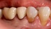
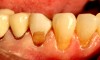
A 70-year-old patient presented with a carious/abfracted lesion on the buccal aspect of tooth No. 12. The tooth was prepared using rotary abrasion with an 888-012 diamond bur (Brasseler). A long bevel of 3 to 5 mm was placed on the superior margin. Next, an infinity bevel of 3 to 5 mm mm was placed, which extended to the line angles and upwards to the occlusal one-third of the tooth with the 888-012 diamond bur. This infinity zone is an interface that is luxury afforded for enamel adhesion,24 with the goal of exposing prismatic enamel to promote micromechanical retention and leverage the bond strength of enamel.25
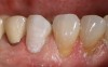
As a restorative dentist, this author studies the dark dentin and translucent enamel (Figure 1) with the goal of employing a one-shade composite that will pick up the color of the environment. For this case, the material should not only mimic its environment but also have enough opacity to block the dark dentin color underneath.
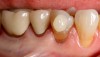
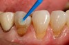
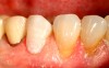
Due to the effect of dehydration, it is important to begin color matching promptly. The esthetic preview in Figure 2 is the beginning of color determination, which is both a visual and an instrumental process. A color swatch is used, and in this case, the esthetic preview matches the canine. This example uses a single-shade omni-chromatic restorative composite material (Admira Fusion x-tra), although other resin composites, including but not limited to the two aforementioned products Beautifil II and OMNICHROMA, could likewise be used.
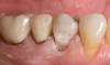
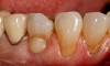
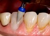
Now that the color is matched, the next step is to understand the stress distribution of this tooth and how that informs preparation design. Figure 3 shows both an esthetic and functional bevel. Bevels require minimal tooth involvement and do not sacrifice the resistance and retention of the restoration.26 With a Class V restoration, the functional bevel will be a gradual transition from the abfraction to the enamel. The relationship between the functional bevel to the exposed dentin of the tooth should be a 1:1 ratio. The radius bevel is where the final blending effect is achieved. Additionally, the ratio of the beveled area to the defect is leveraged; the bevel to untreated ratio is nearly 3:1. There is a definite scalloping in the enamel visible with a gradual transition. Engineering design is crucial because it drives the restoration to achieve the "chameleon effect" goal. This case shows almost an entire overhaul of the facial surface, which helps prevent microleakage.
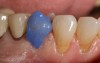
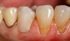
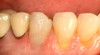
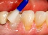
The technique used in this case study is a total acid etch of the whole facial volume according to the manufacturer's directions (Figure 4). Preparation of the enamel beyond the bevel, creating a tooth restoration interface zone, maximizes enamel integration, minimizing the need for mechanical retention.24 Alternatively, a selective etch could have been performed, etching only the enamel and using a universal bonding product. Figure 5 shows the true nature of the phosphoric acid's demineralization. In Figure 6, a dual-cured universal adhesive (Futurabond® U, VOCO) is used, although other products, such as One Coat 7 Universal by Coltene and Clearfil® Universal by Kuraray, would also work. The key is using the right volume of bonding agent on the microbrush so that the entire surface can be covered in one layer. Then the dentin layer bonding agent is agitated, without leaving any debris subgingivally. According to the manufacturer's recommendations, the bonding agent was air-thinned and light-cured for 20 seconds. It is important to floss the proximal joints before light-curing to ensure there is no excess bonding agent or debris in the interproximal areas.27
For material placement, the author recommends placing the gingival layer first and tapering into the upper part of the preparation in a triangular form, careful not to leave any excess or debris. This technique should guarantee a total adhesive seal of the gingival layer. Figure 7 shows the author's technique of placing a small layer first, tapered in a triangular fashion, making sure it is definitively seamed, and then light-curing it so that there is nothing pulling against it. This technique reduces the risk of shrinkage that can lead to microleakage,25,28 the clinically undetectable passage of bacteria, fluids, molecules or ions between a cavity wall and the restorative material applied to it.29 One of the main factors associated with marginal shrinkage and gap formation is the resin composite shrinkage at the tooth-restoration interface, a causative factor in bond failure.30-32 Continuing the material placement in Figure 8, the second layer has been placed and sculpted into the first layer. The image shows the total surface volume placed up to the radius bevel and overbuilt about 5% to 10% over grade. The layering demonstrated no imperfections, following the confines of the whole tooth to decrease the amount of finishing and polishing. The tooth should be uniform, without any irregularities, before light-curing. Each layer was light-cured for 60 seconds according to the manufacturer's instructions (VALO™, Ultradent).
Light-curing is a critical step for a successful restoration, especially when working with a bulk-fill in layers of up to 4 mm. According to the recommendations from the Northern Light Conference,33.34 an appropriate lamp with a sufficient output (irradiance) between 1000 and 1500 mW/cm² should be used. However, lights with a higher output ("power" curing units, > 1500 mW/cm²) create higher temperatures and therefore show an increased risk of irreparable damage to the pulp and surrounding tissue.
Sufficient curing lights have a tip diameter of at least 8 mm. While curing, the tip should be positioned rectangular to the restoration, as close as possible.
It is further agreed that the minimum curing time for every restorative must be at least 10 seconds for resin-based material to receive the required energy dose to be fully cured. Many LCUs are marketed as requiring only a short exposure time (3 seconds or less) because they deliver high irradiance. Although these power curing units deliver the same required energy dose in a shorter time period, there is concern that rapid light-curing of composites may increase shrinkage stress, and as a result decrease the bond strength of resin and adhesives to the tooth.35 Short curing advices incorrectly assume that similar material properties can be achieved when the same radiant exposure is delivered, regardless how high the irradiance or how short the exposure time, but this is not always the case.35
After light-curing, the author uses a dull red tapered diamond to lightly touch all the restored surfaces in a windshield-wiper fashion to ensure the lamination is correct. Any potholes or air pockets should be found and corrected before moving forward. Figure 9 shows the striations from the diamond, with no imperfections nor bleeding. This is the pre-finishing. Figure 10 is the set-up, the precursor to polishing. All margins of the restoration have been examined to make sure there are no irregularities that would create microleakage, staining, or chipping.
One-step polishers such as Dimanto® by VOCO, OneGloss®by Shofu, and A.S.A.P.® by Clinician's Choice are diamond-impregnated silicone polishing systems that can be used wet or dry as pre-polish and final polisher. These polishers have assorted shapes and sizes to polish the hard-to-get-at surfaces. Manufacturers recommend using them at about 12,000 to 15,000 rpm with a fine water spray to observe the pre-luster and final luster. One-step polishers are dependent on how much manual pressure is applied.
The main purpose of finishing and polishing composite restorations is to create a restoration that is smooth, uniform, and easily cleaned (Figure 11). Roughness of restoration surfaces caused by wear and chemical degradation may also affect gloss and consequently increase extrinsic staining.36,37 The structure of the resin composite and characteristics of the particles have a direct impact on the surface smoothness and susceptibility to extrinsic staining.38,39
Figure 12 shows the final restoration immediately postoperative, demonstrating no gingival irritation. This image clearly demonstrates the physics of the restoration, showing off its light dynamics. The highlights that were created in the previous step break up the light, creating a mirroring effect off the adjacent teeth. The chameleon-like material has picked up the color of the environment and of the enamel, while simultaneously masking the dark dentin color.
The final image (Figure 13) shows the last step, using varnish (Profluorid®, VOCO) as a fluoride sealer in case any root surfaces were exposed during the restoration. MI Varnish® by GC America, Embrace™ by Pulpdent, and Clinpro™ 5000 by 3M are examples of other varnishes that would work. These varnishes act as a fluoride bandage to prevent any transient sensitivity issues. Dentin sensitivity issues are not limited to recently restored teeth; discomfort from dentin sensitivity is a common finding in adult populations, with available prevalence data ranging from 8% to 57%.40,41 Through the deposition of calcium fluoride on the tooth surface and with the formation of fluoroapatite, the dental tubules are completely sealed to promote a secondary dentin surface.42 The restoration is now complete.
Conclusion
The development of dentistry has been marked by an ever more pronounced interdisciplinary character. An elite dental restoration involves a process that incorporates both engineering and art. Ceramic-based restoratives are attractive dental restoration materials because of their esthetics, biological acceptance, and chemical durability.
The artistic side of restorative dentistry is evident in color matching, beveling, and polishing. Designing and crafting a restoration requires an artistic eye. Shade matching is inherently complex and involves understanding the science of color, as well as determining the shade and surface characteristics of teeth.43However, composites that exhibit a higher propensity to blend (or chameleon-like qualities) can simplify shade matching. Developing an understanding of the optical characteristics of teeth, such as color and translucency, as well as the structural characteristics of teeth, such as stress distribution, is essential for esthetically pleasing, enduring restorations.
Thus, an approach to restorations that incorporates art and engineering decreases risk and ensures a strategy for enhanced patient satisfaction and long-lasting restorations.
References
1. Radz GM. Composite resins in 2013: an update on their progress. Compend Contin Educ Dent. 2013;34(1):48-51.
2. Sakaguchi LR, Powers JM. Restorative materials-composites and polymers. In: Sakaguchi R, Powers JM, eds. Craig's Restorative Dental Materials. 13th ed. Philadelphia, PA: Elsevier; 2011:161-182.
3. Pitel ML. Low-shrink composite resin: a review of their history, strategies for managing shrinkage, and clinical significance. Compend Contin Educ Dent. 2013;34(8):578-590.
4. Margeas RC. Composite restoration esthetics. Academy of Dental Therapeutics and Stomatology. November 2009.
5. Bhattacharya M, Seong WJ. Carbon nanotube-based materials-preparation, biocompatibility, and applications in dentistry. In: Nanobiomaterials in Clinical Dentistry.2nd ed. Elsevier; 2019:37-67.
6. Paravina RD, Westland S, Johnston WM, Powers JM. Color adjustment potential of resin composites. J Dent Res. 2008;87(5):499-503.
7. VOCO. Admira Fusion. https://www.voco.dental/au/portaldata/1/resources/products/folders/gb/admira-fusion-x-tra-flow-x-base_fol_gb.pdf. Accessed April 23, 2020.
8. Tokuyama Dental. OMNICHROMA Technical Report. https://omnichroma.com/us/wp-content/uploads/sites/4/2019/01/OMNI-Tech-Report-Color-Final.pdf. Accessed April 23, 2020.
9. Gordan VV, Mondragon E, Watson RE, et al. A clinical evaluation of a self-etching primer and a giomer restorative material: results at eight years. J Am Dent Assoc. 2007;138(5):621-627.
10. Gordan VV, Blaser PK, Watson RE, et al. A clinical evaluation of a giomer restorative system containing surface prereacted glass ionomer filler: results from a 13-year recall examination. J Am Dent Assoc.2014;145(10):1036-1043.
11. Paravina R, Westland S, Imai FH, et al. Evaluation of blending effect of composites related to restoration size. Dent Mater. 2006;22(4):299-307.
12. Cal E, Güneri P, Kose T. Comparison of digital and spectrophotometric measurements of colour shade guides. J Oral Rehabil.2006;33(3):221-228.
13. Terry DA, Geller W, Tric O, et al. Anatomical form defines color: function, form, and aesthetics. Pract Proced Aesthet Dent. 2002;14(1):59-67.
14. Boksman L. Shade selection: accuracy and reproducibility. Ontario Dentist. 2007:84(4):24-27.
15. Fondriest J. Shade matching in restorative dentistry: the science and strategies. Int J Periodontics Restorative Dent. 2003;23(5):467-479.
16. Milicich G, Rainey JT. Clinical presentations of stress distribution in teeth and the significance in operative dentistry. Pract Periodontics Aesthet Dent. 2000;12(7):695-700.
17. Goldberg M, Kulkarni AB, Young M, Boskey A. Dentin: structure, composition and mineralization: the role of dentin ECM in dentin formation and mineralization. Front Biosci. 2011;3(1): 711-735.
18. Rainey JT. A sub-occlusal transverse ridge: identification of a previously unreported tooth structure: the Rainey ridge. J Clin Pediatr Dent. 1996;21(1):9-13.
19. Rainey JT. The maxillary molar mesial sub-occlusal enamel web: Identification of a previously unreported tooth structure: the maxillary Rainey web. J Clin Pediatr Dent. 1998;22(3):195-198.
20. Gale EN, Osborne JW. The effects of alloy, cavity width and tooth position on marginal failure of Class II amalgams. J Dent Res.1980;59(special issue A):293:Abstract 101.
21. Gelb MN, Barouch E, Simonsen RJ. Resistance to cusp fracture in Class II prepared and restored premolars. J Prosthet Dent. 1986;55(2):184 -185.
22. Larson TD, Douglas WH, Geistfeld RE. Effect of prepared cavities on the strength of teeth. Oper Dent. 1981;6(1):2-5.
23. Hood JA. Experimental studies on tooth deformation: stress distribution in class V restorations. NZ Dent J. 1972;68(312):116-131.
24. Clark DJ. The injection molded Class II restoration. Dentistry Today.May 2018:109-110.
25. LeSage B, Milnar F, Wohlberg J. Achieving the epitome of composite art: creating natural tooth esthetics, texture, and anatomy using appropriate preparation and layering techniques. J Cosmet Dent.2008;24(3):42-51.
26. Poojary PK, Bhandary S, Srinivasan R, et al. Influence of restorative technique, bevelling and aging on composite bonding to sectioned incisal edges: a comparative in vitro study. J Conserv Dent.2013;16(1):28-31.
27. Gomes Torres CR, Barbe AG, Noack MJ, Wicht MJ. Diagnosis and treatment planning. In: Gomes Torres CR, ed. Modern Operative Dentistry. New York: Springer; 2020.
28. Milnar F. Direct composite and finishing/polishing systems for esthetic anterior restorations. Inside Dent. 2014;10(3):58-62.
29. Arisu HD, Eliguzeloglu E, Uctasli MB, et al. Effect of multiple consecutive adhesive coatings on microleakage of class V cavities. Eur J Dent. 2009;3(3):178-184.
30. Cenci M, Demarco F, de Carvalho R. Class II composite resin restorations with two polymerization techniques: relationship between microtensile bond strength and marginal leakage. J Dent.2005;33(7):603-610.
31. Amaral CM, Peris AR, Ambrosano GM, Pimenta LA. Microleakage and gap formation of resin composite restorations polymerized with different techniques. Am J Dent. 2004;17(3):156-160.
32. Tay FR, Moulding KM, Pashley DH. Distribution of nanofillers from a simplified-step adhesive in acid-conditioned dentin. J Adhes Dent.1999;1(2):103-117.
33. Roulet JF, Price RB. Light curing - guidelines for practitioners - a consensus statement from the 2014 symposium on light curing in dentistry held at Dalhousie University, Halifax, Canada. J Adhes Dent.2014;16(4):303-304.
34. Price R, Roulet JF. The value of consensus conferences: Peer review by 50 key opinion leaders. Stoma Edu J. 2018;5(4):202-204.
35. Price RB, Shortall AC, Palin WM. Comtemporary issues in light curing. Oper Dent. 2014;39(1):4-14.
36. Söderholm KJ, Zigan M, Ragan M, et al. Hydrolytic degradation of dental composites. J Dent Res. 1984;63(10):1248-1254.
37. Yu H, Wegehaupt FJ, Wiegand A, et al. Erosion and abrasion of tooth-colored restorative materials and human enamel. J Dent.2009;37(12):913-922.
38. Deitschi D, Campanile G, Holz j, Meyer JM. Comparison of the color stability of ten new-generation composites: an in vitro study. Dent Mater. 1994;10(6):353-362.
39. van Groeningen G, Jongebloed W, Arends J. Composite degradation in vivo. Dent Mater. 1986;2(5):225-227.
40. Duran I, Sengun A. The long-term effectiveness of five current desensitizing products on cervical dentine sensitivity. J Oral Rehabil.2004;31(4):351-356.
41. Dababneh RH, Khouri AT, Addy M. Dentine hypersensitivity - an enigma? A review of terminology, mechanisms, aetiology and management. Br Dent J. 1999;187(11):606-611.
42. Orchardson R, Gillam DG. Managing dentin hypersensitivity. J Am Dent Assoc. 2006;137(7):990-998.
43. Derbabian K, Marzola R, Donovan TE, Alessandro A. The science of communicating the art of esthetic dentistry. Part III: precise shade communication. J Esthet Restor Dent. 2001;13(3):154-162.