You must be signed in to read the rest of this article.
Registration on CDEWorld is free. Sign up today!
Forgot your password? Click Here!
Dentistry, like many other healthcare professions, is evolving at a rapid pace. In 2010, Daniel Wismeijer, DDS, PhD, a prosthodontist from Amsterdam, made the following statement: "Dentistry is developing to a world where patients and in-mouth products are analog, but everything in between becomes digital."1 At the time, this was a bold statement, yet just 10 years later, it has become a reality. But although Wismeijer was a visionary, he made this statement more than 40 years after the "dawn" of digital dentistry.
Perhaps the first technological, or digital, advancement in dentistry was the introduction of the first commercially available CAD/CAM unit, by Werner H. Mörmann, Dr. Med. Dent., in the mid 1970s.2 Now, more than 40 years later, several manufacturers have joined this trend. Several devices are available that have used the concept Mörmann brought to the industry, and chairside indirect dentistry is now an invaluable part of clinical practice. CAD/CAM has improved greatly over the years, enabling dentists to provide lifelike restorations with incredible accuracy. In 2016, the global CAD/CAM materials and systems market was valued at $1.675 billion, and it is estimated to grow to $3.161 billion by 2023. This represents a compounded annual growth rate (CAGR) of 9.6%.3 Clearly, this advancement is here to stay.
Another technological advancement in dentistry that has proven itself over time is digital radiography. Digital radiography was introduced in 1987 by French dentist Francis Mouyen.4 Digital radiography has been highly beneficial to dentists and patients over the years. It has improved efficiency by decreasing exposure time and, in turn, radiographic exposure to the patient by up to 90% compared with conventional radiography.4 It was not long ago that digital radiography was in its innovation phase; however, over time this efficient way of capturing images has become readily adopted in clinical practice. The combination of benefits to both the practitioner and patient has increased adoption of technology.
The list of dental technological advancements is long, and each advancement has varying degrees of adoption. This article will focus on the benefits of digital intraoral scanners in clinical practice. Intraoral scanning has been a vital part of CAD/CAM systems since their inception. It was not until roughly the turn of the century that the scanners started gaining traction as a stand-alone device. They were previously seen as a component of a system and not as a system in themselves.
Several intraoral scanners exist today, and many others will enter the marketplace in the future. The market has grown substantially in recent years—the scanner market alone is expected to reach $556 million by 2023.5 In addition, the CAGR is anticipated to be 18.3% from 2017 to 2025.6 The conclusion is that the market for this technology, as a stand-alone device, will experience more rapid growth than that of CAD/CAM systems.
Intraoral scanners were initially seen and marketed as a replacement for traditional impression techniques. Although the scanners can be used to take an impression, they are multifunctional and have many capabilities beyond impression taking. Because of these expanded functions, scanners are quickly becoming an integral part of clinical practice. This article will begin to explore some of the capabilities of the intraoral scanner in the clinical setting. The workflows to be discussed are for diagnostics and patient education, restorative dentistry, implant dentistry, orthodontics, and preventive dentistry.
Diagnostics and Patient Education
When assessing a patient at an initial examination, each clinician has a mental checklist of procedures to be performed to ensure they get a complete picture of a patient's overall health, including an in-depth review of the patient's oral health. These assessments are both subjective and objective and allow the clinician to make an informed plan for that patient's dental treatment.
During the initial diagnostic examination, one of the principles that clinicians should keep in mind is the "pictorial superiority effect." According to this principle, the human brain retains more information that is presented visually than information presented through words.7 This concept has been researched extensively over the years. The takeaway is that clinicians should use more visual aids not only for diagnosis, but also for patient education. Digital aids for dental diagnosis come from many sources. Radiographs, intraoral and extraoral photography, educational videos, and other tools represent a portion of the devices that dentistry has in its armamentarium for better diagnoses and to improve clinical acumen. Digital intraoral scanners are included as well.
In the objective portion of an examination, a procedure that is often neglected is taking diagnostic impressions to create models on patients. Although impressions are clinically beneficial, this procedure was not performed frequently in the past for various reasons. The physical impression-making process is time-consuming, including the model pouring and subsequent waiting period for the stone to set. Often a patient would need to return for the dentist to review the findings from the models. This return appointment was considered to be a drain on chairtime and productivity, resulting in a loss of efficiency. In addition to the increased efficiency of an intraoral scanner, the use of such a device for impressions is deemed more comfortable to the patient than traditional analog impressions.8
Incorporating an intraoral scan in the initial examination serves several purposes and aids in providing valuable diagnostic information. With most devices, the scan can be accomplished in a short time. After the scan is acquired, it can be processed within seconds and available for view. The images acquired can then be shared with the patient by enlarging them to the size of the screen provided with the scanner. The scan can then be rotated, inverted, and viewed from all angles. The information gathered from these scans is invaluable. Depending on the practice management software, the scans can become an integral part of a patient's digital chart.
With intraoral scans, various anomalies can be seen, such as abfraction, abrasion, erosion, recession, wear, malalignment, broken teeth, large restorations, caries, and inflammation, among other common dental issues. This information can be shared with patients to better educate them on the current condition of their dentition and periodontium.
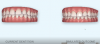
If it is determined that a patient has malalignment and a possible need for orthodontic therapy, an outcome simulator can be used. This simulation is available through various scanners. Although one scanner on the market provides simulation directly, most offer it through a desktop portal that the scans are uploaded to, rather than from the device itself. Through this simulation, a computer-generated proposal is rendered for the doctor and patient to visualize a projected beneficial outcome. This can be done within minutes and is an effective diagnostic, educational, and marketing tool for a clinician. It is important to understand that a simulation is just a proposal; other treatment planning is involved if a patient decides to accept orthodontic treatment (Figure 1).9
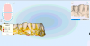
One scanner also has a unique time-lapse feature that enables the clinician to take scans at varying points in time; the scanner will analyze and interpret the data from scans made at two different times for comparison. Again, a similar feature is available through other scanners, but most often through a desktop portal. The scanner then shares both the additive and subtractive changes in time, which makes these changes both visual and measurable to the clinician and patient (Figure 2 and Figure 3).10
Restorative Dentistry
From the outset intraoral scanners have had capabilities for use in restorative procedures. Restorative workflows are perhaps the longest standing in digital and CAD/CAM dentistry. Most scanners have mill connectivity, and the open architecture of the new digital age affords the clinician many options when it comes to buying a scanner and a mill. Today the two pieces of technology often are made by two different manufacturers, in contrast to the previous versions that were manufactured together.
Perhaps the biggest change in intraoral scanning and milling workflows has been the ability to use a scanner and send the scan to the laboratory for the fabrication of a restoration. In today's dental world, this represents a huge shift in the marketplace. Dentists looking to enter into digital dentistry may be interested in a scanner but not necessarily a mill for their practices. Consequently, laboratories have benefitted from the adoption of digital dentistry in private practice. Glidewell Laboratories received more than 40,000 digital impressions in May 2019, compared with just over 1,000 in January 2012.11 This represents tremendous growth in digital scanning over the last decade alone.
In regard to restorations fabricated from digital scans, the capabilities of laboratories are broad. They are fabricating numerous restorations, including crowns, bridges, implant restorations, partial dentures, nightguards, orthodontic appliances, and sleep appliances, among others. By far, the majority of the restorations are zirconia or zirconia based, representing more than 70% of all digital restorations.12
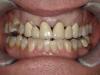
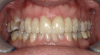
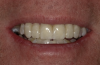
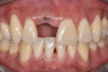
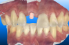
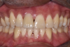
For many years, traditional analog impressions have provided success to clinicians. The efficacy of scanners has been researched and documented, and they have been found to be accurate. It has been determined that for single units, scanners are as good as, if not better than, traditional analog techniques; some discrepancies and inaccuracies can occur on longer spans (more than 4 or 5 teeth) or cross-arch scans.8,13 It should also be understood that digital scanners do not overcome poor impression techniques but rather have the same requirements for making a quality impression as physical impressions. A fundamental understanding of preparation design, isolation, retraction, and moisture control is critical for both digital and analog success (Figure 4 through Figure 7).
Implant Dentistry
The use of an intraoral scanner can also aid in diagnosis, treatment planning, and restoration in the dental implant field. Data captured from an intraoral scan can be used to diagnose and evaluate dentition. The data can then be merged with a cone-beam computed tomography image through implant treatment-planning software. After all the data are combined, the appropriate surgical placement can be determined for the implants. Following this planning, guided implant surgery can be performed. There are many benefits to this process, including a safe and efficacious implant placement, a flapless surgery, and increased potential for immediate loading. Additionally, it provides the clinician with an ideal, prosthetically driven approach to restoring a patient's dentition.14,15
After the implant is placed using the data from the scan, the scanner can also be useful in the restoration for the implant. An image can be captured with a scan body in place. An image with a scan body provides the laboratory with the exact location of the implant abutment interface. The laboratory can then fabricate the type of restoration desired by the clinician. The entire restoration can be fabricated, whether analog or digitally, from this information, depending on the desire of the clinician and material requested (Figure 8 through Figure 10).
Outcomes for implant restorations that incorporate the use of an intraoral scanner have proven positive. Through digital capture and design, the contacts can be more readily controlled. This has resulted in less adjustment at seating both occlusally and interproximally compared with analog methods. In addition, the emergence profile can be created more accurately digitally, resulting in decreased chairtime for the clinician at the seat appointment for digital implant restorations.14
Orthodontics
Another indication for intraoral scanners is in the field of orthodontics. Data from a scan can be useful to the clinician for diagnosis and treatment planning. The data can also be beneficial for patients by allowing them to visualize a proposed orthodontic outcome. The data can be both diagnostic and educational, benefitting any practice that incorporates a scanner. With the use of a simulator function, within minutes after a scan is acquired, a patient can see the benefits of orthodontic therapy and make an informed decision on treatment.
Workflows from scanners also enable the clinician and laboratory to fabricate custom orthodontic appliances, including aligners. With the open connectivity and appropriate software, models and aligners can be fabricated for treatment progression from the initial scan.
Intraoral scanners and incorporated features have increased productivity for both the general dentist and the orthodontist. With certain scanners, the general dentist has the ability to generate 5.92 more clear aligner cases in the first year. The increase in cases can result in an increase in production of more than $30,000 on average.16,17
Preventive Dentistry
Perhaps the most overlooked benefit of intraoral scanners is their preventive capabilities. In dentistry, it could be argued, the main goal is to preserve tooth structure and function. Awareness of the need for prevention in the profession is high.
For prevention, the capabilities of certain scanners are unique. As discussed in the section on diagnostics and patient education, comparing scans from different appointments is invaluable for prevention and patient monitoring. This feature allows the clinician to see correlated data from consecutive scans and make evidence-based treatment decisions for that patient. For example, gingival recession can be monitored with successive scans. A decision to continue to watch the areas of concern or treat them with periodontal procedures to maintain gingival health could be made according to the changes observed. Another example would be monitoring occlusal wear due to bruxism. In this instance, a nightguard may be recommended to prevent further deterioration from the wear. Other changes, such as gingival swelling (possibly due to inflammation) or tooth movement, can be noted. Based on the changes noted in the scans, decisions could be made to treat these concerns.
Finally, most scanners are equipped with an occlusal contact analysis. This tool can be used to determine whether orthodontics would be necessary to treat or whether an occlusal adjustment should be made to equilibrate the patient.
Using scans in a preventive manner should occur in conjunction with other diagnostic measures. Regardless of the indications, the use of the scanner can be highly beneficial to assist the clinician in treatment decisions.
Conclusion
It is evident that digital technology is advancing modern society, including dentistry. Dentists must consider incorporating technology in their practices to stay current. The technological advancements made in the profession have a profound impact on the way that dentists treat patients. Technology provides added benefit not only to productivity, but to quality of care. Education and research will provide clinicians with a better understanding of how technology can work in a specific dental practice.
About the Author
Chad C. Duplantis, DDS
Fellow
Academy of General Dentistry
Private Practice
Fort Worth, Texas
References
1. Wismeijer D. First we replaced the root by an implant, now we need to replace an analogue dental world by a digital one. ACTA Amsterdam. In: Starget 2. 2010;10-16.
2. Mörmann WH, Bindl A. All-ceramic, chair-side computer aided design/computer aided machining restorations. Dent Clin North Am. 2002;46(2):405-426.
3. Milling. Inside Dentistry. 2019;15(7):44.
4. Jayachandran S. Digital imaging in dentistry: a review. Contemp Clin Dent. 2017;8(2):193-194.
5. Intraoral scanning. Inside Dentistry. 2018;14(7):33.
6. Imaging: seeing clearly. Inside Dentistry. 2018;14(7):22.
7. Nelson DL, Reed VS, Walling JR. Pictorial superiority effect. J Exp Psychol Hum Learn. 1976;2(5):523-528.
8. Mangano F, Gandolfi A, Luongo G, Logozzo S. Intraoral scanners in dentistry: a review of the current literature. BMC Oral Health. 2017;17(1):149.
9. iTero. Invisalign integrated solutions. Align Technology. http://www.itero.com/platforms/invisalign_connectivity. Accessed February 6, 2020.
10. iTero. Visualize improved chairside treatment acceptance with iTero Element® TimeLapse technology. Align Technology. http://www.iterotimelapse.com/. Accessed February 6, 2020.
11. Data on file. Glidewell Laboratories.
12. Data on file. Glidewell Laboratories; August 2019.
13. Abduo J, Elseyoufi M. Accuracy of intraoral scanners: a systematic review of influencing factors. Eur J Prosthodont Restor Dent. 2018;26(3):101-121.
14. Lee CYS, Wong N, Ganz SD, et al. Use of an intraoral laser scanner during the prosthetic phase of implant dentistry: a pilot study. J Oral Implantol. 2015;41(4):e126-e132.
15. Colombo M, Mangano C, Mijiritsky E, et al. Clinical applications and effectiveness of guided implant surgery: a critical review based on randomized controlled trials. BMC Oral Health. 2017;17(1):150.
16. Data on file. Align Technology; April 10th, 2018.
17. Mackay MM, Fallah M, Danyal T. Acquisition of a digital intraoral scanning device: an examination of practice volume changes and the economic impact via an interrupted time series analysis. J Clin Dent. 2017;28(suppl):S1-S5.