You must be signed in to read the rest of this article.
Registration on CDEWorld is free. Sign up today!
Forgot your password? Click Here!
Dental agenesis, the failure of a tooth to develop, is a developmental condition with a worldwide prevalence of approximately 5%, and a North American prevalence of approximately 3.9%.1 Reported prevalence levels vary widely, ranging from as low as 0.3% to as high as 36.5%.1 Females are more likely to have dental agenesis than males,1,2 and time trends have also been reported.1 The affected tooth/teeth varies with race/ethnicity as well as locale. For example, European descendants have premolars as the most affected teeth; Japanese descendants have mandibular central incisors as the most affected teeth; and in the United States (U.S.), the maxillary lateral incisors are the most affected teeth.3
The phenotype of dental agenesis is described with the following terminologies: hypodontia, the absence of 1-5 teeth, excluding the third molars; oligodontia, the absence of 6 or more teeth, excluding the third molars, but not all teeth; and anodontia, the absence of all teeth. Since the manifestation of the phenotype occurs after birth, the use of the terminology "congenitally missing tooth/teeth" (indicating occurrence/presence at birth) has been questioned.2,3
Development of the primary teeth begins at approximately 6 weeks gestation with a thickening of ectodermal tissue into dental lamina and its invagination into neural-crest derived mesenchyme.4 Six connected maxillary placodes and six connected mandibular placodes progress through the bud stage, to the cap stage with the development of an enamel knot by 10-13 weeks gestation.5 If the cap is destined to be a primary central incisor or primary first molar, the dental lamina will grow posteriorly from it (continual dental lamina) to develop a primary lateral incisor bud or primary second molar bud. Permanent molar buds develop from the continual dental lamina from the cap of the primary second molars. Successional dental lamina forms from the lingual of the dental lamina near the primary incisors, canines, and molars5 for the eventual development of tooth buds for permanent incisors, canines, and premolars. Genetic and epigenetic influences affect tooth development throughout the stages.3,6-9
Tooth development may be affected by interference in any of the signaling pathways regulating the process. At least 150 syndromes8 and conditions are associated with missing teeth, including ectodermal dysplasias, Rieger syndrome, cleft syndromes,5 taurodontism, and Down.7 Interference can occur if there is a failure to initiate formation, a reduction in the odontogenic potential for the dental lamina, or arrested development during an early stage such that the last tooth in a tooth family fails to develop, explaining the commonly missing second premolars and lateral incisors.5 Factors during pregnancy, such as smoking,10 rubella, maternal diabetes,9 and drugs,7 have been associated with dental agenesis. Additionally, environmental insults from trauma (luxation/avulsion of primary teeth), therapeutic radiation, ingestion of chemicals, prematurity/low birthweight, severe malnutrition, neonatal hypocalcemia, vitamin D deficiency, bilirubinemia, thyroid and parathyroid disturbances, neonatal asphyxia, severe infections, and metabolic disorders have the potential to arrest the development of permanent tooth germs.9
West Virginia is a state with all 55 of its counties included as part of the U.S. Congressionally defined Appalachian region. This 420 county-wide area is served by the Appalachian Regional Commission to increase the overall opportunities and quality of life. West Virginia has many health-related challenges, particularly related to oral health, and current oral health data is limited. Although the 2014 West Virginia Behavioral Risk Factor Surveillance System Report addressed adult oral health need in the area, the oral health needs assessments data concerning children were not available.11 A recent literature search provided one study of oral health needs; however, it was limited to orthodontic need.12
A broader source of current data concerning children in West Virginia is needed for policy makers, funding agencies, professional educational organizations, and oral health professionals to allocate funds, determine the needed size of a dental work force, and target areas of specific needs. An important aspect of oral health is the prevalence of hypodontia/oligodontia/anodontia. Early recognition and treatment planning for dental care is needed for children with hypodontia/oligodontia/anodontia. To the authors' knowledge, there is no such available data on its prevalence in this unique culture. The purpose of this study is to determine the prevalence of permanent tooth hypodontia/oligodontia/anodontia in West Virginia children and to compare the prevalence by sex.
Methods
Study design
A cross-sectional study design was used for this West Virginia University (WVU) Institutional Review Board approved study (Proposal 1709772065). Data were collected from a total of 500 WVU School of Dentistry digital panoramic radiographs of children, ages 6-12 years, taken from August 2, 2010, to September 15, 2017, and captured in the WVU research electronic data capture (REDCap) system.13 The electronic dental chart administrator did not provide a list of electronic dental charts by date of panoramic radiographic imaging, rather by consecutive chart numbers of participants who had a panoramic radiographic code. The list was therefore randomized by date of service and the reviewers divided the list between them. Panoramic radiographs were examined for permanent teeth, or any stage of permanent development from tooth buds onward (excluding third molars), referred to as permanent tooth buds/permanent teeth. Additionally, the presence of retained primary teeth were recorded. The age range (6-12 years) was appropriate for the visualization of the development of permanent tooth buds/permanent teeth on the panoramic radiographs. When a permanent tooth bud/permanent tooth was not visible on a panoramic radiograph, and a primary tooth was present, researchers identified the condition as a retained primary tooth.
The researchers achieved calibration by viewing 10 panoramic radiographs together and had 100% agreement on the presence or absence of permanent tooth buds/permanent teeth on the radiographs. When a researcher had a question about a panoramic radiograph, he or she consulted the other researcher for agreement. The authors also extracted data reported on the electronic dental record concerning age, sex, race/ethnicity, insurance, medications, and recorded American Society of Anesthesiologist status from the medical record (ASA status).
Measures
The key variable of interest was at least one missing permanent tooth bud/permanent tooth versus no missing permanent tooth bud/permanent tooth on a panoramic radiograph (excluding third molars) (yes, no). The variable considered to be associated with at least one missing permanent tooth bud/permanent tooth was sex (male, female). Other variables considered in this study were race/ethnicity (white, black, other, missing data), insurance (Medicaid, private, or none), medications (0, 1, 2, more than 2), and ASA status (1, 2 or 3, missing category).
Statistical methodology
The data were analyzed for the prevalence of at least one missing permanent tooth bud/permanent tooth. Frequency analyses were determined for the examined charts on all of the variables of interest. Chi square analyses were conducted for the bivariate associations of sex and at least one missing permanent tooth bud/permanent tooth, as well as with the other variables. Logistic regression analysis models (unadjusted and adjusted) for the association of sex on at least one missing permanent tooth bud/permanent tooth were developed. Tests were also performed to determine the frequency of the missing permanent tooth bud/permanent tooth most likely to not be present on the panoramic radiograph, and if there were any retained primary teeth. SPSS, version 24 (IBM; Armonk, New York) was used for statistical analyses. An a priori level of significance was set to <0.05.
Results
Variables of interest
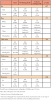
A total of 500 panoramic radiographs and dental records were reviewed (n=500). There were 52.2% female panoramic radiographs examined. A majority of the radiographs were of children who were white, had an ASA of 1, were not taking any medications, and had insurance (Table I). The sample was considered to be reflective of all West Virginia Appalachia children as the West Virginia child population is over 90% white and has access to dental insurance through the Child Health Insurance Program (CHIP), Medicaid, or private insurance.
No child in the sample had anodontia; however, 12.0% (n=60) of the children had at least one missing permanent tooth bud/permanent tooth.
Bivariate analysis on variables versus sex
Bivariate relationships are also presented in Table I. There was a significant association of female sex and at least one missing permanent tooth bud/permanent tooth as compared with males (p=0.027). There were 15.5% of females who had at least one missing permanent tooth bud/permanent tooth as compared to 8.8% of males who had at least one missing permanent tooth bud/permanent tooth. The other variables failed to reach significance.
Logistic regression analyses
In unadjusted logistic regression analyses of sex on at least one missing permanent tooth bud/permanent tooth, females had an odds ratio of 1.91 [95% Confidence Interval: 1.10, 3.32; p = .022] as compared with males (Table II). In adjusted logistic regression analyses of sex on at least one missing permanent tooth bud/permanent tooth, females had an adjusted odds ratio of 2.11 [95% Confidence Interval: 1.18, 3.75; p = .011] as compared with males. Other variables in the analysis failed to reach significance.
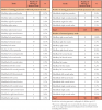
Table III displays the specific missing permanent tooth buds/permanent teeth and specific retained primary teeth. The permanent maxillary left and right lateral incisors had the most missing teeth in the maxilla (14.1%, n=19; n=16, 11.9%, respectively). The permanent mandibular left second molar had the most missing teeth in the mandible (7.4%, n=10). The most common retained primary maxillary tooth was the right second molar (13.8%, n=8) and the most common retained primary mandibular tooth was the left second molar (17.2%, n=10).
Discussion
Early recognition of hypodontia, oligodontia, and anodontia is important for dental practitioners. Such recognition allows for careful assessments and considerations for available treatment options and patient management.14 This is particularly important when patients present with retained primary teeth,16 or have morphological simplifications of their permanent teeth which is commonly associated with hypodontia and oligodontia, including reduced mesiodistal crown lengths, lower/absent cusps or cingula, convergent crowns, and shorter/conical roots.3 Specifically, there are associations of hypodontia and oligodontia and permanent maxillary molars having absent or small cusps of Carabelli, and permanent second molars with only three cusps.3
Current research indicates that, if the root and coronal structures of retained primary teeth are functional and aesthetic, or if aesthetic improvement/restoration/increase in vertical dimension is needed and can be accomplished, it is often beneficial to retain primary teeth as bone and soft tissue is maintained in this circumstance.15 Long-term survival of some primary teeth has been shown to be equivalent to that of implants or other fixed restorations.15 In one study consisting of 20 participants, radiographs were taken at an identified baseline and followed by radiographs taken at a minimum of 5 years from the baseline (and up to 30 years); a total of 28 retained primary mandibular molars without permanent premolars were identified.16 At the end of the study, 86% (n=24) retained primary molars were maintained (mean retention 12.5 years).16 If no retained primary teeth are present, treatment options could include orthodontic treatment, implants, crown and bridge, or partial dental prostheses. In each case, tooth size is important in the treatment planning.17
In this study of hypodontia, oligodontia, and anodontia in permanent teeth of children in West Virginia Appalachia, the prevalence of hypodontia and oligodontia among children ages 6-12 years was 12% (15.5% females; 8.8% males). No child presented with anodontia. This prevalence of hypodontia and oligodontia is higher than reported values for North America (3.9%) and specifically the U.S. (3.6%-5.1%).1 Results of this study indicate that West Virginia Appalachia females are more likely to have hypodontia, and oligodontia. Rolling and Poulsen identified a greater female prevalence in hypodontia, oligodontia, and anodontia in combined studies of Danish school children.18 Brook et al indicated females were 1.5 times more likely than males to have hypodontia.14 Results from this study are consistent with Consolaro et al, in which the authors reported that the most common missing permanent tooth in the U.S. was the maxillary lateral incisor.3
Recent research has been focusing upon genes associated with hypodontia, oligodontia, and anodontia and tooth development in general.5,9,14,17 There are four major signaling pathways in the regulation of tooth development: bone morphogenic proteins (BMPs), fibroblast growth factor (FGFs), sonic hedgehog (SHH), and wingless-related integrated site (WNT) ligands and their receptors.19 The basic genes involved are the homeobox genes, MSX (muscle segment family), DLX (distal less gene), and the PAX (paired box family).19 Tooth development is altered when the genes are mutated.19,20 Epigenetic factors influence genetic expression and may be responsible for higher levels of hypodontia/oligodontia/anodontia in any given population.
Of the many conditions associated with hypodontia/oligodontia/anodontia, perhaps the group of conditions considered the classic example involves ectodermal dysplasias. Ectodermal dysplasias are syndromes in which two or more types of ectodermally derived organs are affected (teeth, hair, sweat glands, etc.). Of the various types of ectodermal dysplasias, hypohidrotic ectodermal dysplasia is considered to be classic, with symptoms involving hypotrichosis (sparse hair), hypohidrosis (reduced ability to sweat), and hypodontia/oligodontia/anodontia.21 The syndrome is inherited as an autosomal dominant, autosomal recessive, or X-linked trait, with the majority of cases being X-linked.21 In a study of ectodermal dysplasias by Thesleff, a novel tumor necrosis factor pathway was discovered involving ectodysplasin, the EDA pathway.22 The EDA regulates ectodermal organ development22 by signaling to its receptor, EDAR, to activate the NF-kappa beta pathway to differentiate epithelium into odontoblasts. The diagnosis of classic hypohidrotic ectodermal dysplasis for males is variations in the genes EDA, EDAR, EDARADD, or WNT10A.22 The diagnosis of classic hypohidrotic ectodermal dysplasis for females is variations in the genes EDAR, EDARADD, or WNT10A.22
Much of the current research of tooth morphogenesis involves learning about the specific genes, signaling pathways, and epigenetic factors. These studies, conducted primarily on mice with various genes associated with tooth development blocked out, are extremely important in understanding the biological details of tooth development. Additional research is also needed to address physiological, psychological, socioeconomic, and ecological factors that influence the oral health quality-of-life of individuals with missing teeth including treatment options. The current study provides information concerning the higher prevalence of hypodontia/oligodontia/anodontia in West Virginia children and serves as a baseline needs assessment.
Limitations and strengths
This study has limitations and strengths. American Dental Association experts created guidelines for radiographs in which panoramic radiographs are recommended to evaluate and monitor dentofacial growth and development, to assess dental and skeletal relationships, and to evaluate craniofacial trauma, based upon clinical judgment.23 Panoramic radiographs reviewed for this study were noted to have been specifically taken to evaluate dentofacial growth and development or assess dental and skeletal relationships. As such, it is possible that the study sample may have had more missing permanent tooth buds/permanent teeth as compared with a general population. There is also the potential bias toward a higher percentage of hypodontia/oligodontia/anodontia due to the populations using a university dental facility. Dentists within the state may be referring children with complex cases to the university pediatric department for care and the population in the university system may be somewhat skewed. However, the sample of panoramic radiographs included over 60% of children who were ASA 1, and were not taking any medications, indicating that the referrals were more likely to be due to the complexity of behavioral issues rather than complex dental procedures.
A strength was the availability of current panoramic radiographs which provided the researchers with a snapshot of current trends. The sample size was also large, providing adequate representation of the area, and the investigators had calibrated to 100% agreement on the presence or absence of permanent teeth or the presence of permanent tooth buds on the radiographs.
Conclusion
In this sample of West Virginia children, females were more likely to have at least one missing tooth bud/permanent tooth than males. Early recognition and treatment planning for dental care is needed for children with hypodontia/oligodontia/anodontia. West Virginia children have a high prevalence of missing permanent tooth buds/permanent teeth as compared with children in the rest of the nation.
Acknowledgements
Research reported in this manuscript was supported by the National Institute of General Medical Sciences of the National Institutes of Health under award number 2U54GM104942-02. The content is solely the responsibility of the author and does not necessarily represent the official views of the National Institutes of Health. The funders had no role in study design, data collection and analysis, decision to publish, or preparation of the manuscript.
Data were collected and managed using REDCap electronic data capture tools hosted at West Virginia University.14
About the Authors
R. Constance Wiener, MA, DMD, PhD is an associate professor, Department of Dental Practice and Rural Health; Christopher Waters, MS is the director of the dental research laboratory, Department of Dental Research; both in the School of Dentistry, West Virginia University, Morgantown, WV.
Corresponding author: R. Constance Wiener, MA, DMD, PhD; rwiener2@hsc.wvu.edu
References
1. Polder BJ, Van't Hof MA, et al. A meta‐analysis of the prevalence of dental agenesis of permanent teeth. Community Dent Oral Epidemiol. 2004 Jun;32(3):217-26.
2. Al-Ani AH, Antoun JS, Thomson WM, et al. Hypodontia: an update on its etiology, classification, and clinical management. BioMed Res Int. 2017 Mar 19; 2017:9378325.
3. Consolaro A, Cardoso MA, Consolaro RB. "Maxillary lateral incisor partial anodontia sequence:" a clinical entity with epigenetic origin. Dental Press J Orthod. 2017 Nov-Dec;22(6):28-34.
4. Tucker A, Sharpe P. The cutting-edge of mammalian development; how the embryo makes teeth. Nat Rev Genet. 2004 Jul;5(7):499.
5. Juuri E, Balic A. The biology underlying abnormalities of tooth number in humans. J Dent Res. 2017 Oct;96(11):1248-56.
6. Dreesen K, Swinnen S, Devriendt K, Carels C. Tooth agenesis patterns and phenotype variation in a cohort of Belgian patients with hypodontia and oligodontia clustered in 79 families with their pedigrees. Eur J Orthodont. 2013 Apr 18;36(1):99-106.
7. Larmour CJ, Mossey PA, Thind BS, et al. Hypodonhia--A retrospective review of prevalence and etiology. Part I. Quintessence Int. 2005 Apr 1;36(4).
8. Yin W, Bian Z. The gene network underlying hypodontia. J Dent Res. 2015 Jul;94(7):878-85.
9. Brook AH. Multilevel complex interactions between genetic, epigenetic and environmental factors in the aetiology of anomalies of dental development. Arch Oral Biol. 2009 Dec 1;54:S3-17.
10. Al-Ani AH, Antoun JS, Thomson WM, Merriman TR, Farella M. Maternal smoking during pregnancy is associated with offspring hypodontia. J Dent Res. 2017 Aug;96(9):1014-9.
11. Yablonsky T. West Virginia behavioral risk factor surveillance system report. Charleston (WV): West Virginia Department of Health and Human Resources; 2014. 197p. Report No.; WV BRFSS 2014 Report.
12. Martin CA, McNeil DW, Crout RJ, et al. Oral health disparities in Appalachia: orthodontic treatment need and demand. J Amer Dent Asso. 2008 May 1;139(5):598-604.
13. Harris PA, Taylor R, Thielke R, et al. Research electronic data capture (REDCap)-a metadata-driven methodology and workflow process for providing translational research informatics support. J Biomed Inform. 2009 Apr 1;42(2):377-81.
14. Brook AH, Jernvall J, Smith RN, et al. The dentition: the outcomes of morphogenesis leading to variations of tooth number, size and shape. Aust Dent J. 2014 Jun;59:131-42.
15. Robinson S, Chan, MF. New teeth from old: treatment options for retained primary teeth. Br Dent J. 2009 Oct;207(7):315.
16. Sletten DW, Smith BM, Southard KA, et al. Retained deciduous mandibular molars in adults: a radiographic study of long-term changes. Am J Orthod Dentofacial Orthop. 2003 Dec 1;124(6):625-30.
17. McKeown HF, Robinson DL, Elcock C, et al. Tooth dimensions in hypodontia patients, their unaffected relatives and a control group measured by a new image analysis system. Eur J Orthod. 2002 Apr 1;24(2):131-41.
18. Rølling S, Poulsen S. Oligodontia in Danish school-children. Acta Odontol Scand. 2001 Jan 1;59(2):111-2.
19. Bei M. Molecular genetics of tooth development. Curr Opin Genet Dev. 2009 Oct 1;19(5):504-10.
20. Vignesh V, Babu NA, Balachander N, Malathi L. Genes in tooth development. Biomed Pharmacol J. Journal. 2015 Oct 1;8(Spec Oct):133-8.
21. Wright JT, Grange DK, Fete M, et al. Hypo-hidroticectodermal dysplasia. GeneReviews [Internet]. 2017 Jun 1[cited 2018 Nov 1] Available from: https://www.ncbi.nlm.nih.gov/pubmed/?term=Wright+JT%2C+Grange+DK%2C+ Fete+M.
22. Thesleff I. The genetic basis of tooth development and dental defects. Am J Medical Genet A. 2006 Dec 1;140(23):2530-5.
23. American Dental Association Council on Scientific Affairs. Dental radiographic examinations: recommendations for patient selection and limiting radiation exposure [Internet]. Chicago: American Dental Association. 2012. [cited 2019 Feb 26] 29p. Available from: https://www.ada.org/~/media/ADA/Publications/ADA%20News/Files/Dental_Radiographic_Examinations_2012.pdf?la=en.