You must be signed in to read the rest of this article.
Registration on CDEWorld is free. Sign up today!
Forgot your password? Click Here!
Dental hygienists do not conduct oral cancer screenings as often as recommended.1 Visual and tactile screenings are the only tests to screen for oral cancer recommended by The Oral Cancer Foundation (OCF), the World Health Organization (WHO), and the National Institute of Dental and Craniofacial Research (NIDCR).2,3 When abnormal lesions are detected, it is recommended they be reevaluated after 2 weeks and then considered for biopsy.3-5 Dental hygienists also need to be aware of the oral cancer screening diagnostic aids and adjunctive techniques that are available; however, these methods are purely an adjunct to oral cancer screenings. This review of the literature discusses the incidence of oral cancer, the significance of early detection, and the types of diagnostic aids and adjunctive techniques available to screen for oral cancer.
Oral cancer is a serious health issue that is globally on the rise.6 The WHO estimates 657,000 new cases of cancer of the oral cavity and pharynx will be diagnosed worldwide each year, and more than 330,000 deaths will occur.7 In the United States, an estimated 53,000 Americans will be diagnosed with oral or oropharyngeal cancer in 2019.6 Oral cancer is twice as likely to occur in male patients as in female patients and usually occurs after the age of 40.6,8 Squamous cell carcinomas account for 95 percent of the malignancies of the oral cavity, with the remainder being adenocarcinomas, adenoid cystic tumors, lymphomas, and melanomas.9
Risk factors include tobacco and alcohol use, exposure to sunlight and x-rays, human papilloma virus (HPV) infections, and poor nutrition.6 Early detection of oral cancer is crucial for patient survival. If not diagnosed in its earliest stages, most oral cancers have a 5-year survival rate of less than 33 percent for blacks versus 55 percent for whites.6 Early detection of oral cancer is the most significant way to improve these survival rates.10 Rates of oral cancer in young adults are on the rise, partially linked to smokeless tobacco use, drug use, and HPV, particularly HPV16.6
Dental hygienists are educated to conduct oral cancer screenings on every patient; however, research has found that less than half of surveyed dental hygienists reported always conducting oral cancer screenings.1 In addition, there are significant knowledge gaps among dental hygienists regarding oral cancer risk factors.
Significance of Early Detection
Detecting oral cancer in its earliest stages increases the 5-year survival rate from less than 30 percent to over 80 percent.11,12 Healthy People 2020 is calling for a 10 to 15 percent reduction in death rates from oral cancer by the year 2020.13 Dental hygienists can play a significant role in detecting early signs of oral cancer and helping to reduce oral cancer death rates; however, they need to be properly educated and trained in screening processes. Delayed detection of oral cancer by dental hygienists can result from the lack of standardized oral cancer examinations, lack of knowledge, and the difficulty of visualizing abnormal tissues in the mouth.14
A variety of diagnostic aids and adjunctive techniques can be used to screen for oral cancer. These tests include the standard screening test, the oral cytology test, vital tissue staining, light-based detection systems and the salivary biomarker test. Each test uses its own unique method to visually screen and/or detect abnormalities in the oral cavity.
Types of Diagnostic Aids and Adjunctive Techniques
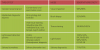
Table I compares the modes, sensitivities and specificities of the types of diagnostic aids and adjunctive techniques described below.
Standard Screening Test
The conventional oral examination is the only recommended test to screen for oral cancer by the OCF, the WHO, and the NIDCR.2,3 The conventional oral examination includes visually inspecting the face, neck, lips, labial mucosa, buccal mucosa, gingiva, floor of the mouth, tongue, and palate, and palpating the regional lymph nodes, tongue, and floor of the mouth.3 Abnormal lesions should be reevaluated after 2 weeks and considered for biopsy.3-5Conventional oral examinations are effective screening methods for melanoma, with sensitivity and specificity as high as 98 percent.15-17 However, they are limited in their ability to detect pre-cancerous or other malignant lesions.15,18-20 Furthermore, they are limited in their ability to detect HPV-related oropharyngeal cancers, since these cancers often originate deep within tissues where they cannot be seen with or without a light.2 A survey found only 66 percent of surveyed dental hygienists conducted conventional oral examinations and only 50 percent performed bimanual neck palpation in their screenings.11,12,21
Established Diagnostic Adjuncts
The oral cytology test, commercially marketed as the OralCDx brush biopsy (BrushTest, CDx, Suffern, New York), uses a brush to collect tissue samples from inside the mouth.22 Samples are sent to a lab and read by a cytopathologist.22 Samples are reported as "negative or benign," "positive," or "atypical."22 When "positive" or "atypical" results are determined, a scalpel biopsy is recommended.22
A study found OralCDx had 100 percent sensitivity and 100 percent specificity with Class I and Class II lesions when positive test results were considered indicative of cancer (see Glossary at the end of this article). This study also reported 92.9 percent specificity with "atypical" or "positive" test results when considered indicative of cancer.15 The study compared 80 patients who had both a brush cytology and a scalpel biopsy and found the brush technique had a sensitivity of 92 percent and a specificity of 94 percent for both "positive" and "atypical" results in detecting dysplasia and oral cancer.15 Research has found the OralCDx brush has a higher sensitivity and specificity in comparison with conventional oral examinations; however, a major limitation of oral cytology tests is that they can detect only abnormal lesions that can be seen by a dental hygienist.23
Vital tissue staining, commercially marketed as toluidine blue staining, methylene blue staining, Rose Bengal staining and Lugol's iodine staining, each use their own dye to stain abnormal tissues in the mouth.22 Abnormal tissues could potentially signify premalignant and malignant lesions, potentially malignant disorders, or both.22,24 Similar to the oral cytology tests, when abnormal tissues are detected, a scalpel biopsy is recommended.22
A study found the sensitivity of toluidine blue staining in detecting high-risk lesions was 94 percent and the specificity was 45 percent. For carcinoma, sensitivity was 100 percent and specificity was 39 percent.25 A systematic review looked at 15 studies that used toluidine blue staining.
Of the 15 studies, there were six studies in which toluidine blue staining was assessed as an adjunctive technique in lower-risk populations. There was one study using toluidine blue staining to survey for mucosal changes in a population with prior treatment for upper aerodigestive cancer with no a priori knowledge of lesions. Investigators in eight studies assessed toluidine blue staining as a diagnostic adjunct in subjects with suspicious lesions or histologically proven dysplasia or cancer in patients at higher risk.22,26-40
This study found the sensitivities varied from 38 to 98 percent, specificities varied from 9 to 93 percent, positive predictive values (PPVs) ranged from 33 to 93 percent, and negative predictive values ranged from 22 to 92 percent.22 According to Messadi, "[The toluidine blue staining] test appears to be highly sensitive (93.5 percent to 97.8 percent) but less specific (73.3 percent to 92.9 percent), mainly because of high false-positive results."10,22,41 Toluidine blue staining does have limitations, including selective staining, which causes some premalignant and malignant lesions to go undetected.25 Furthermore, it has a low specificity rate because it generates false positives.23 As a result of these limitations, a conventional oral examination is recommended prior to all vital tissue staining tests.23
Light-based Detection Systems
ViziLite (Denmat Holdings LLC, Lompoc, California) uses a blue/white light to visualize abnormal cells that have been prepared with acetic acid.22 The patient rinses with acetic acid and the clinician places the ViziLite in the patient's mouth to look for an "acetowhite" change to the tissues.22 An acetowhite change could suggest premalignant and malignant lesions.22 When acetowhite changes are detected, a scalpel biopsy is recommended.22
Two studies using ViziLite found it enhanced the visual parameters of oral lesions in "brightness, sharpness (margin delineation), surface texture and, in some cases, size of lesion compared with results of examination by means of standard illumination" as compared with conventional oral examination.22 In a study using 40 patients with a previous history of oral cancer or premalignancy, ViziLite resulted in a sensitivity of 100 percent and a specificity of 14 percent.15,40 In another study, 501 patients over the age of 40 with a positive history of tobacco use were examined by conventional oral examination. Among the 127 "suspicious" lesions detected, 77 (61 percent) were enhanced by ViziLite examination, while only 21 (5.8 percent) of the 363 "non-suspicious" lesions were ViziLite positive.15,42
The MicroLux DL system (AdDent, Inc., Danbury, Connecticut) uses a "blue-white light-emitting diode (LED)" to detect abnormal tissues.22 The patient rinses with acetic acid, then a clinician places the MicroLux DL system in the patient's mouth to look for any changes in the tissues.22 Tissue changes could suggest premalignant and malignant lesions.22 If tissue changes are found, a scalpel biopsy is recommended.22
A study using 50 patients found that lesion visibility during conventional oral examination ranked an average of 3.28, increasing to 3.66 during Microlux DL examination. This study also determined that the Microlux DL system increased border distinctness from 26 to 35 of the cases.43 Ibrahim et al studied 599 tobacco users and found the sensitivity of the MicroLux DL system to be 94.3 percent and the specificity to be 99.6 percent. This study also found that the MicroLux DL system enhanced detection of lesions, as well as discovered new lesions, as compared with conventional oral examination.44
The VELscope system (LED Dental, Atlanta, Georgia) uses a blue light to look for changes in tissue fluorescence.22 The clinician peers through the device's lens to look for a loss of fluorescence in tissues.22 If a lack of fluorescence is detected, a scalpel biopsy is recommended.22 A total of 44 patients with a history of oral dysplasia or head and neck squamous cell carcinoma were screened by conventional oral examination and then by the VELscope system, which demonstrated a "98 percent sensitivity and a 100 percent specificity for discriminating dysplasia cancers from normal oral mucosa."15,45
A survey was conducted using 30 participants who were either cigarette smokers or cigarette smokers who consumed alcohol. This survey found that there were no statistically significant differences in detecting potentially malignant lesions between using VELscope when compared with conventional oral examinations.14 Research has determined that VELscope has a higher sensitivity (100 percent versus 17 percent) but a lower specificity (74 percent versus 97 percent) when compared with conventional oral examinations.23 Mehrotra et al determined ViziLite and VELscope, in combination with conventional oral examination, were not effective in identifying dysplasia or cancer.46 These false negatives allow for large numbers of premalignant and malignant lesions to go undetected.46
Photodynamic diagnosis uses fluorescence to detect tissue changes in potential premalignant and malignant lesions.24,47 An acid rinse induces fluorescence in tissues in the mouth.24,48 If fluorescence is detected, a scalpel biopsy is recommended.24,49
A meta-analysis of 11 studies found the estimated sensitivity of photodynamic diagnosis was 91 percent and the specificity was 58 percent.24,50 A study looked at 16 patients with neoplastic lesions and determined that fluorescence was detected in the oral mucosa of all patients after the use of an acid rinse.51 Several studies have determined that photodynamic diagnosis has high sensitivity, but limited specificity, as well as a high false-positive rate.24,48,52,53
Light-based detection systems are unable to detect HPV-related oropharyngeal cancers due to the fact "these cancers start deep within the tissues beyond a level that these lights can reach."2
Salivary Biomarkers
Salivary biomarker tests are used to diagnose and predict risk factors for oral potentially malignant disorders.24 Saliva is collected from patients using an oral swab and then sent to a lab for "genomic profiling."54 Genomic profiling includes testing for oral cancer salivary biomarkers including, but not limited to, growth factors, cytokines, and epithelial tumor factors.54 Salivary biomarker testing allows for a noninvasive and cost-effective approach to the diagnosis of oral cancer.46 According to Mehrotra et al, "the six most studied epithelial serum circulatory tumor markers in the saliva of carcinoma patients are Cyfra 21-1, TPS, carcinoembryonic antigen (CEA), SCC, CA125, and CA19-9."46 The presence of Cyfra 21-1, TPS, and CA125 in salivary concentrations was significantly increased and had a sensitivity of 71 percent, a specificity of 75 percent, a negative predictive value of 71 percent, and a positive predictive value of 75 percent.46 Several studies have used salivary proteins as potential diagnostic markers for oral cancer. One study found higher levels of saliva-soluble CD44 in the majority of patients with OSCC.55,56 These higher levels could help differentiate cancer from health with high specificity.55,56
Conclusion
With nearly 657,000 new cases of cancer of the oral cavity and pharynx being diagnosed each year worldwide, and with oral cancer in young adults on the rise, oral cancer is a serious and growing problem.6,7 With early detection, the 5-year survival rate for oral cancer drastically increases from less than 30 percent to over 80 percent.11,12 There are limitations with all the various oral cancer detecting systems available on the market today. While conventional oral examinations have a high sensitivity and specificity, they are limited in their ability to detect premalignant and malignant lesions.15-20 Oral cytology biopsies and vital tissue staining have high sensitivity and specificity, but each has its own major limitations.10,15,22,23,25,41 ViziLite, MicroLux DL, VELscope, and photodynamic diagnosis all show some success in detecting oral cancer lesions; however, a scalpel biopsy is still recommended when abnormal results are detected.22 Salivary biomarker testing is a new technology that shows potential in diagnosing oral cancer.
While each test shows potential in screening for oral cancer, evidence does not support one test over another. Further research is needed to determine a single test for the detection of oral cancer. Until then, it is the professional responsibility of every dental hygienist to conduct thorough conventional oral examinations on every patient and to understand utility and limitations of diagnostic aids and adjunctive techniques.
Glossary
Acetowhite - The areas that stain white after an acetic acid wash is applied are said to have "acetowhite" changes.
Fluorescence - Mucosal tissues have a reflective and absorptive pattern based on naturally occurring fluorophores in the tissue. Exposure to blue light spectra may maximize a differential profile in area undergoing neoplastic change in which a loss of fluorescence visualization is reported.
Sensitivity - The probability that someone who has the target disease (an oral premalignant or malignant lesion [OPML]) will generate a positive result (an OPML as demonstrated by means of gold-standard tissue biopsy).
Specificity - The probability that someone who does not have an OPML will generate a negative test finding.
About the Authors
Heather R. Morse, RDH, BS, is a practicing dental hygienist in Battle Creek, Michigan. She is currently completing her Master of Science in Dental Hygiene degree at the University of Michigan.
Stefanie VanDuine, RDH, MS, graduated from the University of Michigan with a Bachelor of Science in Dental Hygiene degree in 2010 and a Master of Science in Dental Hygiene degree in 2014. She is currently teaching in both the clinical and didactic settings at the University of Michigan.
References
1. Cotter JC, McCann AL, Schneiderman, ED, et al. Factors affecting the performance of oral cancer screenings by Texas dental hygienists. J Dent Hyg. 2011;85(4):326-334.
2. The Oral Cancer Foundation. Newport Beach; 2019: Screening; 2014. Available from: https://oralcancerfoundation.org/screening/. Accessed August 9, 2019.
3. U.S. Preventive Services Task Force. Rockville; 2013: Final recommendation statement: oral cancer: screening; 2013. Available from: https://www.uspreventiveservicestaskforce.org/Page/Document/RecommendationStatementFinal/oral-cancer-screening1. Accessed August 9, 2019.
4. Olson CM, Burda BU, Beil T, et al. Screening for oral cancer: a targeted evidence update for the U.S. Preventive Services Task Force. Evidence Synthesis No. 102. AHRQ Publication No. 13-05186-EF-1. Rockville, MD: Agency for Healthcare Research and Quality; 2013.
5. National Institute of Dental and Craniofacial Research. Bethesda; 2011: Detecting oral cancer: a guide for health care professionals; 2011. Available from: www.nidcr.nih.gov/OralHealth/Topics/OralCancer/DetectingOralCancer.htm. Accessed August 9, 2019.
6. The Oral Cancer Foundation. Newport Beach; 2019: Oral cancer facts; 2019. Available from: https://oralcancerfoundation.org/facts/. Accessed August 9, 2019.
7. World Health Organization. Geneva; 2019: Cancer: oral cancer; 2019. Available from: https://www.who.int/cancer/prevention/diagnosis-screening/oral-cancer/en/. Accessed August 9, 2019.
8. American Cancer Society. Atlanta; 2019: Key statistics for oral cavity and oropharyngeal cancers; 2019. Available from: https://www.cancer.org/cancer/oral-cavity-and-oropharyngeal-cancer/about/key-statistics.html. Accessed August 9, 2019.
9. Cancer Research UK. Oxford: Cancer Research UK; 2014: Mouth and oropharyngeal cancer; 2014. Available at: http://www.cancerresearchuk.org/about-cancer/mouth-cancer/stages-types-grades/types-grades. Accessed August 9, 2019.
10. Messadi DV. Diagnostic aids for detection of oral precancerous conditions. Int J Oral Sci. 2013;5(2):59-65.
11. Barao DMH, Essex G, Lazar AA, et al. Detection of early-stage oral cancer lesions: a survey of California dental hygienists. J Dent Hyg. 2016;90(6):346-53.
12. Forrest J, Horowitz A, Shmuely Y. Dental hygienists' knowledge, opinions, and practices related to oral pharyngeal cancer risk assessment. J Dent Hyg. 2001;75(4):271-81.
13. Weir HK, Thompson TD, Soman A, et al. Meeting the Healthy People 2020 objectives to reduce cancer mortality. Prev Chronic Dis. 2015;2;12:E104.
14. Ayoub HM, Newcomb TL, McCombs GB, et al. The use of fluorescence technology versus visual and tactile examination in the detection of oral lesions: a pilot study. J Dent Hyg. 2015;89(1):63-71.
15. Lingen MW, Kalmar JR, Karrison T, et al. Critical evaluation of diagnostic aids for the detection of oral cancer. Oral Oncol. 2008;44(1):10-22.
16. Whited JD, Grichnik JM. The rational clinical examination. Does this patient have a mole or a melanoma? J Am Med Assoc. 1998;279(9):696-701.
17. Rampen FH, Casparie-vanVelsen JI, van Huystee BE, et al. False-negative findings in skin cancer and melanoma screening. J Am Acad Dermatol. 1995;33(1):59-63.
18. Shugars DC, Patton LL. Detecting, diagnosing, and preventing oral cancer. Nurse Pract. 1997;22(6):105,113-115.
19. Silverman S Jr. Early diagnosis of oral cancer. Cancer. 1988;62(8 suppl):1796-1799.
20. Sandler HC. Cytological screening for early mouth cancer. Cancer. 1962;15:1119-1124.
21. Regezi JA, Sciubba JJ, Jordan RCK. Oral Pathology: Clinical Pathologic Correlations. 5th ed. St. Louis: Saunders Elsevier; 2008:66-68.
22. Patton LL, Epstein JB, Kerr AR. Adjunctive techniques for oral cancer examination and lesion diagnosis: a systematic review of the literature. J Am Dent Assoc. 2008;139(7):896-905.
23. Carreras-Torras C, Gay-Escoda C. Techniques for early diagnosis of oral squamous cell carcinoma: systematic review. Med Oral Patol Oral Cir Bucal. 2015;20(3):e305-315.
24. Liu D, Zhao X, Zeng X, et al. Non-invasive techniques for detection and diagnosis of oral potentially malignant disorders. Tohoku J Exp Med. 2016;238(2):165-177.
25. Chainani-Wu N, Madden E, Cox D, et al. Toluidine blue aids in detection of dysplasia and carcinoma in suspicious oral lesions. Oral Dis. 2015;21(7):879-885.
26. Shedd DP, Hukill PB, Bahn S, et al. Further appraisal of in vivo staining properties of oral cancer. Arch Surg. 1967;95(1):16-22.
27. Vahidy NA, Zaidi SH, Jafarey NA. Toludine blue test for detection of carcinoma of the oral cavity: an evaluation. J Surg Oncol. 1972;4(5):434-438.
28. Mashberg A. Reevaluation of toluidine blue application as a diagnostic adjunct in the detection of asymptomatic oral squamous carcinoma: a continuing prospective study of oral cancer III. Cancer. 1980;46(4):758-763.
29. Mashberg A. Tolonium (toluidine blue) rinse-a screening method for recognition of squamous carcinoma. Continuing study of oral cancer IV. J Am Med Assoc. 1981;245(23):2408-2410.
30. Mashberg A. Final evaluation of tolonium chloride rinse for screening of high-risk patients with asymptomatic squamous carcinoma. J Am Dent Assoc. 1983;106(3):319-323.
31. Warnakulasuriya KA, Johnson NW. Sensitivity and specificity of OraScan® toluidine blue mouthrinse in the detection of oral cancer and precancer. J Oral Pathol Med. 1996;25(3):97-103.
32. Epstein JB, Feldman R, Dolor RJ, et al. The utility of tolonium chloride rinse in the diagnosis of recurrent or second primary cancers in patients with prior upper aerodigestive tract cancer. Head Neck. 2003;25(11):911-921.
33. Myers EN. The toluidine blue test in lesions of the oral cavity. CA Cancer J Clin. 1970;20(3):134-139.
34. Silverman S Jr, Migliorati C, Barbosa J. Toluidine blue staining in the detection of oral precancerous and malignant lesions. Oral Surg Oral Med Oral Pathol. 1984;57(4):379-382.
35. Epstein JB, Scully C, Spinelli J. Toluidine blue and Lugol's iodine application in the assessment of oral malignant disease and lesions at risk of malignancy. J Oral Pathol Med. 1992;21(4):160-163.
36. Epstein JB, Oakley C, Millner A, et al. The utility of toluidine blue application as a diagnostic aid in patients previously treated for upper oropharyngeal carcinoma. Oral Surg Oral Med Oral Pathol Oral Radiol Endod. 1997;83(5):537-547.
37. Onofre MA, Sposto MR, Navarro CM. Reliability of toluidine blue application in the detection of oral epithelial dysplasia and in situ and invasive squamous cell carcinomas. Oral Surg Oral Med Oral Pathol Oral Radiol Endod. 2001;91(5):535-540.
38. Epstein JB, Zhang L, Poh C, et al. Increased allelic loss in toluidine blue-positive oral premalignant lesions. Oral Surg Oral Med Oral Pathol Oral Radiol Endod. 2003;95(1):45-50.
39. Zhang L, Williams M, Poh CF, et al. Toluidine blue staining identifies high-risk primary oral premalignant lesions with poor outcome. Cancer Res. 2005;65(17):8017-8021.
40. Ram S, Siar CH. Chemiluminescence as a diagnostic aid in the detection of oral cancer and potentially malignant epithelial lesions. Int J Oral Maxillofac Surg. 2005;34(5):521-527.
41. Su WW, Yen AM, Chiu SY, et al. A community-based RCT for oral cancer screening with toluidine blue. J Dent Res. 2010;89(9):933-937.
42. Kerr AR, Sirois DA, Epstein JB. Clinical evaluation of chemiluminescent lighting: an adjunct for oral mucosal examinations. J Clin Dent. 2006;17(3):59-63.
43. Mcintosh L, McCullough MJ, Farah CS. The assessment of diffused light illumination and acetic acid rinse (Microlux/DL) in the visualization of oral mucosal lesions. Oral Oncol. 2009;45(12):e227-e231.
44. Ibrahim SS, Al-Attas SA, Darwish ZE, et al. Effectiveness of the Microlux/DL chemiluminescence device in screening of potentially malignant and malignant oral lesions. Asian Pac J Cancer Prev. 2014;15(15):6081-6086.
45. Lane PM, Gilhuly T, Whitehead P, et al. Simple device for the direct visualization of oral-cavity tissue fluorescence. J Biomed Opt. 2006;11(2):024006.
46. Mehrotra R, Gupta DK. Exciting new advances in oral cancer diagnosis: avenues to early detection. Head Neck Oncol. 2011;3:33.
47. Uekusa M, Omura K, Nakajima Y, et al. Uptake and kinetics of 5-aminolevulinic acid in oral squamous cell carcinoma. Int J Oral Maxillofac Surg. 2010;39(8):802-805.
48. Zenk W, Dietel W, Schleier P, et al. Visualizing carcinomas of the mouth cavity by stimulating synthesis of fluorescent protoporphyrin IX. Mund Kiefer Gesichtschir. 1999;3(4):205-209.
49. Driemel O, Kunkel M, Hullmann M, et al. Diagnosis of oral squamous cell carcinoma and its precursor lesions. J Dtsch Dermatol Ges. 2007;5(12):1095-1100.
50. Macey R, Walsh T, Brocklehurst P, et al. Diagnostic tests for oral cancer and potentially malignant disorders in patients presenting with clinically evident lesions. Cochrane Database Syst Rev. 2015;(5):CD010276.
51. Leunig A, Rick K, Stepp H, et al. Fluorescence photodetection of neoplastic lesions in the oral cavity following topical application of 5-aminolevulinic acid. Laryngo Rhino Otol. 1996;75(08):459-464.
52. Leunig A, Betz CS, Mehlmann M, et al. Detection of squamous cell carcinoma of the oral cavity by imaging 5-aminolevulinic acid-induced protoporphyrin IX fluorescence. Laryngoscope. 2000;110(1):78-83.
53. Sharwani A, Jerjes W, Salih V, et al. Assessment of oral premalignancy using elastic scattering spectroscopy. Oral Oncol. 2006;42(4):343-349.
54. Malamud D. Saliva as a diagnostic fluid. Dent Clin N Amer. 2011;55(1):159-178.
55. Liu J, Duan Y. Saliva: a potential media for disease diagnostics and monitoring. Oral Oncol. 2012;48(7):569-577.
56. Franzmann EJ, Reategui EP, Pedroso F, et al. Soluble CD44 is a potential marker for the early detection of head and neck cancer. Cancer Epidemiol Biomarkers Prev. 2007;16(7):1348-1355.