You must be signed in to read the rest of this article.
Registration on CDEWorld is free. Sign up today!
Forgot your password? Click Here!
The introduction of digital technology into the world of dentistry has caused a paradigm shift for doctors and patients alike. Clinicians, faced with a wide range of new tools and methods for use in their practices, are in the position of having to select which changes they may need to make to their workflow and services to keep pace with the times and the competition. Patients, too, are experiencing an influx of information about these new technologies that leads to questions about the value of the treatment they receive and how their available options are changing.
With these concerns in mind, clinicians are in search of the facts behind the marketing of new technology. Although there is little doubt that new digital developments cut down on treatment time and laboratory costs, there are questions regarding the accuracy of these new technologies and what effects, if any, they have on the success of treatment outcomes.
The following article explores two of the most ubiquitous recent developments in digital dentistry: digital impressions taken by intraoral scanning, and chairside CAD/CAM design software. The technology behind these processes is examined and explained, and the accuracy and success of the results achieved with CAD/CAM methods are explored through the use of study data and a comparison with traditional methodology.
Impressions: Issues With Traditional Methods
The process of taking detailed impressions of a patient's dentition was one of the earliest developments in the dental industry. As far back as the 1800s, dentists created negative models of the teeth by filling trays with beeswax and similar malleable materials and placing them in the patient's mouth for a predetermined setting time. The model was then filled with plaster casting to create a replica of the dentition.1
Although impression material has advanced beyond the level of beeswax, the traditional analog impression method has remained largely unchanged. So, too, have the problems inherent in the process, which create inaccuracies that can affect the outcome of the treatment. The problematic situations include:
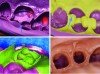
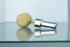
• Issues with the impression material. Most impression materials available today require the clinician to select from various viscosities and to mix the material in a timely manner to create the proper consistency. Errors in mixing are common, leading to improper setting of the material, bubbles, marginal tearing, and other issues (Figure 1).
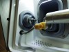
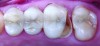
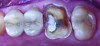
• Lack of proper preparation. Preparation of dentition for the purpose of taking impressions is taught in the earliest stages of dental school (Figure 2). However, according to dental laboratories, preparation is still the step that causes the majority of issues with impressions. Preparation errors include inadequate reduction of the teeth and improper tissue retraction, as well as inadequate removal of saliva or blood from the area. These errors lead to unclear or inaccurate impression results, which can be caused by everything from surface contamination (Figure 3) to failed capture of adequate marginal detail (Figure 4).
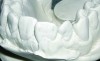
The taking of traditional impressions by the clinician is, of course, only the first step of the analog modeling process. The next step, filling the impression cast with stone to create a model, comes with its own concerns, including stone setting and expansion issues. Any problems with the original impression will also be transferred to the stone model (Figure 5).
These types of issues with traditional impressions, while largely accepted as the nature of the method, are more than just an inconvenience. Remaking a conventional impression doubles the time and discomfort of the patient seated in the chair; remaking a stone model, depending on whether the issues are caught in the practice or at the laboratory, can double the fabrication time of the entire restoration. A survey conducted by Consilium Associates of Irvine, California, found that 36% of dentists must remake impressions at least three or more times per month.2
Digital Impressions: Technology and Performance
Intraoral scanners were first introduced to the commercial market in the 1980s, and digital impressions have been fairly widespread in the industry for the past 10 to 15 years.3 Nonetheless, not all clinicians are familiar with the technology, and many have questions about the ease of taking full and accurate impressions with an unfamiliar method. A basic understanding of how intraoral scanners function is useful for the clinician questioning whether to adopt it into his or her practice.
Intraoral scanners consist of a handheld camera with attached computer and imaging software. The camera, typically in the form of a small wand, is inserted into the patient's mouth. Similar to other 3-dimensional (3D) scanners, the camera projects a light source, sometimes in the form of a laser, while sensors capture the resulting image and transmit it automatically to the computer and imaging software. The software, in turn, processes and compiles the image data from multiple views and then uses a triangulation process to generate a 3D image of the dentition.
Study data comparing the results of digital impressions with analog methods provide immediate information on the benefits of the use of intraoral scanners, including dramatic increases in accuracy:
• A study by Alikhasi et al in 2018 compared conventional impressions (direct and indirect) to digital impressions of a maxillary full arch with tilted implants of two connection types. Digital impressions provided the most accurate results in comparison with both direct and indirect conventional impressions, with minimal linear and angular distortion in all cases.4
• A 2010 study by Syrek et al comparing the fit of all-ceramic crowns made from digital impressions to that of crowns created from conventional impressions found that crowns made using digital impressions had significantly lower median marginal gaps and better interproximal fit.5
Any clinician can likely identify some other obvious and immediate benefits to the digital impression process. For example, compared with the use of traditional trays and impression material, digital scanning minimizes patient discomfort by a noticeable margin. In a 2014 study, Yuzbasioglu, Kurt, Turunc, and Bilir found that patients' stress levels were significantly reduced when their impressions were taken using a digital process rather than a conventional one.6
Another advantage to the digital impression process is the reduction in both messiness and room for error on the part of the clinician or assistant. This is largely due to the fact that the digital impression is processed and immediately visible to the clinician while the patient is still in the chair. Any errors or incomplete areas of the scan will be highlighted by the scanner software so that they can be corrected immediately. The need to reseat the patient for a new impression, or the possibility of sending an incomplete or inaccurate impression to the laboratory, is essentially eliminated.
As with conventional impressions, proper preparation of the teeth is a necessity for capturing an accurate digital impression. Lack of proper retraction, an excess of fluid in the area, or improper reductions can cause the same issues for the final outcome as they do when they occur on an analog impression. Intraoral scanner software may include a range of tools for the clinician to use to make spot corrections to the digital models and detailed evaluation of clearance and undercuts. Before submitting to the laboratory or an in-office CAD station, clinicians should fully examine the digital models to ensure that all the necessary data are captured (preparation, margins, contours and contact surfaces of adjacent teeth, opposing dentition, bite registration) and that the preparation and tissue management are adequate.
After the intraoral scan is completed and confirmed, the clinician has a choice of how to proceed depending on equipment and workflow. Many clinicians who have adopted digital scanning into their practices will, at this stage of the process, submit the completed digital impressions to the laboratory for design and fabrication of the restoration. Other clinicians may choose to invest in a chairside CAD/CAM system, in which case the next step will involve the use of digital design software to create a restoration proposal.
Digital Design Software: Creating Proposals Chairside
Chairside CAD/CAM systems, similar to intraoral scanners, are becoming more widespread and common in the dental industry with each passing year. However, many clinicians still hesitate to take the step and invest in a system, in no small part due to concern over lacking the skills required to create an accurate proposal design without the help or education of a laboratory technician.
Digital design software, which represents the "CAD" (computer-aided design) portion of the CAD/CAM designation, includes a learning curve for its users, as any technology does. However, the latest software takes full advantage of new developments in the fields of artificial intelligence (AI) and machine learning to make the process as foolproof as possible and limit the amount of time and training required by the clinician to create accurate proposals.
After the intraoral scan is completed, it can be sent seamlessly from the scanner to the design software. Digital impression files are typically saved in either STL or PLY file format; the type of scanner used will determine whether the files created are open (designed for use across all platforms and equipment) or closed (for use only with specific equipment from a particular manufacturer).
Although features vary between manufacturers, the general workflow to design a single-unit restoration using chairside CAD/CAM software is as follows:
- Mark restoration margins.
If proper preparation guidelines were followed, most design software will complete this step automatically with a high degree of accuracy. Lack of proper retraction may lead to uncertainty on the part of the software algorithm regarding exact margin lines; in these cases, the clinician must use the built-in software tools to drag, adjust, and redraw the margin lines.
- Choose path of insertion.
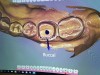
As with the margins, CAD software typically selects the path of insertion automatically based on what it sees within the impression; clinicians may make adjustments as they see fit (Figure 6).
- Generate design proposal.
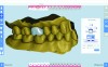
This stage of the process, and its level of accuracy, is determined in part by the specific software the clinician is using. As with other steps of the design process, the clinician is given a set of tools for manual adjustments to the proposal, which can be used as desired to create the best possible outcome (Figure 7).
- Submit proposal for milling.
The completed design proposal, which is used as the template to mill the final restoration, may be either milled chairside using a CAD/CAM system or submitted digitally to the laboratory for fabrication (Figure 8 and Figure 9).
The benefit of time saved by the adoption of a CAD/CAM process is apparent. A clinician can theoretically complete the intraoral scan and design proposal in a single day. Clinicians who own chairside milling systems can complete the fabrication of the restoration itself during one appointment with the patient and deliver it the same day. Even for those clinicians who submit their proposals to an off-site laboratory, significant time is saved by the elimination of the multiple impression steps and waiting for the laboratory to create the design before fabrication.
The question remains, however, whether this type of workflow leads to less-accurate final restorations, particularly among clinicians new to the design process who may compare their own level of knowledge unfavorably with that of the tried-and-true laboratory fabrication team. The answer to that question lies within the technology at work within CAD software. Although the specific features of the software will vary based on manufacturer and type, the majority of available versions rely on some form of AI technology to create proposals. The AI process functions as follows:
• The intraoral scan is analyzed. Specific details of the particular case, ranging from the tooth being replaced and its size and shape to the condition and location of surrounding dentition and tissue, are evaluated by the software to determine the exact needs of the restoration design.
• Data are drawn from outside sources. The most variation between different software types is found within this step. In some cases, proposal software relies on a single standard set of library, or template, tooth designs to create a proposed design for the restoration. Other software uses more advanced technology to draw on large repositories of data from past cases at dental laboratories, evaluating and combining factors from related cases to create unique proposals based on large pools of information.
• Automatic adjustments are made to suit the case specifics. AI technology involves the use of algorithms, which not only pull and analyze source data, but evaluate data against themselves and create new combinations and conclusions. Dental design software typically combines the details of the intraoral scan with the data drawn from its sources and creates a proposal for the case at hand.
CAD software also includes multiple tools for the clinician to use for the purpose of adjusting the proposed design as desired. For those clinicians concerned about their own skill set, or reluctant to invest time in the design process, the more advanced the AI algorithms at work within their software, the easier and smoother this step is because the proposed design may require little or no adjustment before milling.
Conclusion: Accuracy of CAD/CAM Methods
Regarding the accuracy and success of cases treated using CAD/CAM technology (digital impressions, chairside CAD/CAM design and milling, or both), the ever-growing adoption of these tools by clinicians is an undeniable piece of evidence, as are the increasing numbers of studies available showing the accurate and successful outcome of restorations created using CAD/CAM technology:
• Seelbach, Brueckel, and Wöstmann (2013) found that fixed prosthetic restorations manufactured using digital impression systems achieved similar levels of accuracy to conventional techniques and that digital techniques can be regarded as a clinical alternative to conventional methods.7
• In 2004, Sjögren, Molin, and van Dijken reported high patient gratification and satisfactory clinical performance of CAD/CAM-generated inlays after a period of 10 years.8
• A study by Almeida e Silva et al in 2014 indicated that restorations fabricated from digital impressions demonstrated better internal fit than comparison restorations fabricated from conventional impressions.9
• Reich and Schierz (2014), in a study of chairside-generated posterior lithium disilicate crowns over a period of 4 years, recorded a competitive failure-free rate of 93% and a complication-free rate of 83%.10
• A 2014 in vitro study by Tidehag, Ottosson, and Sjögren concluded that the pre-cementation gap width of all-ceramic crowns made with CAD/CAM production demonstrated marginal and internal accuracy on the same levels as those of conventionally manufactured red glass-ceramic crowns.11
• Chochlidakis et al found in a 2016 report that digital impression techniques, used to manufacture fixed restorations, provided better marginal and internal fit than conventional techniques.12
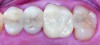
As digital dental technology continues to evolve, a growing number of dental laboratories—and patients—will come to expect the advantages these new methods have to offer. Clinicians today, as they begin the process of incorporating this new technology into their practices, are in an advantageous position: because the technology itself is expanding and developing at such a rapid pace, there is currently no shortage of educational information and research material available to help guide their decisions. This leads to increased confidence in the adoption of these new methods and tools, which in turn leads to excellent case outcomes where digital techniques are put to use. Figure 10 through Figure 12 show a successful digital case study where a fractured all-ceramic restoration on tooth No. 14 was replaced with a fully sintered zirconia crown using a fully digital process, including in-office milling of the restoration.
If the successful outcomes and promising studies cited above are any indication, digital technology will have a lasting positive impact on the dental industry and allow clinicians to offer a wider range of services to their patients at a higher level of quality than ever before.
References
1. Papadiochos I, Papadiochou S, Emmanouil I. The historical evolution of dental impression materials. J Hist Dent. 2017;65(2):79-89.
2. Misch CE. Dental Implant Prosthetics. 2nd edition. St. Louis, MO: Elsevier Mosby; 2015:705.
3. Nayar S, Mahadevan R. A paradigm shift in the concept for making dental impressions. J Pharm Bioallied Sci. 2015;7(suppl 1):S213-S215.
4. Alikhasi M, Siadat H, Nasirpour A, Hasanzade M. Three-dimensional accuracy of digital impression versus conventional method: effect of implant angulation and connection type. Int J Dent. 2018;2018:3761750.
5. Syrek A, Reich G, Ranftl D, et al. Clinical evaluation of all-ceramic crowns fabricated from intraoral digital impressions based on the principle of active wavefront sampling. J Dent. 2010;38(7):553-559.
6. Yuzbasioglu E, Kurt H, Turunc R, Bilir H. Comparison of digital and conventional impression techniques: evaluation of patients' perception, treatment comfort, effectiveness and clinical outcomes. BMC Oral Health. 2014;14:10.
7. Seelbach P, Brueckel C, Wöstmann B. Accuracy of digital and conventional impression techniques and workflow. Clin Oral Investig. 2013;17(7):1759-1764.
8. Sjögren G, Molin M, van Dijken JW. A 10-year prospective evaluation of CAD/CAM-manufactured (Cerec) ceramic inlays cemented with a chemically cured or dual-cured resin composite. Int J Prosthodont. 2004;17(2):241-246.
9. Almeida e Silva JS, Erdelt K, Edelhoff D, et al. Marginal and internal fit of four-unit zirconia fixed dental prostheses based on digital and conventional impression techniques. Clin Oral Investig. 2014;18(2):515-523.
10. Reich S, Schierz O. Chair-side generated posterior lithium disilicate crowns after 4 years. Clin Oral Investig. 2013;17(7):1765-1772.
11. Tidehag P, Ottosson K, Sjögren G. Accuracy of ceramic restorations made using an in-office optical scanning technique: an in vitro study. Oper Dent. 2014;39(3):308-316.
12. Chochlidakis KM, Papaspyridakos P, Geminiani A, et al. Digital versus conventional impressions for fixed prosthodontics: a systematic review and meta-analysis. J Prosthet Dent. 2016(2):184-190.