You must be signed in to read the rest of this article.
Registration on CDEWorld is free. Sign up today!
Forgot your password? Click Here!
One of the most valuable technologies available in dentistry today is the dental operating microscope (DOM). Although many general dentists have historically viewed it as a necessity only for endodontic procedures, the DOM is beginning to gain wider acceptance. Universities have begun incorporating DOMs into programs outside endodontics, and it is becoming more common for undergraduate programs to feature access to DOMs. They provide numerous benefits to doctors, including the flexibility to adjust focal distance without contorting the body. This allows the doctor to remain comfortable in a more ergonomic position. The DOM dramatically increases visibility, with multiple levels of magnification at the turn of a dial. The enhanced visibility offered by the DOM allows shadow-free lighting. Finally, the DOM facilitates communication between the doctor and patient, as well as the doctor and assistant, through digital documentation and imaging capabilities.
The History of Dental Microscopy
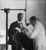
The concept of using microscopes in dentistry is not new. Shirley W. Bowles, DDS, first suggested the use of microscopes in dentistry in 19071 (Figure 1). In his landmark publication, Dr. Bowles wrote:
"The instrument is invaluable in finding exposed pulps without shocking the patient by using broaches and explorers [sic] also, in lifting out partially decalcified dentin, which comes away in a leathery mass from deep cavities, [sic] it will prevent injuring the pulp. When a pulp is nearly exposed, it will quicken our judgment as to whether dentin should be left to recalcify. By using this microscope after a cavity has been prepared, any softened structure, which has been left can be detected, and weakened enamel prisms may be clearly distinguished. It is like proving a mathematical problem, and removes most of the uncertainties of cavity preparation.
"It can be clearly seen whether discoloration around a filling is due to faulty margins and recurring caries or whether it is caused by staining of tooth-structure by the filling material. Imperfect margins can be seen when an explorer passes over them, and small approximal cavities can be found in interdental spaces where it is impossible to pass an instrument, and where a slight separation is necessary to make [sic] sure the presence of a cavity. More than this, in interdental spaces where instruments can pass and detect nothing, the microscope will reveal areas in which the cement substance of enamel rods has been dissolved in the first stage of enamel caries."
Dr. Bowles was therefore ahead of his time in his thinking-not only in regard to the use of microscopes in dentistry, but in medicine in general. Dr. Carl-Olof Nylén, considered by some as the father of microsurgery, first used a surgical operating microscope (SOM) for ear surgery in 1921, and many other surgical specialties in medicine have followed.2 It was not until 1981 that Harvey Apotheker, DMD, and Geza Jako, MD, first introduced the use of surgical microscopes in dentistry.3
In 1992, Gary B. Carr, DDS, published an article emphasizing the importance of the surgical microscope in endodontic procedures.4 In 1994, Dennis A. Shanelec, DDS, and Leonard S. Tibbetts, DDS, published an article describing the importance of using a surgical microscope in periodontal microsurgery.5 In 1999, an article entitled "Microscope-Assisted Precision Dentistry," by Friedman, Mora, and Schmidt, was published in Compendium of Continuing Education in Dentistry; it discussed how an SOM could benefit general practitioners in executing operative/restorative and prosthodontic procedures with greater precision.6 Although slow to gain acceptance during the 20th century, the DOM in the 21st century is increasingly gaining momentum in its adoption in all fields of dentistry and is no longer considered an instrument that is only useful in endodontics.7
Dental Operating Microscopes Versus Loupes
Dentists have long understood the importance of magnification, as evidenced by the widespread popularity of loupes. There are many obvious differences of the DOM from loupes, such as size, cost, portability, multiple levels of magnification, image stability, and the ability to integrate cameras. Less obvious but significant differences include the following:
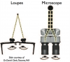
• DOM users have very little eye strain and fatigue compared with loupe users, especially as magnification levels increase. This can be attributed to the difference in optical lens design between the two. The DOM relies on infinity-corrected optics that allow stereoscopic vision, whereas loupes rely on convergent optics8 (Figure 2).
• A DOM offers unobstructed, shadow-free coaxial lighting that is emitted directly from the objective lens parallel to the line of vision. Lighting that is attached to loupes or a headlamp is not coaxial and, therefore, not completely shadow-free.4
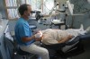
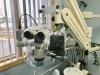
• A DOM enables dentists to operate in an ideal, fully upright postural position (Figure 3), with minimal to no stress or fatigue on the musculoskeletal system, especially the neck and back. Although loupes do help in improving postural ergonomics when compared with unassisted vision, some forward inclination of the head and neck is unavoidable, and the sheer weight of the loupes along with accessory lighting adds additional stressors to the musculoskeletal system.9
Advantages and Benefits of Dental Operating Microscopes
Increased Visualization = Magnification + Illumination
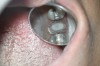
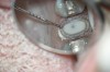
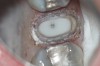
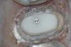
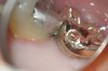
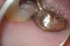
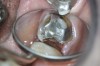
DOMs provide up to six levels of magnification, ranging from 2x to 20x (Figure 4 through Figure 8). Another critical component in increasing visualization is illumination. Most microscopes are equipped with an integrated coaxial light source that allows unobstructed, shadow-free illumination of the operating field. With coaxial illumination, the path of light is directed parallel to the DOM's optical axis, which allows significantly improved visualization of even the most difficult-to-access areas of the oral cavity.8
With enhanced visualization, the clinician's ability to diagnose pathology in the earlier stages of a disease process is possible. Treatments can also be performed with greater precision, thereby reducing the occurrence of failures and the need to redo procedures.10 Enhanced visualization can also allow treatment to be provided more comfortably to the patient because of the clinician's lighter, more refined hand movements, which occur naturally when the clinician is accustomed to operating in a well-illuminated magnified field.4
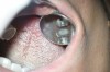
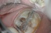
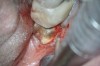
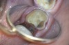
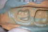
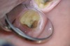
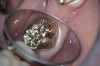
Increased visualization can ultimately result in greater efficiency and productivity because less time is wasted with tactile exploration and checking that all decay has been removed or that a crown margin is fully seated11(Figure 9 and Figure 10). When a clinician can see all areas of the mouth or a tooth perfectly, the level of efficiency and precision in diagnosis and treatment naturally increases. Simply stated, one cannot effectively diagnose or treat what one cannot see.
Communication
Improved communication is perhaps one of the most notable advantages that DOMs offer in comparison with loupes. A significant return on investment can potentially be realized, provided that the DOM is fully integrated and used to its full potential.12 Most DOMs offer options for integrating various types of digital recording devices, such as a digital single-lens reflex camera (DSLR) and/or video camera (Figure 11). Digital documentation capabilities enable the clinician to efficiently capture and share with patients what is seen during an examination preoperatively, during treatment intraoperatively, and postoperatively.12
Images captured during an examination can be used as a communication and education tool in helping patients better understand their diagnostic findings and why certain treatments may be necessary, especially for conditions that produce no obvious symptoms. Allowing a patient to see the images can lead to greater rates of case acceptance and can significantly streamline the amount of time required in gaining it.12
During treatment, images can be efficiently captured, shared, and stored in a patient's digital chart. This is especially useful when unforeseen issues are encountered, such as extensive decay and cracks13 (Figure 12). Images not only help to increase a patient's trust and confidence in the treating doctor, especially for newer patients, but can also aid in reducing one's medico-legal risk.12
Additionally, a live video source can be attached to the microscope and fed to a TV or computer monitor, strategically positioned so the dental assistant can easily view it. When the assistant is able to see exactly what is being performed during a procedure, the assistant's efficiency will increase, and interest and motivation will also rise dramatically because the assistant will tend to feel more involved during the procedure.12
After treatment is completed, a clinician may want to provide patients with preoperative, intraoperative, and postoperative photographs of their treatment to emphasize the results. Giving the photographs to patients may have the added benefit of stimulating referrals of patients' friends and family members. The photographs can be given to patients by either printing or emailing them (Figure 13 through Figure 18).
In addition to improving communication and education for patients, preoperative photographs taken during an oral examination can also be useful in gaining insurance approval for proposed treatment, minimizing or eliminating the need to write narratives.14 Although photographs can also be obtained with an intraoral camera, the quality of the images and the ease in capturing them is more efficient with a DOM.12
Improved Ergonomics
Ergonomic improvement is perhaps the most important benefit of the DOM because it allows the clinician to practice with less pain. The prevalence of neck pain among dentists hovers around 70%.15 Working with a neck posture with only a 20° forward angle has been associated with neck pain.16 By design, DOMs help to facilitate a near neutral head and neck posture.9 This is important because most dentists and hygienists operate with a forward head posture of at least 30° for 85% of their time in the operatory.17
By operating in a more upright, neutral posture position, the operator is less likely to experience fatigue or strain of neck and back muscles and is, therefore, able to work comfortably for extended periods of time. This in turn can enable the practitioner to provide more dentistry in fewer visits, increasing the clinician's productivity and creating happy patients.12
The Learning Curve for Dental Operating Microscopes
Although the learning curve for operating with a DOM may seem challenging and somewhat frustrating at times, especially for the new user, formal training can significantly reduce the time needed to successfully integrate a DOM into a practice.18 Integration time will vary between operators, but after integration is accomplished, the clinician's ability to diagnose problems will dramatically improve.18 Areas of the mouth or a specific tooth that were previously difficult to visualize, even in patients with restricted access, will become easier to see and operate in. A patient's trust level and acceptance of treatment recommendations will likely increase if the DOM is equipped with digital documentation capabilities. Finally, and perhaps most importantly, the clinician will be operating in a comfortable, upright postural position, executing procedures at a level of precision that may have been difficult to imagine previously.
As explained in this article, the attributes of the DOM will ultimately have a significant impact on a clinician's productivity and profitability.12 However, the clinician must realize that improvement does not happen overnight, and that the essential ingredients for integrating any new procedure or technology are patience, practice, and persistence.18
About the Author
Donato (Dino) Napoletano, DMD, graduated from Boston University School of Dental Medicine in 1987 and has been in private practice in his hometown of Middletown, New York, since 1988. He also helps dentists across the country integrate various technologies into their practice.
References
1. Bowles SW. A new adaptation of the microscope to dentistry. In: Kirk EC, ed. The Dental Cosmos: A Monthly Record of Dental Science. Philadelphia, PA: SS White Dental Manufacturing Co.; 1907;49:358-362.
2. Dohlman GF. Carl Olof Nylén and the birth of the otomicroscope and microsurgery. Arch Otolaryngol. 1969;90(6):813-817.
3. Apotheker H, Jako GJ. A microscope for use in dentistry. J Microsurg. 1981;3(1):7-10.
4. Carr GB. Microscopes in endodontics. J Calif Dent Assoc. 1992;20(11):55-61.
5. Tibbetts LS, Shanelec DA. An overview of periodontal microsurgery. Curr Opin Periodontol. 1994:187-193.
6. Friedman M, Mora AF, Schmidt R. Microscope-assisted precision dentistry. Compend Contin Educ Dent. 1999;20(8):723-728.
7. Selden HS. The dental-operating microscope and its slow acceptance. J Endod. 2002;28(3):206-207.
8. Carr GB, Murgel CA. The use of the operating microscope in endodontics. Dent Clin North Am. 2010;54(2):191-214.
9. Valachi B. Magnification in dentistry: how ergonomics features impact your health. Dent Today. 2009;28(4):132-137.
10. Das UK, Das S. Dental operating microscope in endodontics-a review. IOSR J Dent Med Sci. 2013;5(6):1-8.
11. Mamoun J, Napoletano D. Using microscopes in fixed prosthodontics: try-in, adjustment, and insertion of crowns and bridges. Dent Today. 2014;33(4):86-95.
12. van As G, Napoletano D. The video-enabled dental operating microscope (VEDOM) with integrated digital documentation capability (DDC): luxury or necessity? Dental Economics. https://www.dentaleconomics.com/articles/print/volume-97/issue-5/features/the-video-enabled-dental-operating-microscope-vedom-with-integrated-digital-documentation-capability-ddc-luxury-or-necessity.html. Published May 1, 2007. Accessed March 27, 2019.
13. Mamoun JS, Napoletano D. Cracked tooth diagnosis and treatment: an alternative paradigm. Eur J Dent. 2015;9(2):293-303.
14. van As GA. Digital documentation and the dental operating microscope. Oral Health. 91(pt 12):19-30.
15. Lehto TU, Helenius HY, Alaranta HT. Musculoskeletal symptoms of dentists assessed by a multidisciplinary approach. Community Dent Oral Epidemiol. 1991;19(1):38-44.
16. Ariëns GA, Bongers PM, Douwes M, et al. Are neck flexion, neck rotation, and sitting at work risk factors for neck pain? Results of a prospective cohort study. Occup Environ Med.2001;58(3):200-207.
17. Marklin RW, Cherney K. Working postures of dentists and dental hygienists. J Calif Dent Assoc. 2005;33(2):133-136.
18. Napoletano D. The dental operating microscope: basic guidelines for successful dental practice integration. Dentaltown Magazine. https://www.dentaltown.com/magazine/article/5138/the-dental-operating-microscope-basic-guidelines-for-successful-dental-practice-inte. Published November 2014. Accessed March 27, 2019.