You must be signed in to read the rest of this article.
Registration on CDEWorld is free. Sign up today!
Forgot your password? Click Here!
This article will explain the need for clear aligner therapy (CAT) in general dentistry by covering orthodontic basics for general practitioners. Because of the periodontal-orthodontic connection, general dentists should deploy orthodontic therapy in daily diagnoses and treatment planning. The article will cite current research supporting the assertion of a full-body oral health connection and how the mouth functions as a gateway to the body. Long-term oral health solutions using orthodontics will be addressed; when teeth fit together properly, they last longer. Orthodontics enable general dentists to better deliver comprehensive oral healthcare.
Malocclusion Classifications
A comprehensive diagnosis and recommended treatment ideally should include an orthodontic screening on every patient. Malocclusion can lead to detrimental conditions in the mouth, including periodontal disease and broken teeth. Occlusion can be divided among three dimensions: sagittal, transverse, and vertical. Sagittal refers to class I, II, and III molar and cuspid relationships.1 Transverse describes arch width, shape, and form, and the vertical dimension considers the deep bite, open bite, edge-to-edge bite, and normal bite.2
The sagittal dimension refers to how the molars align with each other. Class I, II, and III are not disease classifications; they specify a molar-canine relationship. Although it is common to assume only class I is healthy, there can be healthy class II and III relationships. Even if cuspids and molars align, a class I could be unhealthy if it still causes trauma to the teeth. An anterior-posterior discrepancy in this case is challenging to correct without the assistance of an orthodontist.
The transverse dimension affects decision-making for prevention and treatment planning modalities on a daily basis because it is a primary means of looking at arch form and width.3 An arch can be categorized as having crowding, spacing, or neither crowding nor spacing. Asking why the arch falls into one particular category and finding the answer ensures the ability to fix occlusion and deliver a healthy, "normal" arch. Different arch shapes provide information about occlusion.4,5
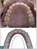
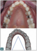
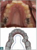
The "U" arch shape is considered healthy (Figure 1). There is plenty of room for the tongue to fit in the palate as well as space for the teeth. Then there is the improper "V" shape, where crowding is common (Figure 2). This arch becomes narrow on the palate, with crowding or flaring of teeth, especially in the interior. There is also the improper "omega" shape, often seen with premolar extractions for orthodontics or when there is lingual inclination (tilting in) of the premolars (Figure 3).6 A very high and narrow palatal vault is common with the omega shape. Sleep apnea is also associated with it.7
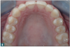
Arch width has an impact on oral health, although the significance and role of asymmetry amid the different classes of arch are still unclear.8 A normal, healthy arch width is approximately 36 mm from first molar to first molar at the gingival level9 (Figure 4). As this width decreases incrementally with a narrower arch shape, crowding increases correspondingly. A simple way to measure is to place a cotton roll in the upper arch from first molar to first molar. If the cotton roll fits from first molar to first molar at the level of the gingiva, then on average there is enough room for teeth to be fit properly. If the roll does not fit into the arch, then the arch is considered narrow. This could be corrected by expansion with clear aligners or interproximal reduction. Expansion is preferred because it causes the least damage to teeth and creates a natural arch width, compared with extraction or interproximal reduction. Digital programs used in CAT have a grid that allows the clinician to measure arch size and determine during treatment planning if there is enough room for proposed interventions.
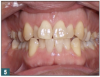
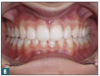
A main factor in occlusion relating to the transverse dimension is buccolingual inclination, which can reveal tooth misalignment, often measured with cone-beam computed tomography (CBCT).10,11 Misalignment is dangerous because it can result in teeth sliding or colliding. There can be loss of hard tissue, such as bone and enamel, in addition to soft tissue, such as clinical attachment. Alternatively, when the long axis of the tooth properly aligns with the long axis of opposing teeth, the periodontal ligament is able to cushion the biting force.12 In Figure 5, the slanted teeth are an indication of poor buccolingual inclination. Visible in this image are both gum recession and chipping of teeth caused by traumatic horizontal forces. Figure 6 shows proper buccolingual inclination of teeth with no visible chipping or periodontal concerns.
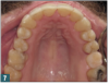
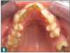
The buccolingual inclination of maxillary and mandibular posterior teeth is assessed using a flat surface extended between the occlusal surfaces of right and left posterior teeth.13 There should be no significant difference between the heights of the buccal and lingual cusps of the maxillary and mandibular premolars and molars, as shown in Figure 7. All tests should be within 1 mm of the straight edge. For the arch in Figure 8, not all cusps would touch, resulting in abfractions and recession caused by poor buccolingual inclination. Clear aligners can be used to control and correct the amount of buccolingual inclination.14
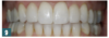
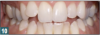
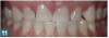
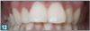
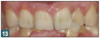
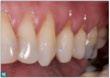
Vertical dimension refers to the amount of open bite or overbite occurring. Figure 9 shows the proper amount of overbite. The teeth align properly instead of collide, and the relationship is functionally stable. When the teeth begin to deviate from this setup, occlusal trauma and periodontal problems may occur. Figure 10 shows an open bite, which can cause difficulty with biting food. Buccolingual inclination is also visible. The edge-to-edge relationship may cause chipping on the interior teeth. Figure 11 shows an edge-to-edge bite, where teeth are shortened. This is a traumatic bite. Figure 12 and Figure 13 show moderate and severe deep bites, respectively, both of which lead to periodontal concerns. With these bites, clinicians often see short teeth because of wear on the anterior.
Why Properly Align Teeth?
Crowding is the most common form of malocclusion. Causes include improper arch form, improper arch width, and buccolingual inclination. The development of malocclusion may have a genetic component.15 A healthy occlusion includes proper arch form, width, and buccolingual inclination; proper amount of overbite and overjet; canine guidance/protection; and posterior intercuspation without interferences. An occlusion with all these features will be healthy, stable, functional, comfortable, and esthetic. When considering orthodontic therapy in a general dentistry setting, it is important to know that malocclusion is considered a disease, which is why all patients should receive an orthodontic screening at their dental examinations.
The World Health Organization (WHO) has two different disease classifications for malocclusion. Classification K07.2 refers to crossbites, open bites, deep bites, and overjet, whereas K07.3 refers to crowding, diastema, spacing, and rotations.16 Adult patients will sometimes say that they do not care about having "straight" teeth, but these classifications go beyond esthetics by indicating how properly aligned teeth impact health. Patients uncertain about orthodontia should be made aware of the consequences of malocclusion, such as improper bite forces, premature wear, fractures, abfractions, gingival recession, and mobility concerns. Figure 14 shows an example of abfractions, attrition, and chipping resulting from malocclusion. Patients with abfractions may believe the manifestations resulted from brushing too hard, but the real cause may be an improper bite. Similarly, patients who have chipped teeth may assume the chipping resulted from eating, but the actual cause may be an edge-to-edge bite and improper overbite.
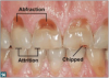
Abfractions are non-carious class V lesions. Rather than resulting from brushing too vigorously, abfractions occur due to tooth flexion from improper forces and occlusal stress when teeth are not aligned correctly.17 Figure 15 illustrates how teeth that align improperly can cause an abfraction. The teeth "hit" at an angle that is not at the proper vertical long axis of the tooth. Lateral (horizontal) forces cause premature wear, chipping, or abfractions by the gum line. They can eventually even lead to tooth loss.18
Oblique or horizontal loading on non-carious cervical lesions results in greater stress concentration than vertical loading. Non-carious cervical lesions with acute angles display higher stress concentrations at the depth of the lesion. If the teeth are properly aligned on the vertical axis, stress fractures will not occur at the gum line. The theory of non-carious cervical lesions suggests that tooth flexure arising from cyclic eccentric occlusal forces causes their formation and progression in vulnerable cervical regions of teeth.19,20
Possible Solutions for Malocclusion
Short-term and long-term solutions are available for conditions caused by malocclusion. Conditions that can be treated include abfractions, anterior incisal fracturing, worn or fractured posterior teeth, and grinding.
For abfractions, a short-term option is a class V buccal fill or gingival graft, but the damage will continue if the affected teeth are kept in the same position. Such interventions will fail if the patient is kept in trauma. A long-term solution would be uprighting teeth to correct the vertical alignment and take teeth out of trauma.21
Incisal fracturing occurs when teeth do not hit properly in edge-to-edge or deep bites. Short-term options for repairing these chips or fractures would be restorative treatments such as veneers, crowns, or occlusal adjustments. However, if the clinician does a filling, crown, or veneer on a patient with interior incisal fracturing due to improper bite, these restorations will likely fail in the future. The long-term solution would be to correct malocclusion by taking the teeth out of trauma and uprighting them.19
For worn or fractured posterior teeth, a nightguard and sometimes crowns are recommended. However, if the fracturing is caused by malocclusion, the long-term solution to prevent further damage would be to correct the malocclusion and provide canine guidance.22
Grinding is usually caused by misalignment of the temporomandibular joint due to malocclusion. Nightguards and occlusal adjustments are short-term options that may take patients out of pain temporarily. However, if the grinding is from malocclusion, patients should be put in proper occlusion with canine guidance to promote pain reduction and relieve trauma to the dentition.23
The Oral-Systemic Connection
On further investigation in regard to abfractions, the short-term solution of a class V buccal composite has been shown to have a low retention rate.24 Onal and Pamir found that after 2 years, one-third of composites in their study had been lost. When loss occurred, patients tended to blame their clinicians for poor-quality composites. Fixing the occlusion can help prevent loss.
Malocclusion also has been shown to increase the risk of periodontal disease. When teeth are crowded, the individual has a decreased ability to clean the teeth properly. Results of a study by Staufer and Landmesser showed an increased risk of periodontal disease as well as an increase in the number of periodontopathogens with malocclusion.25 According to the study, more than 3 mm of crowding can be a host factor for periodontal disease. This amount may seem minor, but the study authors recommend preventive treatment to avoid periodontal concerns. This treatment could include placing the teeth in proper alignment to reduce the amount of crowding.
Another study, by Chung et al, compared the microbial composition of subgingival plaque in crowed versus non-crowded dental regions.26The study showed significantly greater plaque accumulation in crowded areas. It also indicated that the bacteria present in crowded regions consistently represented more species of periodontopathogens. These bacteria are more virulent because of their anaerobic capabilities.
Notably, there is an oral-systemic health link that connects teeth crowding, levels of inflammation, and types of bacteria. Inflammation in the mouth has been linked to many other diseases that are preventable and treatable. Inflammation and bacteria in the mouth have been connected to heart disease,27 diabetes,28chronic obstructive pulmonary disease,29 low birth weight in pre-term babies,30and elevated cholesterol levels.31 Consequently, there are also health benefits to properly aligned teeth. Alignment can decrease bacteria and inflammation, thereby reducing the risk of other oral-systemic problems.
Implementing Clear Aligner Therapy
For the reasons discussed above, general practitioners should discuss orthodontics with patients. According to the American Academy of Periodontology, orthodontia is a basic standard of care.32 An orthodontic problem should be treated with an orthodontic solution. In the long term, it is more effective to treat the cause rather than the symptoms of malocclusion. Restoring teeth that remain in malocclusion will damage the teeth and decrease the lifespan of dental restorations.
CAT is a simple solution for the general practitioner to manage most cases of malocclusion.33The aligners are clear, comfortable, and convenient. This therapy is recommended because it is more esthetic than brackets and wires, and research has shown that clear aligners have less bacterial buildup and gum inflammation.34 The removable aligner does not impede proper brushing and flossing.
Screening for CAT patients involves evaluating for multiple conditions: incisal wear, abfractions, lack of canine guidance, poor buccolingual inclination, crowding or spacing, and orthodontic relapse. The latter usually is signaled by mild to moderate lower anterior crowding.35
It is beneficial to train hygienists and assistants in how to spot these concerns, as well as how to deliver the conversation so that patients can understand the causal relationship between malocclusion and future consequences. Communicating how a solution can be provided is essential to helping patients understand how malocclusion impacts their overall quality of life and how CAT can support their interests.36
General practitioners should in general avoid cases that involve auxiliary appliances, including any case that could need elastics, temporary anchorage devices, or expanders. These cases should be referred to an orthodontist, who will have increased experience and knowledge in proper use of auxiliaries. Aligner-only cases are simple and predictable. Case selection for CAT can be accomplished using decision-making tools provided by the aligner manufacturer. Class I and II are common cases for general practitioners to treat with a predictable outcome, whereas class III should be referred to an orthodontist.
Conclusion
Adhering to the basic rules on which patients should be treated and which should be referred to an orthodontist will enable clinicians to achieve success with CAT and provide healthier dentition. In summation, every patient should be screened for orthodontic conditions as a standard of care and part of a comprehensive examination. Malocclusion can have wide-ranging impacts on patient health; therefore, it is critical to treat the underlying source of poor alignment.
About the Author
Nicole Smith, DDS
Private Practices
Huntington Beach, California
References
1. Batista KB, Thiruvenkatachari BE, Harrison JD, O'Brien KD. Orthodontic treatment for prominent upper front teeth (class II malocclusion) in children and adolescents. Cochrane Database Syst Rev. 2018;3:CD003452.
2. Phulari BS, Naik P. Classification of malocclusions. In: Phulari BS. Orthodontics: Principles and Practice. Jaypee Brothers Medical Publishers; 2017.
3. Banker AM, Pillai JP, Patel KD. Determination of normal maxillary transverse dimension by using intercanine width and interpalatal first molar width. Indian J Dent Res. 2016;27(5):468-472.
4. Slaj M, Spalj S, Pavlin D, et al. Dental archforms in dentoalveolar class I, II and III. Angle Orthod. 2010;80(5):919-924.
5. Zou W, Wu J, Jiang J, et al. Archform comparisons between skeletal class II and III malocclusions. PLoS One. 2014;9(6):e100655.
6. Dahiya G, Masoud AI, Viana G, et al. Effects of unilateral premolar extraction treatment on the dental arch forms of class II subdivision malocclusions. Am J Orthod Dentofacial Orthop. 2017;152(2):232-241.
7. Liu SY, Guilleminault C, Huon LH, Yoon A. Distraction osteogenesis maxillary expansion (DOME) for adult obstructive sleep apnea patients with high arched palate. Otolaryngol Head Neck Surg. 2017;157(2):345-348.
8. Škrinjarić A, Šlaj M, Šlaj M. Fluctuating dental arch asymmetry in different malocclusion groups. Acta Stomatol Croat. 2018;52(2):105-113.
9. McNamara JA. Maxillary transverse deficiency. Am J Orthod Dentofacial Orthop. 2000;117(5):567‑570.
10. Alkhatib R, Chung CH. Buccolingual inclination of first molars in untreated adults: a CBCT study. Angle Orthod. 2017;87(4):598-602.
11. Shewinvanakitkul W, Hans MG, Narendran S, Martin Palomo J. Measuring buccolingual inclination of mandibular canines and first molars using CBCT. Orthod Craniofac Res. 2011;14(3):168-174.
12. Rangarajan V, Padmanabhan TV. Textbook of Prosthodontics. 2017;490.
13. Shu R, Han X, Wang Y, et al. Comparison of arch width, alveolar width and buccolingual inclination of teeth between class II division 1 malocclusion and class I occlusion. Angle Orthod. 2013;83(2):246-252.
14. Sfondrini MF, Gandini P, Castroflorio T, et al. Buccolingual inclination control of upper central incisors of aligners: a comparison with conventional and self-ligating brackets. BioMed Res Int. 2018;2018:9341821.
15. Weaver CA, Miller SF, da Fontoura CS, et al. Candidate gene analyses of 3-dimensional dentoalveolar phenotypes in subjects with malocclusion. Am J Orthod Dentofacial Orthop. 2017;151(3):539-558.
16. World Health Organization. ICD-11: International Classification of Diseases 11th Revision. 2018.
17. Lee WC, Eakle WS. Possible role of tensile stress in the etiology of cervical erosive lesions of teeth. J Prosthet Dent. 1984;52(3):374-380.
18. Michael JA, Townsend GC, Greenwood LF, Kaidonis JA. Abfraction: separating fact from fiction. Aust Dent J. 2009;54(1):2-8.
19. Antonelli JR, Hottel TL, Garcia-Godoy F. Abfraction lesions--where do they come from? A review of the literature. J Tenn Dent Assoc. 2013;93(1):14-19.
20. Soares PV, Santos-Filho PC, Soares CJ, et al. Non-carious cervical lesions: influence of morphology and load type on biomechanical behaviour of maxillary incisors. Aust Dent J. 2013;58(3):306-314.
21. Grippo JO, Simring M, Coleman TA. Abfraction, abrasion, biocorrosion, and the enigma of noncarious cervical lesions: a 20-year perspective. J Esthet Restor Dent. 2012;24(1):10-23.
22. Manns A, Miralles R, Valdivia J, Bull R. Influence of variation in anteroposterior occlusal contacts on electromyographic activity. J Prosthet Dent. 1989;61(5):617-623.
23. Manns A, Chan C, Miralles R. Influence of group function and canine guidance on electromyographic activity of elevator muscles. J Prosthet Dent. 1987;57(4):494-501.
24. Onal B, Pamir T. The two-year clinical performance of esthetic restorative materials in noncarious cervical lesions. J Am Dent Assoc. 2005;136(11):1547-1555.
25. Staufer K, Landmesser H. Effects of crowding in the lower anterior segment-a risk evaluation depending upon the degree of crowding. J Orofac Orthop. 2004;65(1):13-25.
26. Chung CH, Vanarsdall RL, Cavalcanti EA, et al. Comparison of microbial composition in the subgingival plaque of adult crowded versus non-crowded dental regions. Int J Adult Orthodon Orthognath Surg. 2000;15(4):321-330.
27. Oral health and heart disease. Harv Heart Lett. 2001;11(7):1-3.
28. Grossi SG. Dental plaque attack: the connection between periodontal disease, heart disease and diabetes mellitus. Compend Contin Educ Dent. 2001;22(1):13.
29. Healthy gums may lead to healthy lungs. American Academy of Periodontology. https://www.perio.org/consumer/healthy-lungs. Published January 18, 2011. Accessed January 28, 2019.
30. López NJ, Smith PC, Gutierrez J. Periodontal therapy may reduce the risk of pre-term birth weight in women with periodontal disease: a randomized controlled trial. J Periodontol. 2002;73(8):911-924.
31. Katz J, Flugelman MY, Goldberg A, Heft M. Association between periodontal pockets and elevated cholesterol and low density lipoprotein cholesterol levels. J Periodontol. 2002;73(5):494-500.
32. Cobb CM, Carrara A, El-Annan E, et al. Periodontal referral patterns, 1980 versus 2000: a preliminary study. J Periodontol. 2003;74(10):1470-1474.
33. Rossini G, Parrini S, Castroflorio T, et al. Efficacy of clear aligners in controlling orthodontic tooth movement: a systematic review. Angle Orthod. 2015;85(5):881-889.
34. Azeem M, Ul Hamid W. Incidence of white spot lesions during orthodontic clear aligner therapy. JWFO. 2017;6(3):127-130.
35. Kahl-Nieke B, Fischbach H, Schwarze CW. Post-retention crowding and incisor irregularity: a long-term follow-up evaluation of stability and relapse. Br J Orthod. 1995;22(3):249-257.
36. De Abreu MHNG. Some malocclusion traits significantly reduce quality of life among adults. J Evid Based Dent Pract. 2017;17(3):287-289.