You must be signed in to read the rest of this article.
Registration on CDEWorld is free. Sign up today!
Forgot your password? Click Here!
Successful endodontic treatment is predicated on obtaining a pretreatment pulpal and periradicular diagnosis. The pretreatment pulpal and periradicular diagnosis of a tooth begins with a review of the patient's medical and dental history. Pretreatment also includes taking a patient's blood pressure, pulse, and temperature (if indicated). If a patient presents in pain, the etiology of the pain must be identified before any emergency dental treatment is performed. The first step in determining this etiology is listening to the patient's perception of the problem, followed by a dentist's objective clinical testing to reproduce the patient's subjective pain symptoms.
If a patient presents with an asymptomatic dental condition, as often occurs in restorative dentistry, the same objective tests described below must be completed to properly make a pretreatment pulpal and periradicular diagnosis. To arrive at a proper pretreatment pulpal and periradicular diagnosis, clinicians may be uncertain of which test to perform. The following are the five objective clinical tests that a dentist must use to determine the pulpal and periradicular diagnosis.
Clinical Tests
1. Cold Test, EPT, and/or Heat Test for Pulp Sensibility
Pulp sensibility tests (thermal and electric) have been used to indirectly determine the state of pulpal health by assessing the condition of the dental pulp nerves. Pulp vitality, on the other hand, is the direct assessment of pulp blood flow.1 This assessment is obtained with laser doppler flowmetry (LDF) or pulse oximetry (PO). The reason clinicians perform sensibility tests rather than pulpal vitality tests is that LDF and PO applications in dentistry are limited (ie, they have not been designed for specific usage in dentistry).
Heat and cold tests do not jeopardize the health of the pulp.2 Additionally, teeth with porcelain or metal crowns conduct temperature and, therefore, can be tested for pulpal sensibility with cold or heat.3
With an electric pulp test (EPT), the clinician should understand what the numerical readings represent. Although the use an EPT can establish pulp sensibility, the numerical readout should not be used to determine the overall health of the pulp.4 For example, if tooth No. 8 has an EPT reading of 12 and tooth No. 9 has an EPT reading of 24, it does not mean tooth No. 8 is twice as vital as tooth No. 9. The EPT is used to determine whether the pulp is vital. In addition, when using an EPT, the clinician must be aware that teeth with metal restorations can give false-positive or false-negative responses.
Weisleder et al5 reported that the cold test and EPT used in conjunction resulted in a more accurate method for proper pulpal diagnostic testing. In another study, Jespersen et al6 reported that a pulp-testing spray and EPT are accurate and reliable methods for determining pulpal sensibility.
2. Percussion Tests for Determining the Status of the Periodontal Ligament
Percussion tests may be considered mistakenly to directly correlate to a pulp's sensibility. Although a tooth's sensitivity to percussion tests may be due to a pulpitis or pulpal necrosis, they are only indirectly associated. This specific test aids only in determining the status of the periodontal ligament. A bite test may also be necessary if a patient complains about pain while masticating.
3. Palpation of the Buccal and Lingual/Palatal Gingival Tissue of the Tooth
A palpation examination tests for sensitivity of the gingival tissue and for infection or inflammation of the cortical and medullary bone. Even when there is no radiographic evidence of an apical infection, an infection may be present clinically. A study by Bender et al7 reported it is not uncommon to have extensive disease of the bone without evidence on a radiograph.
4. Periodontal Examination Including Periodontal Probing and Tooth Mobility
Periodontal disease can develop anywhere around a tooth; therefore, the entire circumference of the tooth, or teeth, must be probed.
When evaluating tooth mobility, the clinician must remember that movement may be endodontic or periodontal in nature. In the case of periodontal disease, the tooth begins to become mobile and loosens as the attachment apparatus and surrounding bone are destroyed. With an acute endodontic infection, mobility is generally associated with an isolated tooth, but when there is generalized mobility involving multiple teeth, mobility suggests a periodontal origin.
5. Current Radiographic Examination Including Periapical, Bitewings, and/or CBCT
Uraba et al reported that cone-beam computed tomography (CBCT) imaging is effective at detecting approximately 20% more periapical lesions than are periapical radiographs, particularly in the maxillary anterior and posterior teeth.8
When a patient presents for restorative treatment and reports that a tooth is asymptomatic, a dentist may assume that the pulpal and periradicular diagnosis is within normal limits and hence may skip the above objective clinical tests, with the possible exception of taking a radiograph. However, using only a dental radiograph to determine the etiology of tooth pain and the pretreatment pulpal and periradicular status may lead to a pulpal and periradicular misdiagnosis. Therefore, a clinician must perform all five objective tests to obtain an accurate pretreatment pulpal and periradicular diagnosis.
Pulpal Diagnosis
The pulpal nerve fibers, A-delta (which respond to cold and the EPT) and C-fibers (which respond to heat and elicit the nerve response when a patient reports spontaneous tooth pain), are nociceptors. Nociceptors are sensory receptors that respond to stimuli by sending nerve signals to the brain. This stimulus can cause the perception of pain in an individual.9 By objectively testing the pulpal nerve fibers, a dentist can best determine pulpal status. Below are the current pulpal diagnosis terminologies.10
Normal pulp tests within normal limits to cold. Clinically, a patient will respond to a cold stimulus, and after the stimulus is removed, the cold sensation will dissipate immediately. The length of time it takes for a patient to respond to cold has no correlation to the diagnosis and therefore does not need to be recorded.
Reversible pulpitis is pain from an inflamed pulp that can be treated without the removal of the pulp tissue. It is not a disease, but a symptom. Classic clinical symptoms are sharp, quick pain that subsides as soon as the stimulus is removed. Physiologically, it is the A-delta fibers that are firing, not the C-fibers of the pulp.11 A-delta fibers are the myelinated, low-threshold, sharp/pricking pain nerve fibers that reside principally in the pulp-dentin junction. They can be stimulated by cold and the EPT and cannot survive in a hypoxic (low oxygen) environment. Reversible pulpitis also does not involve an unprovoked (spontaneous) response.
Symptomatic irreversible pulpitis is an inflamed pulp that cannot be treated except by the removal of the pulp tissue. Classic clinical symptoms are lingering of cold/hot stimulus greater than 5 seconds and/or patient reporting of spontaneous tooth pain. Physiologically, the A-delta fibers and/or the C-fibers can fire the neural impulses. C-fibers are the unmyelinated, high-threshold, aching-pain nerve fibers. They are distributed throughout the pulp. They are stimulated by heat and can survive in a hypoxic environment.
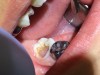
Asymptomatic irreversible pulpitis is a vital pulp that is incapable of healing, and endodontic treatment is consequently indicated. Although asymptomatic irreversible pulpitis is actually a histologic diagnosis to determine the inflammatory extent of the pulp, clinical examples of this diagnosis include a pulp polyp and internal resorption (Figure 1).
Pulpal necrosis can result from an untreated irreversible pulpitis or immediately after a traumatic injury that disrupts the vascular system of the pulp. A necrotic pulp does not respond to cold tests, EPT, or heat tests.
Previously treated: A tooth that has already been endodontically treated.
Previously initiated therapy: Endodontic treatment was started on a tooth but not completed with obturation.
Periradicular Diagnosis
When clinicians perform restorative or endodontic treatment, they do not often obtain a periradicular diagnosis. However, making a periodontal diagnosis is especially helpful when a patient presents in pain. A study by McCarthy et al12 demonstrated that patients presenting with periradicular pain can localize the painful tooth 89% of the time and that patients who present with tooth pain without periradicular pain can localize the tooth only 30% of the time.
By objectively testing the periradicular tissue, a dentist can best determine its gingival and periradicular status. Below are the current periradicular diagnosis terminologies.10
Normal periodontal tissue: Not sensitive to percussion or palpation testing. Also, radiographically, the lamina dura surrounding the root is intact.
Symptomatic apical periodontitis: The tooth has a painful response to biting and/or percussion. This may or may not be accompanied by radiographic periradicular changes.
Asymptomatic apical periodontitis: The tooth has no pain on percussion or palpation. Radiography reveals apical radiolucency.
Chronic apical abscess: Radiography typically reveals a radiolucency. Clinically, there is a sinus tract present on the gingival tissue. The draining sinus tract should be traced with a gutta-percha cone and then confirmed radiographically.
Acute apical abscess is an inflammatory reaction to pulpal infection and necrosis characterized by rapid onset, spontaneous pain, extreme tenderness of the tooth to pressure, pus formation, and swelling of associated tissues. There may be no radiographic signs of destruction, and the patient often experiences malaise, fever, and lymphadenopathy.
Condensing osteitis is a diffuse radiopaque lesion in the periapical region. The opacity represents a localized osseous reaction to a low-grade inflammatory stimulus.
Local Anesthesia
A dentist must obtain profound anesthesia when providing endodontic treatment. A common mistake that clinicians may make when attempting to get a patient "numb" is to not objectively test whether pulpal anesthesia has been achieved before initiating endodontic treatment. Often, the only determination of whether a patient is properly anesthetized is the "subjective" anesthesia level as reported by the patient. Studies have demonstrated that inferior alveolar nerve (IAN) anesthetic blocks administered to patients with mandibular teeth diagnosed with irreversible pulpitis on average had only a 55% incidence of profound pulpal anesthesia, even in the presence of 100% lip numbness as reported by the patient.13,14
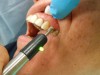
Therefore, before giving local anesthesia for endodontic treatment, the dentist should objectively test the treatment tooth with a cold test and/or EPT. With a preoperative baseline of the pulp sensibility level, after anesthesia is "onboard," the level of anesthesia can be accessed by re-testing the treatment tooth with cold or EPT (Figure 2). If the post-anesthesia tests are either negative to cold or reveal no response to EPT, there is a high likelihood that profound pulpal anesthesia has been achieved. It is important to note that teeth with metal restorations can provide a false-positive result when using the EPT. Additionally, a study by Fuss et al15 reported that in young patients, the EPT was less reliable than cold tests.
Regional and Supplemental Local Anesthesia
Another common clinical mistake that may be made when trying to achieve profound pulpal anesthesia is only giving an infiltration around the treatment tooth. This may be effective for treating a small cavity, but not for endodontic treatment. The dentist should first administer a regional block for local anesthesia. The inferior alveolar nerve block (IANB) is a regional block for the mandibular region; for maxillary teeth, a superior alveolar nerve block (SANB) is used. If profound anesthesia cannot be achieved with a regional block alone (as determined from objective testing), supplemental anesthesia should be administered. Examples of supplemental local anesthesia injections are long buccal nerve blocks (mandibular molars), periodontal ligament, intraosseous, and intrapulpal. If supplemental anesthesia is administered before a regional block, it will either be short-acting or not effective enough to provide pulpal anesthesia. In addition, re-injection of local anesthesia in the same regional or supplemental site has shown an increased success rate in achieving pulpal anesthesia.13
About the Author
James Bahcall, DMD, MS, FICD, FACD
Clinical Associate Professor, Department of Endodontics
University of Illinois-Chicago
College of Dentistry
Chicago, Illinois
References
1. Alghaithy RA, Qualtrough AJ. Pulp sensibility and vitality tests for diagnosing pulpal health in permanent teeth: a critical review. Int Endod J. 2017;50(2):135-142.
2. Rickoff B, Trowbridge H, Baker J, et al. Effects of thermal vitality tests on human dental pulp. J Endod. 1988;14(10):482-485.
3. Miller SO, Johnson JD, Allemang JD, Strother JM. Cold testing through full-coverage restorations. J Endod. 2004;30(10):695-700.
4. Lado EA, Richmond AF, Marks RG. Reliability and validity of a digital pulp tester as a test for measuring sensory perception. J Endod. 1988;14(7):352-356.
5. Weisleder R, Yamauchi S, Caplan DJ, et al. The validity of pulp testing: a clinical study. J Am Dent Assoc. 2009;140(8):1013-1017.
6. Jespersen JJ, Hellstein J, Williamson A, et al. Evaluation of dental pulp sensibility tests in a clinical setting. J Endod. 2014;40(3):351-354.
7. Bender IB, Seltzer S. Roentgenographic and direct observation of experimental lesions in bone: I. 1961. J Endod. 2003;29(11):702-706.
8. Uraba S, Ebihara A, Komatsu K, et al. Ability of cone-beam computed tomography to detect periapical lesions that were not detected by periapical radiography: a retrospective assessment according to tooth group. J Endod. 2016;42(8):1186-1190.
9. Mattscheck D, Law AS, Nixdorf DR. Diagnosis of nonodontogenic toothache. In: Hargreaves KM, Berman LH, Rotstein I, eds. Cohen's Pathways of the Pulp. 11th ed. St. Louis, MO: Mosby Elsevier; 2016.
10. Glickman GN. AAE Consensus Conference on Diagnostic Terminology: background and perspectives. J Endod. 2009;35(12):1619-1620.
11. Kim S. Neurovascular interaction in the dental pulp in health and inflammation. J Endod. 1990;16(2):48-53.
12. McCarthy PJ, McClanahan S, Hodges J, Bowles WR. Frequency of localization of the painful tooth by patients presenting for an endodontic emergency. J Endod. 2010;36(5):801-805.
13. Cohen HP, Cha BY, Spångberg LS. Endodontic anesthesia in mandibular molars: a clinical study. J Endod. 1993;19(7):370-373.
14. Nusstein J, Reader A, Nist R, et al. Anesthetic efficacy of the supplemental intraosseous injection of 2% lidocaine with 1:100,000 epinephrine in irreversible pulpitis. J Endod. 1998;24(7):487-491.
15. Fuss Z, Trowbridge H, Bender IB, et al. Assessment of reliability of electrical and thermal pulp testing agents. J Endod. 1986;12(7):301-305.