You must be signed in to read the rest of this article.
Registration on CDEWorld is free. Sign up today!
Forgot your password? Click Here!
The clinical goal of any restorative treatment should be to maintain dental pulp vitality. The pulp tissue is necessary for tooth nutrition, innervation, and immunocompetency.1 Maintaining the vitality of the dental pulp increases a tooth's mechanical resistance and long-term survival.2 Vital pulp therapy is performed clinically to preserve and maintain the health of the pulpal tissue in cases of pulp exposure due to trauma, caries, or restorative procedures.3 Pulp capping, pulpotomies, and apexogenesis are clinical examples of vital pulp therapy. Over the last several years, vital pulp treatment has changed in regard to procedure and materials used. This CE article will provide the clinician with details regarding these changes in clinical vital pulp therapy.
Traditional Vital Pulp Therapy
For many dental practitioners, the treatment technique used for exposed dental pulps during caries excavation goes back to dental school training. This treatment technique does not include obtaining a pretreatment pulpal and periradicular diagnosis. Therefore, if the pulp is exposed during treatment, the dentist does not have any diagnostic basis concerning the health status of the pulp or periradicular tissue. When the pulp is exposed during treatment while under a rubber dam, calcium-hydroxide usage for pulp-capping has traditionally been the material of choice.4 This procedure is then followed by temporarily restoring the tooth with an intermediate restorative material.5 After this vital pulp treatment, the patient is usually put on 2-week recall for the placement of a permanent restoration if the tooth is asymptomatic.
Current Vital Pulp Therapy
Unfortunately, there are several concerns with the above-stated traditional vital pulp therapy. To address these concerns, in current vital pulp therapy, a pretreatment pulpal and periradicular diagnosis is determined, bioceramic is used as a pulp-capping material, a glass-ionomer is placed over the bioceramic material, and a permanent restoration is placed.
Concern No. 1
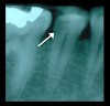
First, it is paramount for the clinician to obtain a pretreatment pulpal and periradicular diagnosis. The value of obtaining a pretreatment pulpal and periradicular diagnosis before performing restorative treatment on a carious vital tooth is that it will allow the clinician to better understand how to treat the dental pulp, especially if it is exposed during caries excavation (Figure 1). A study by Ricucci et al6 found that a clinical pulpal diagnosis of a normal pulp or reversible pulpitis had a 96.6% histologic match to the actual pulp tissue in a tooth.
A normal pulp diagnosis tests within normal limits to cold. Clinically, a patient will respond to cold stimulus, and after this stimulus is removed, the cold sensation will dissipate immediately. A reversible pulpitis diagnosis is pain from an inflamed pulp that can be treated without the removal of the pulp tissue. The diagnosis is not a disease, but a symptom. The classic clinical symptom is sharp, quick pain that subsides as soon as stimulus is removed. Physiologically, it is the A-Delta fibers that are firing, not the C-fibers of the pulp.7 A-Delta fibers are the myelinated, low-threshold, sharp/pricking pain nerve fibers that reside principally in the pulp-dentin junction. They are stimulated by cold and electric pulp testing (EPT) and cannot survive in a hypoxic (low oxygen) environment. Reversible pulpitis also does not involve unprovoked (spontaneous) response. Therefore, with a pretreatment pulpal diagnosis of normal pulp or reversible pulpitis, the treatment of choice is pulp capping or pulpotomy.4
Furthermore, if the pretreatment pulpal diagnosis before a restorative treatment is necrotic or symptomatic/asymptomatic irreversible pulpitis, a pulpectomy is indicated, followed by the placement of a permanent restoration. In teeth with signs and symptoms suggestive of a pretreatment pulpal diagnosis of symptomatic irreversible pulpitis, the pulp has less than a 15.6% chance of reverting to normal through a pulp with a pulp-cap or pulpotomy treatment alone.6
Concern No. 2
Another problem with the traditional procedure is that although for years calcium hydroxide has been the material of choice for vital pulp therapy, it is now contraindicated.8,9 Calcium hydroxide can cause inflammation and necrosis of the pulp surface after pulp capping.4 Calcium hydroxide also has high solubility in oral fluids, will degrade over time, and has a low mechanical resistance.4 Therefore, it should be replaced with a bioceramic material for coverage of the exposed pulp tissue.10 In a randomized clinical trial conducted by Hilton et al,11 it was reported that mineral trioxide aggregate (MTA), a bioceramic material, performed significantly better than calcium hydroxide as a direct pulp-capping agent. Bioceramics or calcium silicate-based materials are biocompatible, nontoxic, nonshrinking, and usually stable within a biologic environment. Further advantages of bioceramic materials are their ability to form calcium hydroxide and hydroxyapatite.12 Many commercial bioceramics are available for vital pulp therapy.
Concern No. 3
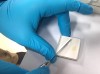
Lastly, after performing a bioceramic pulp cap, the dentist should place a light-cured glass-ionomer material (Figure 2) over the bioceramic pulp cap.13 After this step, a permanent restoration such as a composite or amalgam should be placed. Restoring the tooth with a temporary filling and then placing a permanent filling at a subsequent appointment is not recommended. The reason a permanent restoration should be placed instead of a temporary filling is so that, if the tooth remains asymptomatic after treatment, the clinician does not risk the chance of causing further inflammation of the pulpal and periradicular tissue by replacing the temporary filling with a permanent filling at a subsequent appointment.7 There is always the chance of a patient developing a symptomatic irreversible pulpitis after the pulp-capping procedure, hence requiring a pulpectomy. If this development occurs, having a permanent restoration in place will allow a better access seal compared with a temporary restoration.
In a study by Linu et al,13 it was reported that all pulp-capping failure occurred in the first 2 weeks after treatment. The study also noted that the teeth that were asymptomatic at the 2-week failure point remained asymptomatic, vital, and functional over the follow-up period of 12 to 18 months. Early clinical failures (within 2 weeks) are multifactorial but certainly can be related to an improper pretreatment pulpal diagnosis.2
Summary
Obtaining a pretreatment pulpal and periradicular diagnosis before performing restorative treatment, especially when removing caries, will allow the clinician to make the correct restorative treatment plan in regard to the dental pulp. If this diagnosis is either normal pulp or reversible pulpitis, vital pulp therapy of pulp capping or pulpotomy should be performed. This vital pulp therapy should include using a bioceramic material directly over the pulp, followed by placing a light-cured glass-ionomer over the bioceramic material. Finally, a permanent restoration should be placed instead of a temporary restoration after completion of the vital pulp therapy.
About the Authors
James Bahcall, DMD, MS, FICD, FACD
Clinical Associate Professor, Department of Endodontics
University of Illinois-Chicago
College of Dentistry
Chicago, Illinois
Seema Ashrafi, DDS, MS
Clinical Associate Professor, Department of Periodontics
University of Illinois-Chicago
College of Dentistry
Chicago, Illinois
Qian Xie, DDS, PhD
Assistant Professor, Department of Endodontics
University of Illinois-Chicago
College of Dentistry
Chicago, Illinois
References
1. Zanini M, Meyer E, Simon S. Pulp inflammation diagnosis from clinical to inflammatory mediators: a systematic review. J Endod. 2017;43(7):1033-1051.
2. Reeh ES, Messer HH, Douglas WH. Reduction in tooth stiffness as a result of endodontic and restorative procedures. J Endod. 1989;15(11):512-516.
3. Hilton TJ. Keys to clinical success with pulp capping: a review of the literature. Oper Dent. 2009;34(5):615-625.
4. Brizuela C, Ormeño A, Cabrera C, et al. Direct pulp capping with calcium hydroxide, mineral trioxide aggregate, and biodentine in permanent young teeth with caries: a randomized clinical trial. J Endod. 2017;43(11):1776-1780.
5. Taha N, Khazali M. Partial pulpotomy in mature permanent teeth with clinical signs indicative of irreversible pulpitis: a randomized clinical trial. J Endod. 2017;43(9):1417-1421.
6. Ricucci D, Loghin S, Siqueira JF Jr. Correlation between clinical and histological diagnosis. J Endod. 2014;40(12):1932-1939.
7. Kim S. Neurovascular interactions in the dental pulp in health and inflammation. J Endod. 1990;16(2):48-45.
8. Nowicka A, Wilk G, Lipski M, et al. Tomographic evaluation of reparative dentin formation after direct pulp capping with Ca(OH)2, MTA, biodentine, and dentin bonding system in human teeth. J Endod. 2015; 41(8):1234-1240.
9. Aeinehchi M, Eslami B, Ghanbariha M, Saffar AS. Mineral trioxide aggregate (MTA) and calcium hydroxide as pulp-capping agents in human teeth: a preliminary report. Int Endod J. 2003; 36(3):225-231.
10. Li Z, Cao L, Fan M, Xu Q. Direct pulp capping with calcium hydroxide or mineral trioxide aggregate: a meta-analysis. J Endod. 2015;41(9):1412-1417.
11. Hilton J, Ferracane JL, Manci L, et al. Comparison of CaOH with MTA for direct pulp capping: a PBRN randomized clinical trial. J Dent Res. 2013;92(7 suppl):16s-22s.
12. Wang Z. Bioceramic materials in endodontics. Endod Topics. 2015;32(1):3-30.
13. Linu S, Lekshmi MS, Varunkumar VS, Sam Joseph VG. Treatment outcome following direct pulp capping using bioceramic materials in mature permanent teeth with carious exposure: a pilot retrospective study. J Endod. 2017;43(10):1635-1639.