You must be signed in to read the rest of this article.
Registration on CDEWorld is free. Sign up today!
Forgot your password? Click Here!
Few would argue that dental caries is the most common condition seen in dental practice. In fact, it is the single most common chronic childhood disease worldwide.1 The World Health Organization (WHO) reports that over half of all children aged 5 to 9 have at least one filling or carious lesion.1 More alarming is that caries rates increase with age, jumping to a staggering rate of 78% in 17-year-olds.1
Caries prevention is a professional responsibility. Exactly how dental hygienists approach this with patients can depend on challenges that include patients’ financial limitations, dentists’ preferred treatment methods, and services covered by insurance companies. This article explores both dental sealants and preventive resin restorations (PRRs) as treatment options. Evidence provided includes treatment indications, types of materials, retention rates, and insurance reimbursement.
Sealants and PRRs
Whether to treat teeth with a dental sealant or PRR depends on accurate assessment of the presence and extent of decay. The most widely accepted and scientifically proven interventions for caries prevention are fluoride and dental sealants.2 Although fluoride has been proven to reduce caries rates through tooth structure remineralization, its efficacy is limited to smooth surfaces.3 For caries prevention in pits and fissures, dental sealants have been the “gold standard” since 1955.4 Dental sealants are most often indicated on newly erupted molars that commonly have deep grooves inaccessible to tooth brushing and are more susceptible to decay due to increased plaque bacteria retention.5
Sealants are recommended especially for patients with high to moderate caries risk and when there is no decay or incipient decay present that does not extend into the dentin.5,6 Dental sealants involve placing a resin- or glass-ionomer-based material into the pits and fissures of permanent posterior teeth that adheres to tooth structure by means of a micromechanical bond.3 Sealants are also effective when placed over incipient lesions due to the fluoride release of glass ionomers and some resin materials.5,7-9 This is believed to cut off the nutrient supply to existing microorganisms, preventing further cavitation.7-10
In the 1970s, use of the PRR emerged as a conservative approach to dental amalgam.9 Recently, however, it is sometimes marketed in dental practices as an alternative to dental sealants, with the premise that it has higher retention rates and serves as a means of caries prevention.3,11 PRRs are indicated for those with moderate to high caries risk and active decay that has not extended into the dentin.5,12
Types of Materials
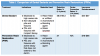
There are two primary classes of sealant materials: filled or unfilled resin-based sealants and glass-ionomer cements (Table 1).7,8,13 Prior to placement of resin-based sealants, acid-etchant is placed to create an environment that will support a micromechanical bond between the sealant and the tooth surface. Bonding agent is then placed to increase resin retention. The resin is then placed and cured either chemically or with a specialized light that polymerizes the material into its hardened form.14
Glass ionomers consist of a basic glass-containing powder combined with an acidic liquid to create a neutral sealant material that releases fluoride into the tooth structure on which it is placed.15 A systematic review by Ahovuo-Saloranta and colleagues found lower retention rates with glass ionomers compared with resin-based materials.13 Although long-term retention may not be achieved with glass-ionomer sealants, they have merit in public health or school settings because the added step of acid-etchant is not required, and they are not as moisture-sensitive as resin-based sealants.11,16,17 These properties allow clinicians to treat a high volume of patients in such settings, and this option is useful as a temporary preventive agent.
Materials used for PRRs, as shown in Table 1, are resin composites.9 After carious lesions are removed from isolated pits and fissures, acid-etchant is applied to the prepared surface, followed by a bonding agent. The resin composite is then placed and light-cured.5 Unlike sealants, glass ionomers are not a consideration for PRRs due to their inferior retention rates.9
Retention and Caries Reduction
Numerous studies have been conducted on sealant retention rates.3,5,8,15,18 The National Institutes of Health (NIH) conducted a 15-year study, finding that unsealed first permanent molars are seven-and-a-half times more likely to develop caries than sealed teeth.4 Results showed a rate of complete sealant retention in 27.6% of sealed teeth after 15 years, with 35.4% of the sealants partially retained. No surfaces of teeth with fully or partially retained sealants experienced caries. Complete sealant loss occurred in 10.9% of the teeth. Of those, 26% required retreatment with a sealant or restoration. It was concluded that sealants are an essential means of preventing future restorative needs.4
The American Academy of Pediatric Dentistry (AAPD) identifies the placement of sealants as more effective in caries reduction than leaving teeth unsealed.7,8 This is based on a Cochrane review that reports a 48% caries reduction rate in sealed first permanent molars.7,13 This significant reduction was enough for AAPD to recommend sealant placement on all newly erupted permanent teeth even if the patient is unlikely to receive follow-up care to monitor sealant retention.7 Furthermore, there is no evidence suggesting that placing sealants over existing non-cavitated lesions leads to further bacterial growth.7,8 In fact, the literature points to a bacterial decrease of 100-fold and a 50% reduction in number of lesions present.7,8
Retention rates of PRRs are less consistent, and fewer studies are available. However, a study by Lekic and colleagues compared retention rates of sealed teeth involving no tooth preparation with two other types of restoration prior to placement of PRRs: minimally prepared (widening of grooves, also known as enameloplasty), and prepared teeth for caries removal.3 The authors found little variance in retention rates of sealants versus the teeth that underwent enameloplasty prior to sealant placement.3 This suggests that widening pits and fissures may not lead to increased sealant retention. Teeth that received PRRs following caries removal yielded significantly lower retention rates and higher rates of caries recurrence.3 These findings are consistent with other studies indicating that removal of tooth structure should be done only to remove active decay, preserving as much tooth structure as possible.9,11,19 A summary of retention and caries reduction rates can be found in Table 1.
AAPD is not in favor of enameloplasty for sealant placement.7 Literature does not provide concrete evidence that this increases retention rates, and, in fact, suggests not only that it increases the risk of recurrent caries, but that it also jeopardizes the tooth’s structural integrity.3-5,7 Walker and colleagues concluded that placement of acid-etchant and bonding agent prior to the sealant material has eliminated the need for enameloplasty altogether.11 Although the PRR is an effective conservative approach to Class I restorations, research does not substantiate its use as a more effective alternative to the dental sealant.
Clinician technique is the most crucial way to maximize sealant success. This is key in extending the life of a sealant, therefore increasing its ability to prevent decay.3,5,7,17,20 When using resin material, a dry field must be meticulously maintained throughout the procedure or retention rates will be compromised.8,11
Dental Insurance
Specific Current Dental Terminology (CDT) codes for procedures are in place to ensure accurate reporting by providers and uniformity across insurance companies. Dental sealant, CDT code D1351, is defined as “a sealant placed on the enamel surface to prevent decay. The enamel surface is noncarious.”6 The CDT code for the PRR (D1352) defines the procedure as “conservative restoration of an active cavitated lesion in a pit or fissure that does not extend into dentin; includes placement of a sealant in any radiating noncarious fissures or pits.”12 According to these definitions, a sealant is not considered a PRR if mechanical preparation or enameloplasty is necessary to improve flow of resin. Active decay must be present to perform a PRR. Essentially, a PRR is used when treatment involves caries but requires a more conservative approach than a Class I restoration.
According to the 2013 ADA Survey of Dental Fees, the average fees charged by dental providers for sealants range from $44 to $57, while the fees associated with PRRs range from $78 to $96 (Table 1).21 In working with patients to obtain insurance coverage for dental services rendered, PRRs may be offered over sealants when sealants are not a covered benefit. However, it is important to base treatment on the evidence rather than insurance coverage parameters.
AAPD recognizes that insurance companies traditionally offer low reimbursement rates for sealants and is committed to raising awareness of their preventive benefits, encouraging insurance plans to change their coverage guidelines.22 Their intent is to increase the acceptance of sealants by making them a more substantially covered benefit, reducing the possibility that non-evidence-based treatment is delivered and reported to the insurance companies.22
Conclusion
Dental sealants are still deemed the most effective means of caries prevention for pits and fissures.2,7-11 Sealants are recommended in non-decayed pits and fissures, or for teeth with incipient lesions.5,6 PRRs are used for actively cavitated lesions that do not extend into the dentin.6,12 Careful radiographic and clinical assessment of the presence and extent of decay are crucial to making an accurate diagnosis to determine which treatment to render and the appropriate CDT code to report. Evidence-based decision-making should be practiced to deliver the highest standard of care, always making the patient’s health and treatment needs top priority.
ACKNOWLEDGMENTS
The author thanks Bridget Beattie-Smith, RDH, MS, and Connie MacKinnon, RDH, MS, for their support in the development of this article.
ABOUT THE AUTHOR
Sabrina Kruger, RDH, BSDH, is a 2016 graduate of the University of Michigan dental hygiene degree completion program. She has been in clinical dental hygiene practice since graduating from the Oakland Community College (OCC) dental hygiene program in 2014. She served as OCC’s American Dental Hygienists’ Association Student Chapter representative from 2012 to 2014. She currently practices in Dexter, Michigan.
Faculty mentors were Anne Gwozdek, RDH, BA, MA, and Iwonka Eagle, RDH, MS. Gwozdek is a Clinical Assistant Professor in the Department of Periodontics and Oral Medicine at the University of Michigan School of Dentistry in Ann Arbor, Michigan. She is the director of the dental hygiene graduate and degree completion programs. Eagle is an Adjunct Clinical Lecturer, also in the Department of Periodontics and Oral Medicine at the University of Michigan School of Dentistry.
REFERENCES
1. World Health Organization. Oral health (fact sheet). April 2012. http://www.who.int/mediacentre/factsheets/fs318/en/. Accessed May 12, 2016.
2. Shulman JD, Cappelli DP. Epidemiology of dental caries. In: Cappelli DP, Mobley CC. Prevention in Clinical Oral Health Care. St. Louis, MO: Mosby Inc.; 2008:2-13.
3. Lekic PC, Deng D, Brothwell D. Clinical evaluation of sealants and preventive resin restorations in a group of environmentally homogenous children. J Dent Child. 2006;73(1):15-19.
4. Simonsen RJ. Retention and effectiveness of dental sealant after 15 years. J Am Dent Assoc. 1991;122(10):34-42.
5. Corona SA, Borsatto MC, Garcia L, et al. Randomized controlled trial comparing the retention of a flowable restorative system with a conventional resin sealant: one-year follow up. Int J Paediatr Dent. 2005;15(1):44-50.
6. Blair C. Eight new codes introduced in CDT 2011-2012. Sidekick. Spring 2011. http://sidekickmag.com/dental-continuing-education/eight-new-codes-introduced-in-cdt-2011-2012/. Accessed May 12, 2016.
7. American Academy of Pediatric Dentistry Clinical Affairs Committee – Restorative Dentistry Subcommittee, American Academy on Pediatric Dentistry Council on Clinical Affairs. Guideline on restorative dentistry. Pediatr Dent. 2014;37(6):232-243.
8. Beauchamp J, Caufield CW, Crall JJ, et al. Evidence-based clinical recommendations for the use of pit-and-fissure sealants: a report of the American Dental Association Council on Scientific Affairs. J Am Dent Assoc. 2008;139(3):257-268.
9. Simonsen RJ. From prevention to therapy: minimal intervention with sealants and resin restorative materials. J Dent. 2011;39 suppl 2:S27-S33.
10. Fontana M, Platt JA, Eckert GJ, et al. Monitoring of sound and carious surfaces under sealants over 44 months. J Dent Res. 2014;93(11):1070-1075.
11. Walker J, Floyd K, Jakobsen J, Pinkham JR. The effectiveness of preventive resin restorations in pediatric patients. ASDC J Dent Child. 1996;63(5):338-340.
12. The Council on Dental Benefit Programs. Optimize your practice: understanding the “CODE.” Chicago, IL: American Dental Association; 2011.
13. Ahovuo-Saloranta A, Forss H, Walsh T, et al. Sealants for preventing dental decay in the permanent teeth (review). Cochrane Database Syst Rev. 2013;4(3):CD001830.
14. Gladwin M, Bagby M. Clinical Aspects of Dental Materials: Theory, Practice, and Cases. 4th ed. Philadelphia, PA: Lippincott, Williams, and Wilkins; 2001:302-308. chap 25.
15. Croll TP, Nicholson JW. Glass ionomer cements in pediatric dentistry: review of the literature. Pediatr Dent. 2002;24(5):423-429.
16. Lindemeyer RG. The use of glass ionomer sealants on newly erupting permanent first molars. J Can Dent Assoc. 2007;73(2):131-134.
17. Hevinga MA, Opdam NJ, Bronkhorst EM, et al. Long-term performance of resin based fissure sealants placed in a general dental practice. J Dent. 2010;38(1):23-28.
18. Simonsen RJ. Preventive restorations and sealants in light of current evidence. Dent Clin N Am. 2005;49(4):815-823.
19. Houpt M, Fuks A, Eidelman E. The preventive resin (composite resin/sealant) restoration: nine-year results. Quintessence Int. 1994;25(3):155-159.
20. Feigal RJ. Sealants and preventive restorations: review of effectiveness and clinical changes for improvement. Pediatr Dent. 1998;20(2):85-92.
21. American Dental Association, Health Policy Institute. 2013 survey of dental fees. Chicago, IL: American Dental Association; 2014.
22. American Academy on Pediatric Dentistry Clinical Affairs Committee — Restorative Dentistry Subcommittee, American Academy on Pediatric Dentistry Council on Clinical Affairs. Policy on third party reimbursement of fees related to dental sealants. Pediatr Dent. 2011;37(6):99-100.