You must be signed in to read the rest of this article.
Registration on CDEWorld is free. Sign up today!
Forgot your password? Click Here!
Non-carious class V lesions are a familiar yet perplexing patient concern. Dentists frequently encounter such lesions and are faced with the challenge of diagnosing and treating them. How these lesions are diagnosed directly affects their treatment and prognoses. What may come as a surprise to many dental practitioners is that non-carious class V lesions are quite possibly being misdiagnosed, interfering with successful (long-term) treatment and prevention of future lesions.
Patients frequently come to the dental office concerned about “notched out” areas near the gumline. In many cases, the first response would be assessing oral hygiene practices for sources of abrasion, including applying too much pressure and/or scrubbing while brushing, using a firm toothbrush, or using too abrasive a dentifrice. If everything in the home care regimen seems correct, the next move is most likely to assess the patient’s diet and oral environment for erosive factors, such as a high sugar or acid diet, the chronic use of lozenges, or acidic saliva. The patient is then sent home with a new tooth brushing method, an extra soft toothbrush, a sample of non-abrasive gel toothpaste and dietary recommendations. Six months later, the patient returns and the clinician notes two more cervical lesions. Is the patient not complying, or are these lesions being cause by something else altogether?
There are patients with these lesions who admittedly hardly ever brush their teeth, thus ruling out toothbrush abrasion. Dentifrices are no longer made from crushed oyster shells, eggshells and bones. Additionally, patients are using soft toothbrushes. Is it really plausible they are still managing to brush away the hardest substance in the human body? Even using firm brushes, is it conceivable that brushing back and forth could cause these lesions, which often affect areas of the teeth notoriously missed when patients brush? Would a toothbrush head be able to cause these lesions, which range from broad and shallow to very narrow, deep and angular? A review of the literature reveals little to no evidence that toothbrushes and dentifrice are capable of causing significant damage to teeth. There are patients with these lesions who have never experienced a cavity and have very low sugar/acid intakes, making diet an unlikely culprit. Finally, these lesions have been found on the roots of extracted teeth with no gingival recession, making abrasion and erosion both virtually impossible. Recent dental research investigating non-carious cervical lesions has resulted in the theory of abfraction, where forces cause flexure and deformation of the teeth, usually at the cervical.1
Characteristics of Non-Carious Class IV lesions
Non-carious class V lesions, or cervical lesions, are losses of tooth structure (enamel and dentin) without the presence of dental caries.2 The exposed dentin is often smooth and shiny. The patient may or may not experience increased sensitivity in these areas. There are two morphological types of these defects: concave and wedge-shaped. Concave lesions are smaller and shallower than wedge-shaped lesions, which are deep, with sharp angles, and result in a greater loss of tooth structure. The concave lesions are generally much less severe than wedge-shaped, and it has been hypothesized that wedge-shaped lesions are caused by more stressful, destructive forces than the concave lesions.2 It is this severe and rapid destruction that has caused many researchers to investigate the potential causes and treatments of cervical lesions.
Wedge-shaped cervical lesions are often found on teeth with prominent occlusal wear.3 Interestingly, these lesions are most frequently found on surfaces opposite the surface with the most severe wear facet/attrition. Literature as early as the 1977 study by Xhonga documented higher prevalence of cervical lesions in bruxers.4 In 1987, Lambrechts et al. also concluded bruxism, in addition to malocclusion, were also seemingly associated with abfractive lesions.5 In agreement was the study by Burke et al. in 1995, in which the authors noted cervical lesions in teeth subjected to lateral forces while the adjacent teeth (not subjected to lateral forces) presented with no lesions.6 Cervical lesions are rarely found on calm, low-stress individuals with ideal (class I) bites. More often, they are found in individuals with malocclusions resulting in mal-aligned cusps subject to heavy and/or oblique occlusal loads.3
A Theory of Physics and Engineering
What if repeated forces, namely lateral and oblique forces, could cause stress points on teeth that eventually break loose? These forces could be due to malocclusion or tongue thrust. At first, the idea of a tongue movement causing tooth breakage may seem unrealistic. An analogy would be a new dining room table with a pedestal base, made of solid and very strong maple wood. Imagine the person sitting at the “head” of the table pushes his hands against the table top to help him stand up from sitting in a chair. This action alone is not likely to break the table. However, if this person does this repeatedly, hundreds or even thousands of times per day (as often as humans swallow), it is plausible this force could cause such stress on the table that it breaks. The table top is analogous to the crown of a tooth, and the pedestal base is analogous to the root. Repeated pressure on the crown, albeit slight and undamaging as a single incident, may accumulate as chronic stress that eventually manifests as fractures.
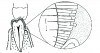
Abfraction is based on principals in physics and engineering. Ideally, occlusal forces are transmitted vertically along the axis of the tooth and absorbed by the periodontium. In the table analogy, this would be similar to a person pushing their bodyweight perfectly downward on the very middle of the dining table where the pedestal base attaches beneath – the force would pass through the pedestal to the floor beneath with no damage done. If forces are applied in a non-axial direction as lateral or oblique pressure, or if the forces are repeated excessively, they may concentrate in the cervical area of the tooth and cause enamel and dentin destruction (Figure 1).
Strains on teeth are concentrated into the cervical region, particularly forces not directed onto the occlusal surface along the axis of the tooth.7 Cervical enamel is inherently weaker, with poorer structure and greater pore volume, and with fewer areas of gnarled enamel (interwoven enamel rods that produce stronger enamel).8 Cervical enamel also has less compressive strength than enamel found on cusps9 and is considerably thinner and more brittle.10
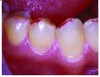
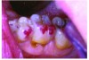
Occlusal loads, particularly lateral forces, may cause the teeth to flex. This flexure may in turn break the bonds in the hydroxyapatite, which leads to cracks and fractures in the enamel and underlying structures. Oblique stresses applied to the cuspal inclines, rather than cusp tips, put more stress on the tooth structure (Figure 2).3 Teeth are better able to withstand direct vertical pressure than lateral or oblique pressures. Heavy occlusal contact areas, determined by heavy markings using pressure-detecting sheets, have been significantly linked to the occurrence of cervical lesions (Figure 3).2
Several studies have revealed the impact chewing has on cervical enamel. Extracted teeth subjected to cycles of occlusal loading began to suffer fractures in the cervical enamel after only 2.5 months’ worth of chewing (200,000 cycles).11 These fractures worsened as more cycles were applied. Networks of microcracks have been noted near the CEJ when on teeth subjected to cyclic occlusal pressures.12 Occlusal forces pass through the teeth into the periodontium, where the supporting structures help to absorb the forces and provide a cushion. Mobile teeth are less prone to cervical concentration of these forces.10 A relationship between mobility and cervical lesions was discovered where less mobile teeth (with more rigid support) exhibited higher rates of cervical lesions.13
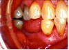
If no heavy attrition, wear facets, or markings on cuspal inclinations are present, pressure from tongue movements may be the culprit (Figure 4 and Figure 5). An ideal swallow involves placing the tip of the tongue on the palate, behind the anterior teeth. As the swallow proceeds, the body of the tongue continues to press upward against the palate, pushing the food towards the throat in a peristaltic manner.14 The tongue should not press against any teeth during the swallowing action. A tongue-thrust swallow, one where the tongue pushes the teeth in order to force the food towards the throat, may be the result of habit, an abnormally large tongue or a congested airway.14 It has been theorized that abnormal swallows in infants may actually cause malocclusions such as narrow arches, crowding and overjets.15 The repeated pressing of the tongue against the tooth acts as a person pushing against the edge of a table when standing up; over time, it may cause enough stress for a fracture to occur. These lesions are most common on anteriors and premolars, the teeth most affected by abnormal tongue movements.14
Abfraction may also serve as a co-contributor to cervical lesions. One possible hypothesis is that non-carious class V lesions could occur when erosive dietary fluids (which affect buccal surfaces more than lingual surfaces) leak into the microscopic cracks in the hydroxyapatite that resulted from occlusal loads concentrated into the cervical areas. A 2004 study examined abfraction in teeth with cervical enamel that has been undermined (the dentine beneath has begun to erode).16 The researchers applied 100 Newtons of oblique load pressure (bruxers may apply as much as 500 Newtons) to intact teeth and teeth with varying degrees of undermined enamel and measured the resulting levels of stress on the buccal enamel near the amelo-dentine junction. Teeth with cervical enamel defects exhibited much higher stress values; the larger the enamel defect, the higher the stress value. Enamel defects as small as 0.34 mm in length resulted in stress values above the known fracture limit. Gingival crevicular fluid has been shown to be acidic and may contribute to enamel undermining.17
In addition to the corrosion-abfraction theory, there are several other types of possible multifactorial causes of cervical lesions.10 Attrition-abfraction may occur in bruxers, where tooth-to-tooth contact causes friction, which combines with the concentration of the stress in the cervical area. In abrasion-abfraction, tooth structure is lost when an external friction (such as partial denture clasp) is applied to an area where occlusal loading has caused stress concentration, making it more susceptible to abrasion. Attrition-corrosion is the result of corrosive agents damaging areas already worn by attrition. Abrasion-corrosion, likewise, combines corrosive chemical exposures with external friction sources. Biocorrosion-abfraction is the damaging of enamel due to a combination of caries and stress concentration.10 To complicate the issue, cervical lesions may involve more than two factors. The clinician needs to keep all of this in mind when trying to identify the cause(s) of the patient’s lesions.
Patient Assessment
As stated earlier, a tongue-thrust swallow results in the tongue pressing against the lingual surfaces of the teeth rather than against the palate. Assessing patients for tongue-thrust is quite simple. While retracting the patient’s cheeks, the clinician should ask the patient to swallow. If any teeth are missing, or if large interproximal spaces are present, the clinician can watch for the tongue to push into these gaps while the patient swallows. If dentition is full and contacts are closed, a tongue-thrust swallow will usually result in saliva and/or bubbles being forced through the interdental spaces.14 The patient may need to swallow more than once for this to be seen and/or heard by the clinician.
The typical occlusal assessment during office exams consists of noting Class I, II and III malocclusions, over/underbite, overjet, crossbite, openbite and end-to-end relationships. These indeed are important factors for many aspects of dental health. However, when evaluating for the potential of abfractive lesions, more detective work is necessary. Articulating paper, occlusal indicator wax, pressure detecting sheets and other occlusal pressure analyzers are useful tools for assessing the risk of abfraction. A healthy occlusion should result in small areas of contact on cusp tips or in occlusal fossae. The clinician should look for large areas of contact, particularly on cuspal inclinations and on buccal and lingual surfaces. Canine guidance is another key element of the occlusal exam. While the patient is in centric occlusion, ask him to very slowly slide their mandible to the right (and then left), until the buccal surfaces of the mandibular and maxillary molars are flush with one another. At this point, only the patient’s canines should be in contact. Similarly, when the patient extrudes their mandible forward, the incisors should first guide the movement, followed by the canines alone being in contact with one another. If non-canine teeth are guiding occlusion movement, they are experiencing more occlusal load than they are designed to handle and therefore may be at risk for periodontal damage, excessive wear and abfraction.
Assessing the patient’s diet and oral environment for sources of erosive elements is also necessary, as erosion and occlusal/tongue forces may serve as co-contributors to cervical lesions. Saliva pH and salivary flow rates can be tested in the office. Interviewing the patient can reveal dietary sources, such as carbonated beverages, fruit juices and alcohol. Foods and beverages high in sugars also need to be identified as contributors to erosion due to the well-known oral “acid attack” that occurs after consuming fermentable carbohydrates. Frequency of consumption will be the key to determining which dietary factors are likely to be contributors. In addition to assessing for corrosion-abfraction, other possible co-contributors should be identified. Areas of severe attrition also need to be noted, as well as abrasion from partial denture clasps, habits such as excessive toothpick use, parafunctional habits or any other finding that may weaken the tooth structure.
Treatment of Abfractions
There are mixed viewpoints about restoring these lesions. Resin-based composites are frequently used to reduce sensitivity and improve esthetics. Many dental practitioners do not restore abfractions for two main reasons: the lesions are non-carious, and they are likely to return if the cause of abfraction has not been addressed. The longevity of resin class V restorations is questionable in patients who experience these lesions due to occlusal stresses. In 2009, Friscisconi et al. investigated the effects of occlusal pressure on resin Class V restorations.18 The researchers created class V lesions in 40 extracted teeth, restored them with resin composites, and then subjected the teeth to 150 Newtons of occlusal pressure on the buccal cusps, lingual cusps and central fossae. The restoration margins were inspected using fluorescein to evaluate defects. Occlusal pressure, regardless of location, contributed to the formation of margin gaps. This occurred on resins of varying sizes and depths. The researchers concluded that occlusal loading must be addressed in patients being treated for abfraction lesions to ensure that resin margin integrity is maintained.
Treating the cause of abfraction may require retraining the individual to swallow correctly. Patients may be referred to speech therapists for help in breaking the tongue thrust habit. Bruxers should be fitted with splints or guards. The clinical work of Robert Palmer, DDS, has demonstrated success with creating equilibrium. Palmer performs fine modification of the contact areas, reducing their size (Figure 6 and Figure 7). Palmer’s patients reported immediate relief of sensitivity, indicating that the sensitivity may be caused by flexure resulting from occlusal load.14 Occlusal therapy may be useful in treating gingival clefts, which Palmer identified as a precursor to abfraction.14,19 In 1983, Solnit and Stambaugh reported that refining contact points on teeth with gingival clefts actually resulted in a partial to complete reversal of the clefts in all 25 cases studied.19 It is Palmer’s standard to refine occlusion for teeth with abfractions that are being restored with resin-based composites to prevent recurrence.14
If co-contributors are identified, they will also need to be addressed as part of the treatment plan. Sources of erosion need to be reduced or eliminated. Salivary replacement products should be recommended for those with xerostomia. Fluoride rinses, gels or varnishes are appropriate for patients who are at high risk for erosion, though research has produced mixed results regarding fluoride’s ability to prevent erosion in highly acidic environments.20-22 Products that contain the casein phosphopeptide-amorphous calcium phosphate (CPP-ACP) complex have been shown to reduce acidity and strengthen the tooth surface.23-26 Xylitol products, such as chewing gums and mints, inhibit microbial metabolism, thus lowering oral acidity; research suggests this effect may persist after using the products.27,28 Orthodontia may be recommended to reduce end-to-end occlusion areas or other malocclusion. If abrasion is being caused by partial denture clasps, the dentist may need to make adjustments to the partial or consider recontouring and/or restoring the abutment tooth.
Conclusion
As with any theory, there are conflicting viewpoints that must be considered. Abfraction is a theory based on engineering principals of occlusal stress being concentrated at the cervical portions of teeth. While the evidence is compelling, there is limited conclusive research on occlusal forces causing cervical lesions, indicating a need for further research.29 Rees’ 2002 studies demonstrated occlusal loads cause stress on the lingual cervical areas also, yet lesions are rarely found here, suggesting a multifactorial nature.3 It is difficult to “prove” abfraction, as the lesions resemble those thought to be caused by toothbrush abrasion and erosion, though research has historically failed to prove toothbrushing and dentifrice are causes of significant cervical wear.30-35 Many practitioners feel more definitive data is needed to attribute these lesions solely to abfraction and caution against making permanent changes to patients’ teeth based on abfraction theory alone. When patients present with non-carious class V lesions, the clinician may be faced with a complex mystery to solve, one with a multifactorial answer. It is in patients’ best interest for the clinician to be open-minded and thoroughly investigate all possible causes, as a treatment is only as good as the diagnosis.
About the Author
Nichole Dicke, LDH, BS, works part time as a dental hygienist in private practice in Indiana. She graduated with her associate’s degree in 2003 and completed her bachelor degree in 2008 from Indiana University - Purdue University in Fort Wayne, Ind., where she currently also works as a clinical instructor. She is finishing her thesis research through Idaho State University and will earn a Master of Science in dental hygiene with an emphasis in education.
References
1. Owens BM, Gallien GS. Noncarious dental “abfraction” lesions in an aging population. Compend Contin Educ Dent. 1995; 16: 552-8.
2. Takehara J, Tomotsugu T, Akhter R, et al. Correlations of noncarious cervical lesions and occlusal factors determined by using pressure-detecting sheet. J Dent. 2008; 36: 774-9.
3. Rees J. The effect of variation in occlusal loading on the development of abfraction lesions: a finite element study. J Oral Rehabil. 2002; 29: 188-93.
4. Xhonga FA. Bruxism and its effect on the teeth. J Oral Rehabil. 1977; 4:65.
5. Lambrechts P, Braem M, Vanherle G. Evaluation of clinical performance for posterior composite resins and dentine adhesives. Oper Dent. 1987; 12:53.
6. Burke FJT, Whitehead SA, McCaughey AD. Contemporary concepts in the pathogenesis of the Class V noncarious lesion. Dental Update. 1995; 22:28.
7. Nohl FS, McCabe JF, Walls AWG. The Effect of Load Angle on Strains Induced in Maxillary Premolars in vitro. British Society of Dental Research Meeting. University of Leeds. April 12-15 1999; Abstract no. 200.
8. Scott JH, Symons NBB. Introduction to Dental Anatomy, 9th ed. 1982. Churchill Livingstone, Edinburgh, UK.
9. Stanford JW, Paffenbarger GC, Kampula JW. Determination of some compressive properties of human enamel and dentine. J Am Dent Assoc. 1958;57: 487-95.
10. Grippo JO, Simring M, Schreiner S. Attrition, abrasion, corrosion, and abfraction revisited. J Am Dent Assoc 2004; 135: 1109-18.
11. Palamara D, Palamara JE, Tyas MJ, et al. Effect of stress on acid dissolution of enamel. Dent Mater. 2001; 17(2):109-15.
12. Hanaoka K, Magao D, Mitusi K, et al. A biomechanical approach to the etiology and treatment of non-carious dental cervical lesions. Bull Kanagawa Dent Coll. 1998; 26(2) 103-11.
13. Kuroe T, Itoh H, Caputo AA, et al. Potential for load-induced cervical stress concentration as a function of periodontal support. J Esthet Dent. 1999;11: 215-22.
14. Palmer B. The significance of lateral forces to the development of dental abfractions. Available at http://www.brianpalmerdds.com/lateralforce_abfract.htm. Accessed Jan 27, 2011.
15. Tepper HW. Tongue thrust correction in one easy lesson. Functional Orthod. 1986; March/April: 40-3.
16. Rees J, Hammadeh M. Undermining of enamel as a mechanism of abfraction lesion formation: a finite element study. Eur J Oral Sci. 2004; 112:347-52.
17. Bodecker CF. Local acidity: a cause of dental erosion-abrasion. Ann Dent. 1945; 4(1): 50-55.
18. Francisconi LF, Graeff MS, Martins LM, et al. The effects of occlusal loading on the margins of cervical restorations. J Am Dental Assoc. 2009; 140:1275-82.
19. Solnit A, Stambaugh R. Treatment of gingival clefts by occlusal therapy. Int J Periodont Rest. March 1983:38-55.
20. Hughes JA, West NX, Addy M. The protective effects of fluoride treatments against enamel erosion in vitro. J Oral Rehab. 2004; 31: 357-363.
21. Ganss C, Schlueter N, Hardt M, et al. Effect of fluoride compounds on enamel erosion in vitro: a conmparison of amine, sodium, and stannous fluoride. Caries Res. 2008; 42: 2-7.
22. Larsen MJ, Richards A. Fluoride is unable to reduce dental erosion from soft drinks. Caries Res. 2002; 36: 75-80.
23. Tantbirojn D, Huang A, Ericson MD, Poolthong S. Change in surface hardness of enamel by a cola drink and a CPP-ACP paste. J Dent. 2008; 36:74-9.
24. Ramalingam L, Messer LB, Reynolds EC. Adding casein phosphopetideamorphous calcium phosphate to sports drinks to eliminate in vitro erosion. Ped Dent. 2005; 27: 61-7.
25. Piekarz C, Ranjitkar S, Hunt D, McIntyre J. An in vitro assessment of the role of Tooth Mousse in preventing wine erosion. Aust Dent J. 2008; 53:22-25
26. Rose RK. Effects of an anticariogenic casein phosphopeptide on calcium diffusion in streptococcal model dental plaques. Arch Oral Biol. 2000; 45:569-75.
27. Fraga CP, Mayer MP, Rodriguez CR. Use of chewing gum containing 15% of xylitol and reduction of mutans streptococci salivary levels. Brazilian Oral Res. 2010; 24: 142-146.
28. Burt BA. The use of sorbitol- and xylitol-sweetened chewing gum in caries control. J Am Dent Assoc. 2006; 137: 190-196.
29. Litonjua LA, Andreana S, Bush PJ, et al. Noncarious cervical lesions and abfractions. J Am Dent Assoc. 2003; 134, 845-50.
30. Radentz WH, Barnes GP, Cutright DE. A survey of factors possibly associated with cervical abrasion of tooth surfaces. J Periodontol. 1976; 47:148-54
31. Sangnes G, Gjermo P. Prevalence of oral soft and hard tissue lesions related to mechanical toothcleansing proceedures. Community Dent Oral Epidemiol. 1976;4:77-83
32. Saxton CA, Cowell CR. Clinical investigation of the effects of dentifrices on dentin wear at the cementoenamel junction. J Am Dent Assoc. 1981; 10:38-43.
33. Sognnaes R, Wolcott R, Xhonga F. Dental erosion: erosion-like patterns occurring in association with other dental conditions. J Am Dent Assoc. 1972; 84: 571-82.
34. Volpe A, Mooney R, Zumbrunnen C, et al. A longterm clinical study evaluating the effect of two dentifrices on oral tissue. J Periodont. 1975; 46:113-8.
35. Joiner A, Pickles MJ, Tanner C, et al. An in situ model to study the toothpaste abrasion of enamel. J Clin Periodontol. 2004; 31: 434-8.