You must be signed in to read the rest of this article.
Registration on CDEWorld is free. Sign up today!
Forgot your password? Click Here!
Bad breath, halitosis and oral malodor are labels placed on an unpleasant smell or odor that may emanate from the oral cavity. With up to 50% of people worldwide assessing themselves as having frequent or constant incidents of malodor, it is a common complaint of many adults.1 Advertisements in today’s media focus on the American culture and its obsession with fresh smelling breath. The American public spends between $1 billion and $3 billion annually on gum, mints and breath fresheners.2 Along with public concern, the American Dental Association Council on Scientific Affairs concludes that oral malodor is an identifiable condition that should be treated by the dental professional.3
The etiology of halitosis can be of systemic (extra–oral) or intra–oral origins. Halitosis is often caused by food debris and biofilm buildup on the teeth and tongue. The odor emanating from the oral cavity is produced by microbial putrefaction of the debris left in the mouth, resulting in the production of malodorous volatile sulfur compounds (VSCs). Systemic or extra–oral conditions may also produce volatile compounds that are eliminated through exhaled air, contributing to halitosis.1
A literature search was conducted to assess the scientific community’s recent (2000 to 2009) recommendations on classification, assessment, diagnosis, contributing factors, associated bacteria and treatment of halitosis. In this review, classification, assessment, diagnosis, treatment and management and contributing factors, both intra–oral and systemic, will be discussed. Based on current literature and research, this review will help the reader bridge information into clinical application by suggesting protocols developed to assist patients in overcoming halitosis.
The term halitosis, also referred to as oral malodor, fetor ex ore, bromopnea and bad breath, is generally used to describe breath odor regardless of its origin. The term oral malodor describes odor from the oral cavity. In this review, the term halitosis will be used to describe odor originating from the oral cavity and other origins as well.
Classification of Halitosis
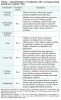
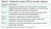
The International Society for Breath Odor Research established a method of classifying halitosis through scientific analyses.4,5 The classification system allows the dental team to identify causative factors and establish potential treatment protocols (Table 1 and Table 2). The significance of these categories and recommended path of treatment will assist the dental hygienist with treatment planning and prioritization of care.
Assessment
There are three primary assessment measurements for genuine halitosis5,6:
1. Organoleptic: a sensory test that is scored by a trained judge or clinician based on the perception of the judge or clinician
2. Gas chromatography: considered the method of choice for researchers, it makes a distinction between VSCs that contribute to halitosis and helps the clinician determine intra– or extra–oral origin
3. Sulfide monitoring: a portable device for monitoring VSCs. These monitors are better at measuring total VSCs instead of determining individual compounds
Organoleptic
There are several ways to perform this subjective measurement. One method is to insert a tube into the patient’s mouth and while the patient exhales slowly, the examiner smells from the other end of the tube. Often, confidentiality is maintained through use of a screen. The assessment can also be performed by scraping the posterior dorsum of the tongue with a spoon and smelling the contents. Various scoring systems have been designed, however, most are based on a numerical scale of 1 to 5, with 1 being barely noticeable odor and 5 being extremely foul odor.4,6 Morning appointments for assessment are preferred. Participants are encouraged to arrive without having had anything to eat or drink, performed oral hygiene or used perfume or tobacco products. Examiners are also encouraged to refrain from drinking coffee, tea or juice and abstain from using tobacco or perfume.5
Gas Chromatography
With this device, the measurement of VSCs can be obtained and differentiated with samples from saliva, tongue coating and breath. This assists in determining the origin of halitosis.6 Tangerman and Winkel state that without this device, extra–oral blood–borne halitosis may never have been identified.7 While it is a highly objective measurement device, it is expensive and not financially feasible for most dental practitioners. New, more affordable portable devices are being developed.6
Sulfide Monitoring
This portable monitor measures VSCs by an electrochemical reaction with sulfur compounds found within the breath, which is generated from a tube in the patient’s mouth. Electrical current that is generated is directly proportional to the levels of VSCs.6 The Halimeter® (Interscan Corporation, Chatsworth, Calif.) is the most recognized device for sulfide monitoring. Limitations include an inability to accurately estimate levels of dimethyl sulfide, the compound shown to be most evident in extra–oral halitosis.7 It is most sensitive for hydrogen sulfide and less sensitive for methyl mercaptan. Also, if VSCs are shown to be low by the monitor, it may not accurately determine halitosis when other factors are involved such as alcohols, phenyl compounds and polyamines.6
Other Methods
1. BANA test: an operator–friendly test that detects gram–negative anaerobes and short–chain fatty acids on the dorsum of the tongue. However, the specific role of different bacteria in the production of VSCs cannot be fully determined by this method.6
2. Chemical sensors: this gives the clinician the ability to measure VSCs from the periodontal pocket and on the tongue. A sulfide sensing element on the probe recognizes sulfide ions and measures their concentration.6
Another option for assessing halitosis is to have the patient report what they are experiencing. In a study to determine a patient’s ability to self–assess, researchers had patients self–evaluate using questionnaires and organoleptic scores and then compared them to more objective methods with a portable sulfide monitor. A significant correlation (p<0.001) was established between patient self–assessments and the Halimeter® results of VSC levels.8
Diagnosis
In order for the proper treatment and management of halitosis to occur, an accurate diagnosis must be obtained. Steps towards an accurate diagnosis include a thorough medical history complete with dietary analysis and identification of personal habits. The patient’s chief complaint should be understood and the dental and halitosis history, if any, recorded. Clinical observations of the tongue, teeth (including large carious lesions and faulty restorations),3 periodontal tissues and upper respiratory tract, along with a complete extra–oral exam, must be included as part of patient assessment.9 Once a thorough assessment has been completed, the dental hygienist can then classify the halitosis as genuine (extra–oral or intra–oral origins), pseudo or, in rare cases, as halitophobia. Understanding extra– oral and intra–oral origins is important for determining the appropriate course of treatment.
A study by Tangerman and Winkel7 was conducted to differentiate extra– and intra–oral halitosis. Analytical techniques were employed to identify the volatile compounds associated with odor and their emanating origin. The diagnostic tools used were full–mouth and nose organoleptic odor assessments using a 0 to 5 scale of VSCs by means of the Halimeter®, and the Winkel Tongue Coating Index (WTCI). Results showed clear distinctions in concentrations and location of VSCs between extra– and intra– oral halitosis. Subjects with intra–oral halitosis had odor stemming from the oral cavity but not the nose, whereas subjects with extra–oral halitosis had blood–borne odor that was measurable from both the mouth and nose. Dimethyl sulfide was the only malodorous compound found in significant levels for extra–oral halitosis, whereas methyl mercaptan and hydrogen sulfide were compounds most associated with intra–oral halitosis.
Intra–Oral Contributing Factors and Bacteria Associated with Halitosis
Intra–oral contributing factors account for 90% of cases of halitosis in dental patients3 and are most often evident upon arousing from sleep.10 This malodor is usually caused by low salivary flow, lack of oral hygiene and/or breathing through the mouth. However, for most individuals, the odor has no special significance, as it is resolved with brushing, flossing, eating and/ or drinking water.10 Patients who experience halitosis of intra–oral etiology, not resolved by simple personal hygiene habits, usually have an infection in the mouth (caries or periodontal disease)10 or other factors such as gross dental neglect, smoking or xerostomia.2
As previously discussed, VSCs are produced in the mouth by bacterial putrefaction, which is the breakdown of substances such as food debris, cells, saliva and blood by enzymes produced from the bacteria. Amino acids are metabolized through this process, creating malodorous gases. Most common compounds are hydrogen sulfide, methyl mercaptan and dimethyl sulfide.1,3,10 The most common bacteria to produce these compounds are gram–negative anaerobic bacteria, such as Porphyromonas gingivalis, Prevotella intermedia, Fusobacterium nucleatum, Bacteroides forsythus and Treponema denticola.1,10 Many sites harbor these bacteria, such as teeth, buccal mucosa, periodontal pockets, faulty restorations and removable partial dentures. However, the posterior dorsal surface of the tongue is considered the primary site in cases of halitosis.1,3,10
To determine the relationship of VSC concentrations to tongue coating and periodontal health, Lee et al11 used gas chromatography to sample mouth air prior to tongue scraping and after prophylaxis and tongue scraping. With a population of 40 subjects, mouth air was sampled for a baseline, and then tongue scraping was performed with another mouth air testing. This was followed by a prophylaxis with mouth air testing performed again, totaling three different times that mouth air was tested. Each tongue scraping was also evaluated by weight. The subjects were divided by levels of methyl mercaptan into high and low halitosis groups. VSC concentrations were higher in those with high methyl mercaptan levels prior to tongue scraping. However, both groups had decreased levels of methyl mercaptan after tongue scraping. Evaluation of periodontal health showed that 73% of the high methyl mercaptan group had one or more periodontal pockets greater than 4 mm as compared to 38.1% of the low methyl mercaptan group. Each group showed significant differences in all measurements except for tongue coating weight. It was concluded that the tongue is a strong contributor to halitosis by harboring bacteria that produce VSCs and that periodontal disease can also contribute to VSC production. This study supported earlier findings of Morita and Wang that intra–oral malodor and VSC levels significantly correlated with tongue coating and periodontal disease condition.12
Under magnification, the tongue is compared to the “surface of the moon after a rain shower.”2 Craters, fissures and peaks are covered with a fine sticky substance that harbors the malodorous bacteria. Researchers reported that a single epithelial cell in the oral cavity can harbor up to 25 bacteria, whereas one epithelial cell on the dorsum of the tongue can harbor up to 100 bacteria.13 Crevices of the tongue create an ideal environment for the bacteria to proliferate and produce VSCs. The dorsum of the tongue and its bacteria has been the subject of several studies.13-15
A microbiological analysis of the tongue was conducted on patients with and without halitosis by Donaldson et al.14 The experimental group (halitosis patients) and control group (patients without halitosis) had samples taken from the posterior dorsum of the tongue. The subjects with halitosis were classified using an organoleptic assessment and a Halimeter®. After incubation under anaerobic conditions, the samples were analyzed. Both groups had a predominant level of Veillonella, Prevotella and Fusobacterium species. The halitosis group, however, presented with more diverse species. Many of these species were unidentifiable gram–negative and gram–positive rods, along with gram–negative coccobacilli. The researchers concluded that halitosis is a result of multifaceted interactions between diverse species of bacteria.
The correlation between diverse, unidentifiable bacteria and halitosis was supported in a study designed to identify bacterial species on the tongue associated with halitosis.13 Researchers took samples from eight adult subjects with halitosis and five control subjects who did not have halitosis. The samples were taken from the dorsum of the tongue by scraping an area of about 2 square cm. Halitosis assessments occurred by organoleptic means and a portable sulfide detector. Bacterial species were identified, and the thickness and extent of tongue coating was determined. The results of the study indicated that both common and uncommon species were present in the experimental and control groups. Most prevalent bacterial species in both groups were Streptococcus salivarius, Prevotella melaninogenica, Streptococcus parasanguinis, Campylobacter concisus and Streptococcus mitis. However, bacteria were present in greater number and with greater diversity in the halitosis or experimental group. Among the bacteria identified, 32 were present solely in the halitosis group (84 bacterial species in the halitosis group, with 16 to 23 per subject, 69 in the control group, with 11 to 19 species per subject). Thirteen of these species were unidentifiable or uncultured. Solobacterium moorei was the key species unique to all halitosis subjects. S. moorei is a gram–positive bacteria first noted in human feces and has been linked to bacteremia, septicemia and refractory endodontic infections. The findings of this study confirmed the importance of the presence of specific bacterial species associated with halitosis and the differences between patients.
Riggio et al15 confirmed earlier findings of Donaldson et al.14 A diversity of bacteria, unidentifiable species and a greater abundance of bacteria were again identified in the halitosis subjects. The bacterial species Veillonella, Prevotella and Fusobacterium were identified in both test groups. However, no significant periodontal pathogens were observed. It was recommended that further studies investigate the process and amount of VSC production by individual bacterial species.
As the understanding of the role played by bacteria grows, researchers are examining other areas of the oral cavity as potential sources of VSCs. It has long been accepted that there is a link between periodontal disease and oral malodor.16,17 Hydrogen sulfide and methyl mercaptan, both associated with intra–oral halitosis, have been found to potentially facilitate the penetration of lipo–polysaccharide into the gingival epithelium, thus inducing inflammation.16 Hydrogen sulfide and methyl mercaptan are also thought to aid in bacterial invasion of the connective tissue by their toxic effects on epithelial cells.16 Methyl mercaptan was shown to hinder epithelial cell growth and production in a study conducted by Setoguchi et al.18 Researchers were surprised to find that gingival fibroblasts were left unaffected.
Recently, levels of VSCs were evaluated in 72 patients with chronic periodontitis to assess outcomes after tongue scraping, non–surgical periodontal therapy and oral hygiene instruction.19 Pre–treatment measurements were taken by organoleptic test scores and VSCs measured with the OralChroma™ device (Abilit Corp., Osaka City, Japan), a portable gas chromatograph. Periodontal examinations, along with full–mouth radiographs, were completed. Tongue scraping, non–surgical periodontal therapy and oral hygiene instructions, including the use of chlorhexidine rinse (CHX), were each followed with a VSC measurement. For each treatment, a progressive reduction of VSCs occurred during the course of the study. Hydrogen sulfide levels showed the most significant decrease after each treatment, whereas methyl mercaptan decreased only following tongue scraping and oral hygiene instructions that included rinsing with CHX. There was no correlation between pocket depth and concentrations of VSCs. The researchers concluded this contradiction with past research occurred because they measured VSCs indirectly versus measuring levels directly from the pocket. While tongue scraping alone produced the largest decrease of VSCs, the researchers concluded that tongue scraping in conjunction with periodontal therapy significantly reduced oral malodor.
A study was conducted by Awano et al20 to determine the relationship between periodontal disease–associated bacteria and oral malodor production. One hundred and one adults were classified into three groups: patients with halitosis and pocket depths greater than 4 mm, patients with halitosis without pocket depths greater than 4 mm and non–halitosis patients without pocket depths greater than 4 mm. Gas chromatography was used to evaluate hydrogen sulfide and methyl mercaptan concentration levels. Saliva was then collected from each subject to determine levels of periodontal pathogenic bacteria. Subjects with B. forsythus in saliva were shown to have higher levels of VSCs and more severe periodontal conditions compared to those without. Subjects with higher levels of P. gingivalis had higher levels of methyl mercaptan production. Actinobacillus actinomycetemcomitans and P. intermedia presence in saliva did not correlate with VSC production in subjects.
Extra–Oral Factors Contributing to Halitosis
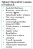
Approximately 10% of halitosis cases originate from systemic conditions or a location other than the oral cavity. Such cases are referred to as extra–oral halitosis.3,7 Therefore, the dental hygienist must be diligent in completing a thorough medical history to understand all possible origins.3 Possible systemic contributors associated with extra–oral halitosis are identified in Table 3.
Extra–oral halitosis can be further categorized by origin, either respiratory tract or blood–borne. Tangerman reported that upper and lower respiratory tract origins usually result from anaerobic infections, ulcerations and/or cancer. Confirmation of upper and lower respiratory tract halitosis is largely based on medical assessments of these systems.21 Infections of the respiratory tract create discharge from the nasal and sinus cavities, which in turn can contribute to halitosis and tonsillitis.1
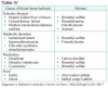
Table 4 presents the causes of extra–oral halitosis with blood– borne origins and the associated odorants.21 Odorants are produced in the blood and transported to the lungs. Pulmonary emissions of these odorants and their associated toxins are exhaled through the nose and mouth.2,21
Extra–oral halitosis from blood–borne sources may originate from any compound. However, the most identifiable odorant is dimethyl sulfide.2 For example, trimethylamine has been described as the substance contributing to Fish–odor Syndrome or Trimethylaminuria. This disorder is differentiated by greater than normal levels of trimethylamine in the body and is distinguished by the smell of rotting fish emanating from breath, sweat and urine.22 Gene mutations and the body’s inability to produce enzymes to break down the compound account for most cases. However, it has also been noted in individuals with kidney or liver disease, a small number of premature babies and, in a few cases, women at the start of menstruation. This condition should no longer be considered rare as more cases are being recognized.22
Moshkowitz et al studied the relationship of halitosis and upper gastrointestinal diseases.23 One hundred and thirty–two patients complaining of upper gastrointestinal symptoms were included in the study. Each patient completed a questionnaire that included questions about bad breath. The study was designed to measure the severity and presence of reflux and other gastrointestinal diseases. Subjects were then given an upper gastrointestinal endoscopy. The final diagnoses of these patients revealed no significant relationship or correlation between patient–perceived (self–assessed) halitosis and gastrointestinal diseases such as functional dyspepsia, peptic ulcer or Helicobacter pylori infection. However, there was a significant association between patient–perceived (self–assessed) halitosis and gastroesophageal reflux disease (GERD) (p=0.002). Researchers suggested that halitosis caused by GERD resulted from direct damage to the oropharyngeal mucosa, causing inflammation. While the study was limited to patient–perceived halitosis, the findings concluded that it is important to recognize halitosis as a symptom of GERD, and physicians and dental practitioners should consider it a manifestation of the disease.
In a case study by Murata et al,24 a 33–year–old Japanese woman’s chief complaint was bad breath of about 1 month duration. She had a previous diagnosis of asthma and had received periodic examinations. Medications for the treatment of asthma included suplatast tosilate administered after each meal for treatment of asthma. Her VSC levels were measured with a gas chromatograph. An attempt to remove intra–oral odor was completed with tooth brushing, flossing, inter–dental brushing and tongue cleaning. Prophylaxis by a dentist was completed twice a week for the first 2 weeks and then periodic check–ups were executed every 3 months. No disinfectants were used before measurements of VSC levels were obtained. The results showed levels of methyl mercaptan and hydrogen sulfide were significantly lower following treatment, but levels of dimethyl sulfide remained stable. The examiners suspected that dimethyl sulfide was a side effect of the asthma medications. Upon discontinuation of the medication, dimethyl sulfide was not detected. This case study emphasizes the need for the dental hygienist to recognize the extra–oral manifestations of halitosis, such as patient medications, so that referral to an appropriate physician occurs.
Treatment and Management Mechanical Reduction
As in most oral diseases, mechanical removal of biofilm and microorganisms is the first step in control of halitosis.9 Brushing and flossing of teeth are important, but tongue cleansing is paramount for halitosis reduction. It is estimated that approximately 60% of VSCs originate on the surface of the tongue.9,25 In a study conducted to compare the effects of polystyrene tongue scrapers and toothbrush bristles on the surface of the tongue against measurable VSCs, the tongue scraper performed at 75% reduction while the toothbrush bristles reduced levels of VSCs by 45%. Patients reported they preferred the tongue scraper over the toothbrush.26
In a Cochrane systematic review of tongue scrapers, researchers conducted a database search for randomized clinical trials.27 Researchers concluded that, although tongue scrapers produced a reduction in VSCs when compared to tooth brushing, they did not have a long–term effect and were only slightly more effective than tooth brushing alone. Limited evidence of tongue trauma with aggressive use was also reported.
Recently, manufacturers have included a tongue cleansing device on the back of toothbrush heads. Researchers wanted to determine if these devices were as effective as conventional tongue scrapers.28 Using a Halimeter® to score breath air and non–stimulated saliva for microbial analysis, it was determined both methods of cleansing the tongue were equally effective in reducing the number of bacteria on the tongue and VSCs.
To understand how different methods of oral hygiene reduced halitosis and VSC concentrations in morning breath, Faveri et al conducted a cross–over study of 19 volunteers who were divided into four groups.29 Baseline and end–of–study VSC concentrations were determined with a Halimeter®, and organoleptic scores were obtained. Assigned groups were given different oral hygiene regimens: Group I tooth brushing, Group II tooth brushing and inter–dental flossing, Group III tooth brushing and tongue scraping and Group IV tooth brushing, inter–dental flossing and tongue scraping. Subjects performed procedures three times a day for 7 days. Morning breath was evaluated again at the end of the study. The highest mean score for both measurements was found in the two groups that excluded tongue scraping. The two groups that included tongue scraping revealed a statistically significant difference from groups that did not use the tongue scraper (p<0.05). This confirmed prior research that the tongue is the recognized site for most VSC production and tongue scraping results in an improvement in breath quality.14,15
Chemotherapeutic Reduction
Toothpastes and mouth rinses have long been used to help reduce halitosis through chemotherapeutic reduction. The most common active ingredients included in these products are triclosan, essential oils, cetylpyridinium chloride (CPC) and CHX.9 Zinc, another active ingredient in mouthwash, has been shown to be effective by inhibiting bacterial breakdown of proteins, thus inhibiting VSC production.25 Chlorine dioxide solution (0.1% solution) has also been shown to maintain VSCs at lower levels when compared to a placebo mouth rinse.30
Roldan et al31 researched five different commercial mouth rinses, all containing CHX. Each product differed in concentration and additives including alcohol, sodium fluoride, zinc lactate and CPC. The researchers wanted to determine their efficacy in reducing salivary bacterial count and VSCs in expelled air. Methods included a randomized, double–blind, cross–over design and included un–stimulated saliva samples from subjects to determine bacterial count. Halitosis was measured by calibrated examiners with an organoleptic assessment of ratings on a 0 to 5 numerical scale. Bacterial count and VSC levels were recorded for each sampling of time and product used. Results showed that formulations of CHX combined with CPC attained the best results for reduction in both VSCs in expelled air and salivary bacterial count. CHX combined with sodium fluoride was the least effective of the formulations for both bacterial count and VSCs. CHX and zinc lactate had the best effect after 1 hour, but did not sustain this effect at the 5-hour mark. Inability to correlate the results with tongue coating indices was identified as a study limitation.
Thrane et al32 also tested a formula of zinc acetate and CHX in low concentrations against other existing formulations. Researchers hypothesized that the low concentrations would be more effective in reducing hydrogen sulfide in mouth air. The population sample included 10 healthy volunteers in a double–blind clinical study. Baseline hydrogen sulfide levels were standardized by first rinsing with a solution of L–cysteine. A mouth air sample was then obtained and analyzed by a gas chromatograph. The subjects were tested using different mouth rinses containing the following active ingredients: essential oils, CHX combined with CPC, triclosan, CPC alone, zinc gluconate and zinc acetate at 0.3% combined with CHX at 0.05%. Statistically significant results occurred in all 10 volunteers after using low levels of zinc acetate and CHX mouth rinse (p<0.05). The formula not only inhibited hydrogen sulfide, but continued to show reductions at the 3 hour mark. It was speculated that low concentrations of zinc acetate and CHX molecules provide sites for the sulfur ion to bind to. Subjects also reported fewer side effects such as discoloration, metallic taste and mucosal desquamation at the lower concentration level than when stronger concentrations were used.
A study was conducted by Fine et al33 to investigate the efficacy of either essential oil mouth rinse containing 0.09% zinc chloride as an anti–calculus agent (Tartar Control Listerine® Antiseptic) and a rinse containing 5% hydro–alcohol in controlling pathogens associated with halitosis. Baseline bacteria samples were obtained from subgingival buccal surfaces of posterior teeth and the dorsum of the tongue from all participants. All subjects were given an ADA-approved dentifrice and soft toothbrush to use during the trial. Subjects were examined 12 hours after the first rinse and again after 2 weeks of rinsing twice daily, with measurements taken 12 hours after the last rinse. The study was a randomized, double–blind, controlled crossover design. Bacterial samples were taken at the designated 12-hour marks for each time period. Results showed a statistically significant reduction in bacteria both on subgingival buccal surfaces and the dorsum of the tongue after the 12-hour mark of the first rinse containing essential oils and 0.09% zinc chloride (p<0.001). Reductions were even higher after 14 days of use.
A systematic review, published by Cochrane, compared the effectiveness of mouth rinses in controlling halitosis. Baseline characteristics, diversity of subjects and measurement methods prevented the possibility of a meta–analysis between chosen studies. However, the researchers concluded that mouth rinses containing CHX and CPC can inhibit production of VSCs, while mouth rinses containing chlorine dioxide and zinc may neutralize the sulfur compounds producing halitosis.34
A widely used ingredient in many oral health products is triclosan. It is lipid–soluble and recognized for its antibacterial and anti–plaque effects. It has also been acknowledged as having broad–spectrum effects on gram–negative microbes.6 When combined with copolymer, it adheres to soft and hard tissues for up to 12 hours.35
In a study to determine the effectiveness of a triclosan/copolymer/ disodium fluoride dentifrice (0.243%), Hu et al tested the dentifrice against an over–the–counter product containing 0.243% sodium fluoride.36 A 3-week, randomized, double–blind, longitudinal clinical trial was conducted. Organoleptic judges were calibrated to examine the subjects at 1.5, 4 and 12 hours after subjects used their assigned toothpaste. This evaluation was followed each week for 3 weeks to assess odor scores. There was no difference in baseline scores for the two groups. Breath odor scores showed a statistically significant reduction for the triclosan dentifrice of 87.8% to 97.6% at each examination. Percentage ranges for the dentifrice containing only sodium fluoride were 0% to 10%. Researchers concluded that the triclosan/copolymer/sodium fluoride dentifrice reduced oral malodor for up to 12 hours.
A Combined Therapeutic Approach
In an effort to explore combined therapeutic approaches, Roldan et al aimed to treat halitosis by evaluating a mechanical and chemotherapeutic protocol.37 Nineteen patients were followed for 3 months and evaluated with organoleptic and VSC level assessments, tongue coating indices, periodontal variables, bacterial ratios in oral niches and subgingivally and bacterial flora of the saliva and tongue. Treatment for each patient included a prophylaxis, oral hygiene instructions that included tongue scraping and use of a mouth rinse that contained CHX, CPC and zinc lactate. Variables were measured at 1 month and 3 months after the baseline. Results showed that periodontal and halitosis pathogens were reduced at both the 1- and 3-month measurements. Of the microflora evaluated, P. gingivalis was most affected. Mean probing depths and plaque levels decreased significantly after 3 months. Tongue coating indices were reduced significantly along with organoleptic scores (p<0.001) and VSC levels (p<0.05). Researchers concluded and results demonstrated that oral halitosis can be managed.
Clinical Application
The scenario is familiar. A patient enters the dental clinic, hoping for answers to questions that may be difficult to ask: “Do I have bad breath?” and “What can I do about it?” The clinician, in a confident and professional manner, needs to then follow established evidence–based protocols to help the patient.
Ideally, the first step is to establish the origin of the malodor. A thorough medical history along with diet and medications needs to be confirmed.1 Intra–oral and extra– oral halitosis have different treatment protocols with distinguishable VSCs.4,5 However, not all clinicians have access to instruments that document VSCs or exact levels. The organoleptic assessment is the most common method to evaluate halitosis,3 and the research shows that patients are even capable of scoring their own malodor.8 An assessment taken in the morning before eating and oral hygiene procedures is best.4-6
When the clinician follows treatment protocols established by the International Society for Breath Odor Research (Table 2), all patients are instructed in correct oral hygiene habits, including the important step of tongue cleansing.29 Beyond the patient’s ability to cleanse the teeth and tongue, researchers recommend an oral prophylaxis as an important step in mechanical removal of causative volatiles and bacteria and control of halitosis.1-3,9,38 If either hard tissue or periodontal diseases are present, they must be treated as contributors of halitosis. In addition, faulty restorations should be replaced.3,5,38 Chemotherapeutics have demonstrated effectiveness as an adjunct to therapy. Based on current research, a dentifrice with triclosan36 can be recommended along with a mouth rinse that would contain either CHX or CPC to inhibit production of VSCs or chlorine disodium oxide and zinc to neutralize the sulfur compounds.34
If the halitosis is not resolved with the above–mentioned measures, additional assessment needs to be conducted to determine if it is extra–oral and/or blood–borne halitosis. The best method for this is to use gas chromatography as it distinguishes between hydrogen sulfide, methyl mercaptan and dimethyl sulfide.6 Dimethyl sulfide is the VSC most associated with extra–oral halitosis.2,7,21 If measured in high levels, with the reviewed health history considered, the patient would be referred to a physician for further evaluation.7 In rare cases, pseudo–halitosis can be resolved with education, and those patients exhibiting halitophobia will need to be referred to a therapist.4,5,38,39
Conclusion
Fifty percent of the public worldwide suffers from some form of oral halitosis and is looking to the oral health care professional for guidance. Upon satisfactory completion of treatment for halitosis, research has shown that patients recognize an improvement in social life and satisfaction of care.40 Since halitosis is a recognizable condition, and a common chief complaint among patients,3 the clinician should be prepared to diagnose, classify, treat and manage patients that suffer from this uncomfortable and sometimes socially debilitating condition.
About the Author
Brenda L. Armstrong, RDH, MDH
Adjunct Clinical Instructor, University of Minnesota. She is a recent graduate of the Master of Dental Hygiene Program at the University of Minnesota School of Dentistry, Minneapolis, Minnesota
Michelle L. Sensat, BSDH, MS
Adjunct Assistant Professor, Division of Dental Hygiene, University of Minnesota School of Dentistry, Minneapolis, Minnesota
Jill L. Stoltenberg, BSDH, MA, RF
Associate Professor, Division of Dental Hygiene, University of Minnesota School of Dentistry, Minneapolis, Minnesota
References
1. Porter SR, Scully C. Oral malodour 1. (halitosis). BMJ. 2006;23;333(7569):632–635.
2. Lee SS, Zhang W, Li Y. Halitosis update: A review of causes, diagnoses, and treatments. J Calif Dent Assoc. 2007;35(4):258–260, 262, 264–268.
3. ADA Council on Scientific Affairs. Oral malodor. J Am Dent Assoc. 2003;134(2):209–214.
4. Murata T, Yamaga T, Iida T, Miyazaki H, Yaegaki K. Classification and examination of halitosis. Int Dent J. 2002;52(Suppl 3):181–186.
5. Yaegaki K, Coil JM. Examination, classification, and treatment of halitosis; clinical perspectives [see comment]. J Can Dent Assoc. 2000;66(5):257–61.
6. van den Broek AM, Feenstra L, de Baat C. A review of the current literature on aetiology and measurement methods of halitosis. J Dent. 2007;35(8):627–635.
7. Tangerman A, Winkel EG. Intra– and extra–oral halitosis: Finding of a new form of extra–oral blood–borne halitosis caused by dimethyl sulphide. J Clin Periodontol. 2007;34(9):748–755.
8. Iwanicka–Grzegorek E, Michalik J, Kepa J, Wierzbicka M, Aleksinski M, Pierzynowska E. Subjective patients’ opinion and evaluation of halitosis using halimeter and organoleptic scores. Oral Dis. 2005;11(Suppl 1):86–88.
9. van den Broek AM, Feenstra L, de Baat C. A review of the current literature on management of halitosis. Oral Dis. 2008;14(1):30–39.
10. Scully C, Felix DH. Oral medicine––update for the dental practitioner: Oral malodour. Br Dent J. 2005;199(8):498–500.
11. Lee CH, Kho HS, Chung SC, Lee SW, Kim YK. The relationship between volatile sulfur compounds and major halitosis–inducing factors. J Periodontol. 2003;74(1):32–37.
12. Morita M, Wang HL. Relationship between sulcular sulfide level and oral malodor in subjects with periodontal disease. J Periodontol. 2001;72(1):79–84.
13. Haraszthy VI, et al. Identification of oral bacterial species associated with halitosis. J Am Dent Assoc. 2007;138(8):1113–1120.
14. Donaldson AC, et al. Microbiological culture analysis of the tongue anaerobic microflora in subjects with and without halitosis. Oral Dis. 2005;11(Suppl 1):61–63.
15. Riggio MP, et al. Molecular identification of bacteria on the tongue dorsum of subjects with and without halitosis. Oral Dis. 2008;14(3):251–258.
16. Morita M, Wang HL. Association between oral malodor and adult periodontitis: A review. J Clin Periodontol. 2001;28(9):813–819.
17. Sanz M, Roldán S, Herrera D. Fundamentals of breath malodour. J Contemp Dent Pract. 2001;2(4):1–17.
18. Setoguchi T, et al. The effects of methyl mercaptan on epithelial cell growth and proliferation. Int Dent J. 2002;52(Suppl 3):241–246.
19. Tsai CC, et al. The levels of volatile sulfur compounds in mouth air from patients with chronic periodontitis. J Periodont Res. 2008;43(2):186–193.
20. Awano S, Gohara K, Kurihara E, Ansai T, Takehara T. The relationship between the presence of periodontopathogenic bacteria in saliva and halitosis. Int Dent J. 2002;52(Suppl 3):212–216.
21. Tangerman A. Halitosis in medicine: A review. Int Dent J. 2002;52(Suppl 3):201–206.
22. Mitchell SC. Trimethylaminuria (fish–odour syndrome) and oral malodour. Oral Dis. 2005;11(Suppl 1):10–13.
23. Moshkowitz M, Horowitz N, Leshno M, Halpern Z. Halitosis and gastroesophageal reflux disease: a possible association. Oral Dis. 2007;13(6):581–585.
24. Murata T, Fujiyama Y, Yamaga T, Miyazaki H. Breath malodor in an asthmatic patient caused by side–effects of medication: A case report and review of the literature. Oral Dis. 2003;9(5):273–276.
25. Yaegaki K, Coil JM, Kamemizu T, Miyazaki H. Tongue brushing and mouth rinsing as basic treatment measures for halitosis. Int Dent J. 2002;52(Suppl 3):192–196.
26. Pedrazzi V, Sato S, de Mattos Mda G, Lara EH, Panzeri H. Tongue–cleaning methods: A comparative clinical trial employing a toothbrush and a tongue scraper. J Periodontol. 2004;75(7):1009–1012.
27. Outhouse TL, Fedorowicz Z, Keenan JV, Al–Alawi R. A cochrane systematic review finds tongue scrapers have short–term efficacy in controlling halitosis. Gen Dent. 2006;54(5):352–359, 360, 367–368.
28. Casemiro LA, Martins CH, de Carvalho TC, Panzeri H, Lavrador MA, Pires–de–Souza Fde C. Effectiveness of a new toothbrush design versus a conventional tongue scraper in improving breath odor and reducing tongue microbiota. J Appl Oral Sci. 2008;16(4):271–274.
29. Faveri M, Hayacibara MF, Pupio GC, Cury JA, Tsuzuki CO, Hayacibara RM. A cross–over study on the effect of various therapeutic approaches to morning breath odour. J Clin Periodontol. 2006;33(8):555–560.
30. Peruzzo DC, Jandiroba PF, Nogueira Filho Gda R. Use of 0.1% chlorine dioxide to inhibit the formation of morning volatile sulphur compounds (VSC). Braz Oral Res. 2007;21(1):70–74.
31. Roldán S, Herrera D, Santa–Cruz I, O’Connor A, González I, Sanz M. Comparative effects of different chlorhexidine mouth–rinse formulations on volatile sulphur compounds and salivary bacterial counts. J Clin Periodontol. 2004;31(12):1128–1134.
32. Thrane PS, Young A, Jonski G, Rölla G. A new mouthrinse combining zinc and chlorhexidine in low concentrations provides superior efficacy against halitosis compared to existing formulations: a double–blind clinical study. J Clin Dent. 2007;18(3):82–86.
33. Fine DH, Furgang D, Sinatra K, Charles C, McGuire A, Kumar LD. In vivo antimicrobial effectiveness of an essential oil–containing mouth rinse 12 h after a single use and 14 days’ use. J Clin Periodontol. 2005;32(4):335–340.
34. Fedorowicz Z, Aljufairi H, Nasser M, Outhouse TL, Pedrazzi V. Mouthrinses for the treatment of halitosis. Cochrane Database Syst Rev. 2008;(4):CD006701.
35. Ciancio SG. Improving our patients’ oral health: The role of a triclosan/copolymer/fluoride dentifrice. Compend Contin Educ Dent. 2007;28(4):178–180, 182–183.
36. Hu D, Zhang YP, Petrone M, Volpe AR, DeVizio W, Proskin HM. Clinical effectiveness of a triclosan/ copolymer/sodium–fluoride dentifrice in controlling oral malodor: A three–week clinical trial. Compend Contin Educ Dent. 2003;24(9 Suppl):34–41.
37. Roldán S, Herrera D, O’Connor A, González I, Sanz M. A combined therapeutic approach to manage oral halitosis: A 3–month prospective case series. J Periodontol. 2005;76(6):1025–1033.
38. Coil JM, Yaegaki K, Matsuo T, Miyazaki H. Treatment needs (TN) and practical remedies for halitosis. Int Dent J. 2002;52(Suppl 3):187–191.
39. Yaegaki K, Coil JM. Genuine halitosis, pseudo– halitosis, and halitophobia: Classification, diagnosis, and treatment. Compend Contin Educ Dent. 2000;21(10A):880–886, 888–889, 890.
40. Kishi M, Abe A, Yonemitsu M. Relationship between the SF–36 questionnaire and patient’s satisfaction following halitosis therapy.